10
Impact Factor
ISSN: 1449-2288
Int J Biol Sci 2020; 16(2):342-352. doi:10.7150/ijbs.38112 This issue Cite
Research Paper
HCRP-1 regulates cell migration, invasion and angiogenesis via Src/ FAK signaling in human prostate cancer
1. Cancer Institute, Xuzhou Medical University, Xuzhou, Jiangsu, China;
2. Jiangsu Center for the Collaboration and Innovation of Cancer Biotherapy, Cancer Institute, Xuzhou Medical College, Xuzhou, Jiangsu, China;
3. The Affiliated Hospital of Xuzhou Medical College, Xuzhou, Jiangsu, China.
*These authors contributed equally to this work.
Received 2019-7-3; Accepted 2019-9-28; Published 2020-1-1
Abstract

Prostate cancer (PCa) is the third leading malignancy engendering mortality among men globally. The present study aimed at determining the expression of hepatocellular carcinoma-related protein-1 (HCRP-1) in PCa, to explore its potential role in prostate tumorigenesis in vitro and in vivo. We evaluated HCRP-1 protein with immunohistochemistry (IHC) technology and found HCRP-1 expression was significantly low in PCa tissues (PCTs); In addition, the decreased HCRP-1 was significantly associated with TNM (tumor node metastasis) stage, advanced histology grade and gleason score. Transwell, tube formation, Western blot and co-immunoprecipitation (Co-IP) assays were utilized to determine the role of down-regulating HCRP-1 in PCa cell migration, invasion and angiogenesis. Meanwhile, we found HCRP-1 depletion induced Src and focal adhesion kinase (FAK) phosphorylation, which could be reversed by Src inhibitor PP2 or FAK inhibitor. Furthermore, down-regulated HCRP-1 evidently induced lung metastasis of PCa cells in xenograft mode. Taken together, our study indicates HCRP-1 plays an important role in PCa metastasis. HCRP-1 may serve as a potential therapeutic target for PCa.
Keywords: HCRP-1, prostate cancer, migration, invasion, angiogenesis
Introduction
Prostate cancer (PCa) remains the third leading malignancy engendering mortality among men globally. Metastasis is the leading cause for the death of PCa patients, the 5-year survival rate is merely 29% [1].During metastasis process, cancer cells invade into neighboring tissue or lymphatic circulation, blood circulation and survive to form distant metastases[2]. To better understand the mechanistic basis of cancer metastasis, identifying the critical molecules involved in metastasis is necessary.
HCRP-1 is identified as a member of the human endosomal sorting complex required for transport-1 (ESCRT-I) families. It can bind and sort of ubiquitinated receptors to intraluminal vesicles of MVBs (multivesicular bodies), and then degrade them in lysosomes [3, 4]. HCRP-1 has been implicated in cancer progression. HCRP-1 was firstly found significantly lost in hepatocellular cell carcinoma (HCC) tissues but abundant in normal liver tissues [5]. Loss of HCRP-1 is associated with the activation of EGFR (epidermal growth factor receptor), which exerts an influence on EGFR prognosis. Down-regulated HCRP-1 leads to invasive phenotype of ovarian cells and tumor growth in vivo [6]. In addition, it is proved to be of adverse prognostic significance that oropharyngeal squamous cell carcinoma (OOSCC) patients with less HCRP-1 expression received preoperative chemoradiotherapy [7]. Our previous study showed that HCRP-1 was decreased in colorectal cancer (CRC). Reduced HCRP-1 expression caused CRC cancer cells anoikis resistance and surviving cells formed metastasis and resulted in poor prognosis in CRC [8]. These studies indicate that HCRP-1 acts as a tumor suppressor in cancer progression.
In this study, we investigated the relationship between HCRP-1 expression and clinicopathologic features by PCa tissue microarray (TMA). Furthermore, we showed a role for HCRP-1 in regulation of migration, invasion and angiogenesis in human PCa cells. Lastly, our data provided a profound understanding of novel HCRP-1 - Src/FAK signaling in promoting PCa migration, invasion and angiogenesis.
Material and Methods
Patients and samples
A PCa TMA was purchased from Shanghai OUTDO Biotech CO., LTD. (China). Tumors were staged and graded based on 2010 revised TNM system and WHO criteria, respectively [9]. The clinicopathological characteristics of TMA are described in Table 1. Moreover, 90 cases of PCTs were provided.
HCRP-1 staining and patients clinicopathological characteristics.
| Variables | HCRP-1 staining | |||
|---|---|---|---|---|
| Negative(%) | Positive(%) | Total | P-value | |
| Cancer cases | 52 (57.8) | 38 (42.2) | 90 | <0.001 |
| Latero-cancer tissue | 21 (23.3) | 69 (76.7) | 90 | |
| Age | 0.805 | |||
| ≤ 70 years | 26 (50.1) | 18 (40.9) | 44 | |
| >70 years | 26 (56.5) | 20 (43.5) | 46 | |
| Histopathologic Grade | 0.014 | |||
| G1-2 | 13 (40.6) | 19 (59.4) | 32 | |
| G3-4 | 39 (67.2) | 19 (32.8) | 58 | |
| Gleason score | 0.045 | |||
| ≤ 7 | 14 (43.8) | 18 (56.2) | 32 | |
| >7 | 38 (65.5) | 20 (34.5) | 58 | |
| TNM stage | 0.022 | |||
| I-II | 29 (49.2) | 30 (50.8) | 59 | |
| III-IV | 23 (74.2) | 8 (25.8) | 31 | |
*P values are from χ2 test
Antibodies and Reagents
Rabbit monoclonal antibodies to EGFR, p-EGFR (Tyr1173), FAK, p-FAK (Tyr397), Src, p-Src (Tyr416) and CD31 were purchased from Cell Signaling Technology (Beverly, MA, USA). Antibody HCRP-1 was from Proteintech (Wuhan, China). Antibodies against GAPDH were from Santa Cruze (CA, USA). The p-FAK inhibitor, FAK inhibitor 14 and Src inhibitor, PP2 were from Abcam (Cambridge, MA, USA).Cycloheximide (CHX) were ordered from Sigma (Shanghai, China).
Immunohistochemistry
The TMA slide or tissues were incubated with primary antibodies overnight at 4°C, and visualization with diaminobenzidine (DAB; sigma, Shanghai, China). The image was collected by light microscopy (Olympus BX-51 light microscope). The evaluation of HCRP-1 in TMA was performed as previously described [8].
Cell Culture
Human PCa cell lines PC3 and LnCap were cultured in RPMI1640 medium enriched with 10% fetal calf serum (Invitrogen, Shanghai, China). Cells were cultured in a 37℃ humidified incubator with 95% air, 5% CO2.
Transfection and PC3 stable cell line generation
HCRP-1 siRNA were ordered from GenePharma (Shanghai, China). The sequences are as follows: HCRP-1,5′-GACACUGUUUCUUCUUCAACA-3′. Transient transfections were performed using silentFect Lipid Reagent (Bio-Rad, Hercules, CA, USA) following the manufacturer's instructions. GFP-lentivirus-packing HCRP-1 shRNA vector (IBS, Shanghai, China) infectedPC3 cells for two days, and then screened out with 2 μg/ml puromycin (Sigma, Shanghai, China) for 14 days.
Angiogenesis assay
The medium from transfected PC3 and LnCap cells was collected and used as a conditioned medium. 2×10⁴ human umbilical vein endothelial cells in 100 μl conditioned medium were seeded in the Matrigel pre-coated 48-well plate. Three randomly chosen fields were selected to photograph the number of capillary-like tubes using a Nikon inverted microscope (Nikon, Japan).
Cell migration and invasion assay
Cell migration and invasion assay were performed as described previously [10]. Modified two chamber plates with a pore size of 8 μm were employed. The transwell filter that inserts without Matrigel (BD Biosciences) coating was used for migration assay. The transwell filter that coated with Matrigel (BD Biosciences) was used for invasion assay.
Western blot analysis
After transfection, cells were harvested and cell extracts were separated on SDS-polyacrylamide gel. The proteins were blotted on nitrocellulose membrane and incubated with appropriate primary antibodies overnight at 4°C. Secondary antibodies included horseradish peroxidase (HRP)-goat anti-mouse and HRP-goat anti-rabbit (ZSGB-BIO). Protein bands were detected by Tanon5200 automatic chemiluminescence imaging analysis system using ECL reagent (Tanon, shanghai, China).
Co-immunoprecipitation
PC3 cells were lysed and the cell lysates were precleared with mouse normal IgG and protein G-agarose for 30 min. Then, HCRP-1 and HCRP-1-specific IgGs were incubated with the precleared mixture overnight. Then, the immunoprecipitates were washed with cold lysis buffer for five times and applied to Western blotting assay.
In vivo experimental metastasis models
Each group consists of eight male nude mice. 1.0×10⁶ shHCRP-1 stably expressed PC3 cells were injected with mice via tail vein. 8 weeks post-implantation, mice were euthanized and lung tissues were fixed in 10% formaldehyde. HE stained was applied for metastatic nodules. Bioluminescence imaging of the mice were analyzed using Xenogen IVIS200 imaging system (Xenogen Corp.). Animal experiments were performed following protocols approved by the Committee of Xuzhou Medical University for the Use and Care of Animals.
Subcutaneous in vivo experiments
To explore tube formation in vivo, male nude mice were injected with 1.0×10⁶ shHCRP-1 stably expressed PC3 cells in 100 μl PBS/Matrigel (50:50) subcutaneously. The body weight was monitored and tumor volume was calculated by vernier caliper every day. 8 weeks after implantation, mice were euthanized and tumors were surgically removed and fixed in 10% formaldehyde for histology.
Statistical analysis
We used SPSS version 17.0 for Windows (SPSS Inc., Chicago, IL) for all analyses. Data are presented as means ± SD. Dunnett's t test (two-factor) was employed to evaluate differences within treatment groups. HCRP-1 staining in PCa TMA and clinicopathologic parameters was evaluated by χ2 test. A P-value equal to or <0.05 was considered statistically significant.
Results
The expression of HCRP-1 is reduced in human PCa
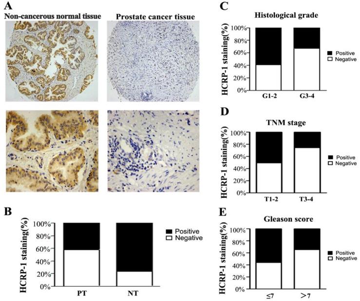
To determine HCRP-1 expression in human PCa, we used TMA slide containing PCTs and paired NTs for immunohistochemistry staining. Data showed that HCRP-1 was mainly localized in the cytoplasmic (Fig. 1A). In NTs, strong cytoplasmic staining was observed in normal prostate epithelia. 76.7% (69 of 90 cases) patients showed positive HCRP-1 staining. In PCTs, strong expression of HCRP-1 was recorded in 42.2% (38 of 90 cases) (Fig. 1B). The expression of HCRP-1 was significant reduced in the PCTs when compared with NTs (P<0.001, χ2 test).
HCRP-1 expression with clinicopathological parameters
We next investigated the correlation between HCRP-1 staining and clinicopathological features in PCa (Table 1). We found that loss of HCRP-1 was significantly correlated with histological grade when compared I-II with III-IV (P<0.05, Fig. 1C). Since TNM stage is a critical prognostic marker for cancer patients, we examined whether reduced HCRP-1 expression correlates with TNM stage. Data displayed that HCRP-1 expression was dramatically reduced in late TNM stages (III-IV) when compared with early stages (I-II) (P<0.05, Fig. 1D). Moreover, decreased HCRP-1 expression was significantly decreased in high gleason score (>7) (P<0.05, Fig. 1E). However, there is no significant correlation between HCRP-1 expression andage.
Loss of HCRP-1 promotes PCa cells migration, invasion and angiogenesis
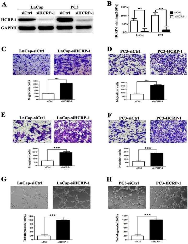
To investigate the effect of HCRP-1 on cell phenotypes, we transiently transfected PC3 and LnCap cells with control siRNA and HCRP-1 siRNA to inhibit HCRP-1 protein levels (Fig. 2A and B). We then assessed whether alteration of HCRP-1 affected the malignant characteristics of the prostate cancer cell lines. We first detected the migration ability of cancer cells and data showed that the thecapacity of cell migration was drastically promoted after knockdown of HCRP-1 expression (Fig. 2C and D). The results from the cell invasion assay were corresponded with the cell migration assay (Fig. 2E and F). Moreover, we evaluated the effect of decreased HCRP-1 expression on angiogenic potential of human PCa cells. As shown in Figure 2G and H, the number of HUVECs formed tubular structures was obviously increased in conditioned medium from HCRP-1 knockdown cancer cells.
Correlation between HCRP-1 expression and clinicopathologic parameters in prostate cancer. A. Representative immunohistochemical photographs were taken at different magnifications in NTs and PCTs (Top panel ×100, bottom panel ×400). B. Compared with that in the tumor adjacent normal prostate tissue, the overall expression level of HCRP-1 in the prostate cancer tissues was significantly lower (P<0.001, χ2 test). C. Decreased HCRP-1 expression was correlated with histological grade (P<0.05, χ2 test, comparing I-II versus III-IV). D. Decreased HCRP-1 expression was correlated with TNM stage (P<0.05, χ2 test, comparing T1-2 versus T3-4). E. Decreased HCRP-1 expression was correlated with Gleason score (P<0.05, χ2 test, comparing≤7 versus >7).
HCRP-1 regulates Src/FAK signaling pathway in PCa cells
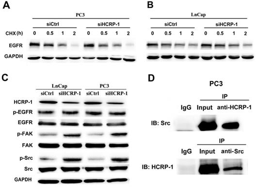
HCRP-1 mediates the internalization and degradation of EGFR. We detected the half-life of EGFR to clarify whether the stability of EGFR exerted influence oncells metastasis phenotypes in HCRP-1 knockdown PCa cells. We observed that the stability was sustained in both two cell lines (Fig. 3A, B). Ineffective receptor sorting could lead to phosphorylated EGFR receptor accumulation in cancer cells. We explored that whether HCRP-1 might regulate EGFR activation. Our data showed that phosphorylation of EGFR remains unchanged (Fig. 3C). It has been well studied that Src/FAK signaling plays an important role in cell migration, invasion and angiogenesis. Therefore, we detected the phosphorylation of Src and FAK in cells transfected with control siRNA and HCRP-1 siRNA. The results showed that knockdown of HCRP-1 expression induced Src and FAK phosphorylation (Fig. 3C). We further performed immunoprecipitation experiments in PC3 cells to investigate the interaction of HCRP-1 and Src. The results showed that HCRP-1 or Src immunoprecipitates were associated with endogenous Src or HCRP-1 protein (Fig. 3D), confirming the specific association between HCRP-1 and Src. There results suggest that HCRP-1 may regulate Src/FAK signaling pathway via interaction of HCRP-1 and Src.
Inhibition of HCRP-1 promotes cells migration, invasion and angiogenesis through Src/FAK signaling pathway
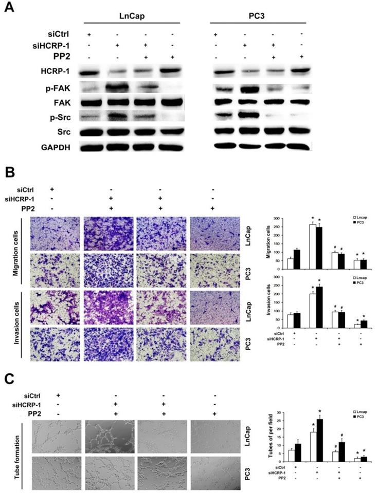
To further investigate whether HCRP-1 regulates cells migration, invasion and angiogenesis through Src/FAK signaling pathway, we firstly added the Src-specific inhibitor, PP2 (5 μM), into PCa cells that was transfected with HCRP-1 siRNA. Western blot data indicated that PP2 obviously suppressed Src and FAK phosphorylation in HCRP-1-knockdown cells (Fig. 4A). The migration and invasion cells were obviously decreased in HCRP-1-deficient cells treated with PP2 (Fig. 4B). In addition, the angiogenesis of PC cells was determined by endothelial cell tube formation assay. The number of HUVECs formed tubular structures was obviously increased in conditioned medium from HCRP-1 knockdown cancer cells. However, this induction was suppressed by Src-specific inhibitor PP2 (Fig. 4C).
Effect of the reduction in HCRP-1 expression on the abilities of cell migration, invasion and angiogenesis in vitro. A,B. Forty-eight hours after transfection, the expression of HCRP-1 in the PC3 and LnCap cells was evaluated by western blotting GAPDH was used as an internal control. C-F. Cell migration and matrigel cell invasion assays were performed in PC3 and LnCap cells after transfection, respectively. Representative fields of migrating or invading cells on the membrane (magnification, ×200). G,H. Representative pictures were taken in situ for tube formation in the supernatant of PC3 and LnCap cells transduced with siRNA foe HCRP-1 or control. The data are presented as mean±SD for triplicate determinations. *** P<0.001.
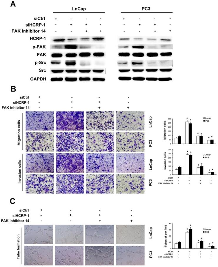
To further explore the functions of Src/FAK signaling pathway in regulating cell migration, invasion and angiogenesis by HCRP-1, we also employed FAK-specific inhibitor 14 (100 mM) to inhibit FAK phosphorylation. Our data testified to that FAK-specific inhibitor 14 lead to the reduction of FAK phosphorylation which was induced by knockdown of HCRP-1 (Fig. 5A). The migration and invasion abilities were suppressed by treatment of PC3 and LnCap cells with FAK-specific inhibitor 14 (Fig.5B). Subsequently, we also found that angiogenesis ability was inhibited by FAK-specific inhibitor 14 (Fig.5C). These data suggest that Src/FAK signaling is involved in the promotion of PCa cancer cells migration, invasion and angiogenesis induced by depletion of HCRP-1.
Endogenous interaction of HCRP-1 and Src contributes to Src/FAK signaling pathway. A,B. Cells were treated CHX for 0, 0.5 h, 1h, 2h after transfected with si-HCRP-1 or control siRNA for 48 h, lysates obtained from these cells were submitted to Western blot detection for the protein expression of EGFR. C. Activities of EGFR, Src and FAK were determined by using Western blot analysis with antibodies specific for total and phosphorylated forms of EGFR, Src and FAK in silencing of HCRP-1 and control group for both PC3 and LnCap cell lines. D. Src were coprecipitated with HCRP-1 in whole-cell lysates of PC3 cells.
Depletion of HCRP-1promoted prostate cancer tumorigenesis, angiogenesis and metastasis in vivo
To evaluate a role for HCRP-1 in tumor metastasis in vivo, an experimental metastasis assay was performed with luciferase-expressing PC3 cells infected with scrambled control or shRNA against HCRP-1. Two months later, a statistical difference was noticed that PCa cells with the HCRP-1-knockdown had a higher signal in the lungs compared with the scrambled control (Fig. 6A). Then, the mice were executed to analyze the lung metastasis status. As shown in Fig. 6B-D, more multiple large metastatic foci in lung were observed in the samples containing PC3-shHCRP-1 cells. Immunohistochemistry staining of metastatic nodules in lungs showed that p-Src and p-FAK expression in the HCRP-1-knockdown group was high compared with the control group (Fig. 6E). Next, we employed xenograft models using nude mice subcutaneously transplanted with PC3 cells that were mock treated or depleted of HCRP-1 to evaluate the effect of HCRP-1 on tumor growth in vivo. As shown in Fig. 6F and G, HCRP-1 knockdown cells formed bigger tumors than the control. Also, the expression levels of HCRP-1, p-Src, p-FAK and CD31 were high in the HCRP-1 knockdown subcutaneous tumors (Fig. 6H). Next, these findings provided compelling evidence for the ability of HCRP-1 to confer tumorigenesis and metastasis in vivo.
Discussion
In our work, we provide a set of data suggesting critical roles for HCRP-1 in PCa angiogenesis and metastasis. First, it was found that in human PCa patients, low HCRP-1 expression correlates/is associated with/related to clinicopathologic parameters, revealing HCRP-1 as a novel candidate tumor-suppressor gene in PCa (Fig. 1). Second, evidence was presented, demonstrating a supporting role for HCRP-1 in prostate cancer cell migration, invasion and angiogenesis (Fig. 2). Third, HCRP-1 was shown to regulate cell migration, invasion and angiogenesis through interacting with Src/FAK signaling (Fig. 3-5). Finally, the results of mouse xenograft experiments revealed a role for HCRP-1 in promoting prostate cancer angiogenesis and metastasis in vivo (Fig. 6).
The vital role for HCRP-1 expression in cancer development has been reported in several malignancies, such as ovarian cancer, HCC and breast cancer [5, 6, 11]. Low HCRP-1 mRNA expression was independently associated with shorter disease-free survival in HCC and breast cancer [5]. We previous found that RCC patients were significantly correlated with decreased overall survival only in tumors with low or missing HCRP-1 expression [10]. In PCTs, we identified decreased expression of HCRP-1 protein in cancer tissues. Moreover, we explored the correlation between HCRP-1 staining and clinicopathological features. Our results displayed that loss expression of HCRP-1 occurred in most of PCa patients, and this reduced expression had significantly correlation with histological grade, TNM stage and Gleason score. Collectively, these observations suggest that HCRP-1 have a potential effect on PCa progression and metastasis.
Inhibition of Src activation blocked HCRP-1-induced cell migration, invasion and angiogenesis. A. PC3 and LnCap cells were pretreated with HCRP-1 siRNA and the Src inhibitor, PP2. The whole cell lysates were analyzed for the protein levels of p-Src, Src, p-FAK, FAK and GAPDH by Western blot. B. Cell migration and Matrigel cell invasion were evaluated after HCRP-1 siRNA treatment and treatment with PP2 (5 μM). Representative fields of migrating or invading cells on the membrane (magnification, ×200). C. Tube formation was explored in the supernatant of PC3 and LnCap cells. The data are presented as mean±SD for triplicate determinations. *P<0.05 versus siCtrl group; #P<0.05 versus siHCRP-1 group.
Inhibition of FAK activation blocked HCRP-1-induced cell migration, invasion and angiogenesis. A. PC3 and LnCap cells were pretreated with HCRP-1 siRNA and the FAK inhibitor, FAK inhibitor 14(200mM). The whole cell lysates were analyzed for the protein levels of p-Src, Src, p-FAK, FAK and GAPDH by Western blot. B. Cell migration and Matrigel cell invasion were evaluated after HCRP-1 siRNA treatment and treatment with FAK inhibitor 14. Representative fields of migrating or invading cells on the membrane (magnification, ×200). C. Tube formationwas explored in the supernatant of PC3 and LnCap cells. The data are presented as mean±SD for triplicate determinations. *P<0.05 versus siCtrl group; #P<0.05 versus siHCRP-1 group.
Distant metastasis has been reported as the main cause of death in PCa patients [12]. A critical step in cancer metastasis is the migration and invasion of cancer cells [13]. Since the invasiveness of cancer cells depends on increased migratory and invasive properties, we explored the potential role of HCRP-1 in suppressing migration and invasion of PCa cells. Knockdown of HCRP-1 protein lead to significant induction and alteration of PCa cells migratory and invasive properties. Similarly, ablation of HCRP-1 obviously promoted tumor progression and metastasis in vivo.
Loss of HCRP-1 induces tumor metastasis and angiogenesis in vivo. A. PC cells were stably transfected with shHCRP-1 lentivirus. Bioluminescence imaging of Balb/c mice were intravenously injected with PC cells stably expressed shHCRP-1. B. Number of metastatic nodules injected with PC cells stably transfected with HCRP-1 lentivirus compared with the control groups. Metastatic nodules were highlighted by arrows. C. H&E staining sections of lungs 2 months after injection of HCRP-1KD-PC3 cell lines in BALB/c nude mouse through tail vein. D. Number of lung metastatic nodules was counted under a dissecting microscope. E. IHC detection of HCRP-1, p-Src and p-FAK protein expression in metastatic nodules. F,G. HCRP-1KD-PC3 cells were subcutaneously injected into BALB/c nude mice (n = 5, for each experimental group), and 8 weeks later, the xenograft tumors were peeled off and weighted. The tumor weights are presented as the means ± S.D. **P<0.01. H. IHC detection of HCRP-1, p-Src and p-FAK protein expression in xenograft tumors formed by PC3-shCtrl/shHCRP-1 cells in BALB/c nude mice.
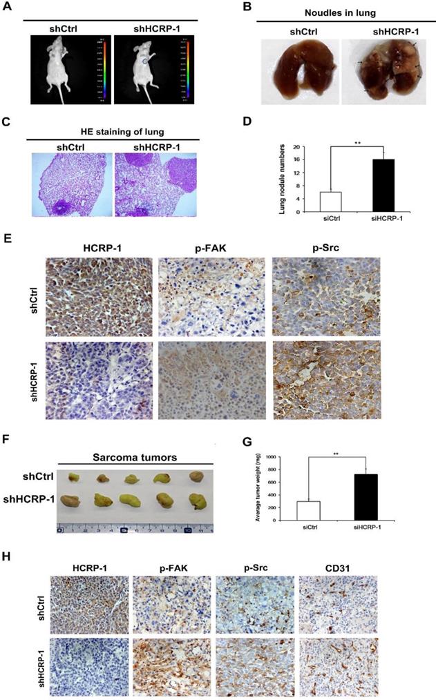
Solid tumors growth and metastasis depends on angiogenesis, including PCa, which is the progression of forming new blood vessels from the existing network of capillaries [14]. Targeting angiogenesis is effective in cancer therapy not only because angiogenesis supplies oxygen and nutrients for the survival of cancer cells, but also provides the way for metastatic spread of these cancer cells [15]. Here, we showed that inhibition of HCRP-1 expression promoted tube formation of HUVECs induced by prostate cancer cells. In vivo, knockdown of HCRP-1 promoted CD31 expression in prostate tumors. These data revealed that angiogenesis serve as a crucial role in promoting cancer metastasis by knockdown of HCRP-1.
In order to further elucidate the mechanisms involved in HCRP-1-induced prostate cancer cell migration, invasion and angiogenesis, PC3 and LnCap cells were transfected with siRNA for HCRP-1, phosphorylation of Src and FAK was detected. We found that knockdown of HCRP-1 induced phosphorylation of Src and FAK. Src is a non-receptor tyrosine kinase, which is induced by various kinds of cellular signal molecules and has a great effect in regulating numerous processes, including cell growth, differentiation, adhesion and the migration signaling pathway [16, 17]. As an other non-receptor protein tyrosine kinase, FAK is widely expressed and regulats several biological processes, including cell adhesion, angiogenesis and migration [18, 19]. At the sites of cell-substratum focal adhesions, FAK can combine withSrc to form a dual-kinase complex[20], which coordinate cell behavior through regulating downstream pathways and molecules, including AKT, p38 and ERK kinase[21, 22]. Autophosphorylation of Src/FAK complexes is a critical event in cancer progression and metastasis [23]. Src/FAK pathway kinases in solid tumors have been viewed as an attractive target in cancer therapy. Inhibition of Src family kinase exerts effects on the tumor microenvironment [24, 25]. In our present study, the Src-specific inhibitor PP2 were used to abrogate the activation of Src, and the results showed that FAK activity and cells migration, invasion and angiogenesis were suppressed by PP2 in PC3 and LnCap cells. Additionally, FAK inhibitor 14 could inhibit the phosphorylation levels of Src and FAK. Combined with our results, we suggested that HCRP-1 knockdown might induce cell migration, invasion and angiogenesis by deregulating Src/FAK-associated signaling pathways in prostate cancer cells.
HCRP-1 is involved in binding and sorting of EGFR and the consequent degradation with lysosomes [4]. Src/FAK and EGFR converge on common downstream pathways [26], which exhibit antitumor activity in patients with advanced NSCLC co-treated with inhibitors of both signaling pathways [27]. We detected the half-life of EGFR to elucidate whether EGFR stability exerted influence on Src/FAK pathway in HCRP ablation cells. The stability was sustained in both WT and HCRP-1 knockdown cancer cells. Next, we investigated the phosphorylation of EGFR and our data showed that reduced HCRP-1 expression did not change EGFR activity. We assumed that HCRP-1 could regulate Src/FAK signaling through other mechanism. As the ESCRT-I subunit, Tsg101 (tumor susceptibility gene101) deletion impaired Src-dependent activation of STAT3 and FAK, resulted in cell migration suppression [28]. Moreover, as other ESCRT-I subunit, Alix binds to Src at two points via SH2 (Src homology 2) and SH3 (Src homology 3) domains. Alix that binds to Src are typically phosphorylated by Src [29]. Therefore, we investigated the endogenous interaction of HCRP-1 and Src. We demonstrated that endogenous Src and HCRP-1 in untransfected cells were associated, suggests that the interaction between Src and HCRP-1 has physiological relevance. We supposed that the interaction of HCRP-1 and Src contributed to the regulation of Src activity.
In conclusion, we have established a vital role for HCRP-1 in PCa and our clinical analyses point the prospect of HCRP-1 as a potential molecular therapeutic target for highly aggressive prostate cancer. We also revealed that HCRP-1 regulated prostate cancer cell migration, invasion and angiogenesis. In mechanism, HCRP-1 may regulate Src/FAK signaling pathway through interaction with Src. Although more details still need to explore the functions of HCRP-1 in a physiologic condition, our data uncover the importance of HCRP-1 in PCa progression and metastasis.
Acknowledgements
This work was supported by the Postgraduate Research & Practice Innovation Program of Jiangsu Province (KYCX19_2213). We thank all the staff of Jiangsu Cancer Biotherapy Institute in Xuzhou Medical University for technical support.
Competing Interests
The authors have declared that no competing interest exists.
References
1. Siegel RL, Miller KD, Jemal A. Cancer Statistics, 2017. CA Cancer J Clin. 2017;67:7-30
2. Zisman A, Pantuck AJ, Wieder J, Chao DH, Dorey F, Said JW. et al. Risk group assessment and clinical outcome algorithm to predict the natural history of patients with surgically resected renal cell carcinoma. J Clin Oncol. 2002;20:4559-66
3. Kostelansky MS, Schluter C, Tam YY, Lee S, Ghirlando R, Beach B. et al. Molecular architecture and functional model of the complete yeast ESCRT-I heterotetramer. Cell. 2007;129:485-98
4. Bache KG, Slagsvold T, Cabezas A, Rosendal KR, Raiborg C, Stenmark H. The growth-regulatory protein HCRP1/hVps37A is a subunit of mammalian ESCRT-I and mediates receptor down-regulation. Mol Biol Cell. 2004;15:4337-46
5. Lai MW, Huang SF, Lin SM, Chen TC, Lin CY, Yeh CN. et al. Expression of the HCRP1 mRNA in HCC as an independent predictor of disease-free survival after surgical resection. Hepatol Res. 2009;39:164-76
6. Wittinger M, Vanhara P, El-Gazzar A, Savarese-Brenner B, Pils D, Anees M. et al. hVps37A Status affects prognosis and cetuximab sensitivity in ovarian cancer. Clin Cancer Res. 2011;17:7816-27
7. Perisanidis C, Savarese-Brenner B, Wurger T, Wrba F, Huynh A, Schopper C. et al. HCRP1 expression status is a significant prognostic marker in oral and oropharyngeal cancer. Oral Dis. 2013;19:206-11
8. Chen F, Zhang L, Wu J, Huo F, Ren X, Zheng J. et al. HCRP-1 regulates EGFR-AKT-BIM-mediated anoikis resistance and serves as a prognostic marker in human colon cancer. Cell Death Dis. 2018;9:1176
9. Chung MS, Lee SH, Lee DH, Chung BH. Evaluation of the 7th American Joint Committee on cancer TNM staging system for prostate cancer in point of classification of bladder neck invasion. Jpn J Clin Oncol. 2013;43:184-8
10. Chen F, Deng J, Liu X, Li W, Zheng J. HCRP-1 regulates cell migration and invasion via EGFR-ERK mediated up-regulation of MMP-2 with prognostic significance in human renal cell carcinoma. Sci Rep. 2015;5:13470
11. Xu J, Yang W, Wang Q, Zhang Q, Li X, Lin X. et al. Decreased HCRP1 expression is associated with poor prognosis in breast cancer patients. Int J Clin Exp Pathol. 2014;7:7915-22
12. Giesing M, Driesel G, Molitor D, Suchy B. Molecular phenotyping of circulating tumour cells in patients with prostate cancer: prediction of distant metastases. BJU Int. 2012;110:E1202-11
13. Rankin EB, Giaccia AJ. Hypoxic control of metastasis. Science. 2016;352:175-80
14. O'Hurley G, Perry AS, O'Grady A, Loftus B, Smyth P, O'Leary JJ. et al. The role of secreted frizzled-related protein 2 expression in prostate cancer. Histopathology. 2011;59:1240-8
15. Folkman J. Angiogenesis and angiogenesis inhibition: an overview. EXS. 1997;79:1-8
16. Thomas SM, Brugge JS. Cellular functions regulated by Src family kinases. Annu Rev Cell Dev Biol. 1997;13:513-609
17. Parsons SJ, Parsons JT. Src family kinases, key regulators of signal transduction. Oncogene. 2004;23:7906-9
18. Parsons JT, Martin KH, Slack JK, Taylor JM, Weed SA. Focal adhesion kinase: a regulator of focal adhesion dynamics and cell movement. Oncogene. 2000;19:5606-13
19. Cox BD, Natarajan M, Stettner MR, Gladson CL. New concepts regarding focal adhesion kinase promotion of cell migration and proliferation. J Cell Biochem. 2006;99:35-52
20. Mitra SK, Schlaepfer DD. Integrin-regulated FAK-Src signaling in normal and cancer cells. Curr Opin Cell Biol. 2006;18:516-23
21. Hynes RO. Integrins: bidirectional, allosteric signaling machines. Cell. 2002;110:673-87
22. Guo W, Giancotti FG. Integrin signalling during tumour progression. Nat Rev Mol Cell Biol. 2004;5:816-26
23. Liang Y, Yi L, Liu P, Jiang L, Wang H, Hu A. et al. CX3CL1 involves in breast cancer metastasizing to the spine via the Src/FAK signaling pathway. J Cancer. 2018;9:3603-12
24. Wu Y, He L, Zhang L, Chen J, Yi Z, Zhang J. et al. Anacardic acid (6-pentadecylsalicylic acid) inhibits tumor angiogenesis by targeting Src/FAK/Rho GTPases signaling pathway. J Pharmacol Exp Ther. 2011;339:403-11
25. Vultur A, Buettner R, Kowolik C, Liang W, Smith D, Boschelli F. et al. SKI-606 (bosutinib), a novel Src kinase inhibitor, suppresses migration and invasion of human breast cancer cells. Mol Cancer Ther. 2008;7:1185-94
26. Leung EL, Tam IY, Tin VP, Chua DT, Sihoe AD, Cheng LC. et al. SRC promotes survival and invasion of lung cancers with epidermal growth factor receptor abnormalities and is a potential candidate for molecular-targeted therapy. Mol Cancer Res. 2009;7:923-32
27. Haura EB, Tanvetyanon T, Chiappori A, Williams C, Simon G, Antonia S. et al. Phase I/II study of the Src inhibitor dasatinib in combination with erlotinib in advanced non-small-cell lung cancer. J Clin Oncol. 2010;28:1387-94
28. Tu C, Ortega-Cava CF, Winograd P, Stanton MJ, Reddi AL, Dodge I. et al. Endosomal-sorting complexes required for transport (ESCRT) pathway-dependent endosomal traffic regulates the localization of active Src at focal adhesions. Proc Natl Acad Sci U S A. 2010;107:16107-12
29. Schmidt MHH, Dikic I, Bogler O. Src phosphorylation of Alix/AIP1 modulates its interaction with binding partners and antagonizes its activities. J Biol Chem. 2005;280:3414-25
Author contact
![]() Corresponding authors: Jin Bai, Address: Jiangsu Center for the Collaboration and Innovation of Cancer Biotherapy, Cancer Institute, Xuzhou Medical College, 84 West Huai-hai Road, Xuzhou, Jiangsu, P.R.China. Tel: +86-0516-85582530; E-mail: bjedu.cn OR Junnian Zheng, Address: Jiangsu Center for the Collaboration and Innovation of Cancer Biotherapy, Cancer Institute, Xuzhou Medical College, 84 West Huai-hai Road, Xuzhou, Jiangsu, P.R.China. Tel: +86-0516-85582530; E-mail: jnzhengedu.cn
Corresponding authors: Jin Bai, Address: Jiangsu Center for the Collaboration and Innovation of Cancer Biotherapy, Cancer Institute, Xuzhou Medical College, 84 West Huai-hai Road, Xuzhou, Jiangsu, P.R.China. Tel: +86-0516-85582530; E-mail: bjedu.cn OR Junnian Zheng, Address: Jiangsu Center for the Collaboration and Innovation of Cancer Biotherapy, Cancer Institute, Xuzhou Medical College, 84 West Huai-hai Road, Xuzhou, Jiangsu, P.R.China. Tel: +86-0516-85582530; E-mail: jnzhengedu.cn

 Global reach, higher impact