Impact Factor ISSN: 1449-2288




Int J Biol Sci 2027; 23(1):288-320. doi:10.7150/ijbs.140085 This issue Cite
Review
Extracellular Vesicle Heterogeneity in Colorectal Cancer: From Biological Diversity to Therapeutic Translation and Precision Intervention
Yining Sun1,3,*, Xian Zhang2,*, Feifan Chen1,3, Binyang Luo1,3, JinLin Yang1,3  , Zhu Wang1,3
1. Department of Gastroenterology and Hepatology, West China Hospital of Sichuan University, Chengdu 610041, Sichuan, China.
2. Department of Pathology, West China Hospital of Sichuan University, Chengdu 610041, Sichuan, China.
3. Laboratory of Gastrointestinal Cancer and Liver Disease, West China Hospital of Sichuan University, Chengdu 610041, Sichuan, China.
*Yining Sun and Xian Zhang contributed equally to this article and should be considered co-first authors.
Received 2026-6-29; Accepted 2026-9-7; Published 2027-1-1
Abstract
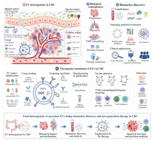
Colorectal cancer (CRC) remains a major global health burden, with persistent challenges in disease monitoring, therapeutic resistance, and durable treatment efficacy. Extracellular vesicles (EVs) are key mediators of intercellular communication and have emerged as promising biomarkers, therapeutic delivery platforms, and intervention targets in CRC. However, EVs are highly heterogeneous in biogenesis, cargo composition, cellular origin, biological source, surface properties, and functional state, leading to context-dependent effects on tumor progression, microenvironment remodeling, immune regulation, metastasis, and therapy response. In this review, we integrate EV heterogeneity with CRC biology, biomarker-guided stratification, and therapeutic translation. We first discuss how distinct EV populations shape CRC-associated signaling, tumor-stromal crosstalk, immune remodeling, metastatic dissemination, and treatment resistance. We then summarize emerging analytical strategies, including single-vesicle profiling, multi-omics integration, and AI-assisted biomarker discovery, that may help resolve EV heterogeneity and identify clinically informative EV subpopulations. Finally, we focus on EV-based therapeutic engineering, including therapeutic cargo delivery, vaccine platforms, immune microenvironment reprogramming, targeting of EV biogenesis, secretion and uptake, biomimetic platform development, standardization, and scalable manufacturing. By linking EV-mediated molecular signaling, cellular phenotype remodeling, biomarker discovery, drug response, and therapeutic engineering, this review proposes a mechanism-informed translational framework for exploiting EV heterogeneity in CRC. Rather than viewing EV heterogeneity solely as a barrier to clinical translation, we highlight its potential as a resource for identifying EV-mediated therapeutic vulnerabilities, improving patient stratification, and guiding the development of EV-based precision intervention strategies.
Keywords: colorectal cancer, extracellular vesicle heterogeneity, tumor microenvironment, biomarker-guided stratification, therapeutic engineering
1. Introduction
Colorectal cancer (CRC) remains a major global health burden, ranking as the third most commonly diagnosed malignancy worldwide. Epidemiological projections further indicate that its incidence will continue to rise, with the global burden expected to increase by nearly 60% by 2030 [1]. Despite advances in screening and treatment, the prognosis of advanced CRC remains poor, with a 5-year survival rate of approximately 10% in patients with distant metastasis [2, 3]. These clinical limitations underscore the urgent need to better understand the molecular mechanisms underlying CRC progression and to develop more effective strategies for early diagnosis and therapeutic intervention.
Extracellular vesicles (EVs) have emerged as key mediators of intercellular communication in CRC. By transferring proteins, nucleic acids, lipids, and metabolites between cells, EVs regulate tumor growth, microenvironment remodeling, immune evasion, metastatic dissemination, and therapy response [4]. However, EVs are not a uniform entity. Their biological activities are shaped by substantial heterogeneity in biogenesis pathways, molecular cargo, cellular origin, and species source. This heterogeneity provides the basis for their functional diversity, but also complicates EV isolation, characterization, mechanistic interpretation, and clinical application [5].
From a translational perspective, EV heterogeneity represents both an opportunity and a challenge. EV-derived molecular signatures offer promising minimally invasive biomarkers for CRC detection, prognosis, and longitudinal monitoring [6-8], while EVs themselves can serve as delivery vehicles, immune modulators, or therapeutic targets [9-11]. At the same time, conventional bulk-level analyses often mask functionally distinct EV subpopulations, reducing biomarker specificity and limiting the precision of EV-based therapeutic strategies. Therefore, the ability to resolve EV heterogeneity is essential for transforming EV biology into reliable clinical tools. Recent methodological advances are beginning to address this challenge. Single-vesicle technologies, multi-omics profiling, and data-driven analytical frameworks enable more precise characterization of EV subpopulations and their disease-specific signatures [12-14]. In parallel, advances in EV engineering, cargo loading, surface modification, and bio-mimetic vesicle design are expanding the therapeutic potential of EV-based platforms [15-18]. Together, these developments are shifting the field from descriptive EV profiling toward functional dissection and rational translational design.
Despite rapid progress, current studies often address EV biology, biomarker development, and therapeutic applications in isolation, lacking a unified framework that links EV heterogeneity to translational outcomes. In this review, we systematically integrate these dimensions by summarizing the origins and functional implications of EV heterogeneity in CRC, and by discussing how this heterogeneity informs biomarker discovery and therapeutic design. We further highlight emerging technologies that enable high-resolution EV analysis and scalable engineering, thereby bridging the gap between fundamental EV biology and clinical application. Together, this review provides a conceptual and translational framework for leveraging EV heterogeneity in precision medicine for CRC.
2. EV Heterogeneity and Functional Consequences in CRC
To clarify how EV heterogeneity shapes CRC biology, this section dissects EV heterogeneity across four interconnected dimensions: biogenesis pathway, molecular cargo composition, cellular origin, and species source. These dimensions are not independent categories, but jointly determine EV identity, functional specificity, and context-dependent effects within the CRC ecosystem. By organizing EV heterogeneity in this way, we provide a framework for understanding how heterogenous EV populations contribute to tumor progression, immune modulation, metastasis, and therapeutic response.
2.1 Biogenesis-Dependent EV Heterogeneity and Nomenclature
EVs are a heterogeneous group of lipid bilayer-enclosed particles released from cells that are incapable of replication. Their heterogeneity arises from distinct biogenesis pathways and molecular packaging mechanisms. Historically, EVs have been classified according to proposed biogenesis pathways into endosome-derived exosomes and plasma membrane-derived ectosomes (also referred to as microvesicles) [19]. In this classical framework, exosomes are generated as intraluminal vesicles (ILVs) within multivesicular bodies (MVBs) and released upon fusion of MVBs with the plasma membrane, whereas ectosomes bud directly from the plasma membrane [19]. However, vesicle size and commonly used markers such as CD9, CD63, and CD81 are not sufficient to establish a specific biogenesis pathway. Accordingly, distinguishing these populations remains challenging in most experimental settings.
Recent MISEV2023 guidelines recommend the use of operational terms based on measurable characteristics, such as size (small EVs <200 nm; medium/large EVs >200 nm), density, biochemical composition, or cellular origin [20]. This framework promotes standardized EV characterization. Consistent with MISEV2023, this review therefore uses the generic term “EV” or, where supported by the reported physical characteristics, operational terms such as “small EV” when subcellular biogenesis has not been experimentally demonstrated. The terms “exosome” and “exosomal” are reserved for studies in which an endosomal origin has been established or are retained when directly reproducing the terminology used in the cited study, a formal assay name, or a commercial product name. In such cases, use of the original terminology should not be interpreted as independent evidence of endosomal biogenesis.
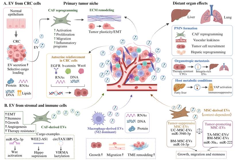
EV-mediated communication in colorectal cancer progression. Extracellular vesicles (EVs) derived from distinct cellular sources mediate diverse and context-dependent functions in colorectal cancer (CRC). (A) CRC cell-derived EVs increase during malignant transformation and selectively carry proteins, RNAs, DNA, and lipids. These EVs remodel the primary tumor niche by reprogramming cancer-associated fibroblasts (CAFs), promoting extracellular matrix (ECM) remodeling, enhancing tumor plasticity and epithelial-mesenchymal transition (EMT), and reinforcing malignant programs through autocrine signaling. They also contribute to premetastatic niche formation, organotropic metastasis, and host metabolism-associated metastatic progression. (B) Stromal and immune cell-derived EVs further shape CRC progression. CAF-derived EVs promote EMT, stemness, angiogenesis, and therapy resistance; macrophage-derived EVs, particularly those from M2-like macrophages, support tumor growth, migration, and tumor microenvironment remodeling; whereas mesenchymal stromal/stem cell-derived EVs (MSC-EVs), including tumor-associated mesenchymal stem cell-derived extracellular vesicles (TA-MSC-EVs), exert source- and context-dependent tumor-suppressive or tumor-promoting effects. Together, the figure highlights cellular origin as a major determinant of EV cargo composition, functional specificity, and biological outcome in CRC. Schematic illustrations were created with BioRender.com.
2.2 Cargo-Level EV Heterogeneity and Functional Output in CRC
Building upon the heterogeneity of biogenesis pathways, EV cargo composition is likewise highly variable and represents a key determinant of their functional diversity. EVs exhibit substantial heterogeneity in molecular cargo, which reflects their biogenesis, cellular origin, and functional state. Based on spatial distribution, EV cargo in CRC can be broadly classified into three categories: (i) internal cargo (e.g., soluble proteins, RNAs, DNAs, and lipids), (ii) membrane-associated components (e.g., transmembrane proteins), and (iii) exofacial molecules located on the outer surface of EVs. These distinct cargo types collectively determine EV-mediated intercellular communication.
2.2.1 Surface Proteins and Biomolecular Corona
Surface-associated proteins regulate EV-mediated communication by mediating signaling and enabling cargo sorting, targeting, uptake, and long-range intercellular communication. EV-associated proteins in CRC span several functional classes, including signaling receptors (e.g., FZD10 and EGFR) [21], immune-checkpoint proteins (e.g., PD-L1), death ligands (e.g., TRAIL) [22, 23], adhesion- and extracellular matrix-related proteins (e.g., ITGBL1) [24], growth-factor ligands (e.g., Gas6 and AREG), stress-response proteins (e.g., HSP70), and intracellular signaling or trafficking proteins associated with EV membranes or cargo (e.g., KRAS and Rab13) [25-29]. Collectively, these protein classes contribute to epithelial plasticity, stromal activation [24], immune regulation [23, 26], and angiogenesis [30] in CRC. In addition to intrinsic membrane components, EV surfaces are dynamically remodeled by a biomolecular corona formed through interactions with extracellular proteins, lipids, proteoglycans, and nucleic acids. This corona represents an acquired layer rather than a passive coating, and can constitute a substantial fraction of EV-associated proteins, in some cases exceeding luminal cargo. Notably, molecules previously regarded as co-isolated contaminants are now recognized as integral components of this structure [31]. Functionally, the biomolecular corona redefines the physicochemical and biological identity of EVs. By altering surface composition, it modulates EV stability, biodistribution, cellular uptake, and immune recognition [32-34], thereby shaping their pharmacokinetics, targeting specificity, and immunoregulatory signaling [35, 36]. In addition, the presence of a corona may introduce biases in nanoparticle-based detection and characterization methods [37]. Despite its emerging importance, the role of the biomolecular corona in CRC remains largely unexplored, warranting further investigation into its regulation of EV behavior within the tumor microenvironment.
2.2.2 RNA Cargo
EVs carry diverse RNA species reflecting the genetic and functional state of their cells of origin. These include small RNAs such as microRNAs (miRNAs), small interfering RNAs (siRNAs), small nuclear and nucleolar RNAs, Y RNAs, and ribosomal RNAs (rRNAs), as well as long noncoding RNAs (lncRNAs), circular RNAs (circRNAs), messenger RNAs (mRNAs), transfer RNAs (tRNAs), and mitochondrial RNAs. These RNAs collectively participate in gene regulation, RNA processing, and intercellular communication, thereby influencing CRC. For example, EV miRNAs promote CRC progression by driving tumor cell proliferation, liver metastasis [38, 39], metabolic reprogramming [40], remodeling of the tumor microenvironment [39], and angiogenesis [41], while siRNAs mediate RNA interference and hold therapeutic potential [42]. circRNAs [43], lncRNAs [44] and tRNA [45] act as epigenetic regulators and molecular sponges, contributing to CRC progression and therapy resistance. The RNA cargo of EVs differs markedly from that of their parental cells, not only in composition but also in size distribution. EVs are typically enriched in small RNA species, whereas longer RNAs (e.g., full-length mRNAs) are often truncated, reflecting the limited nucleic acid capacity of small EVs. Mechanistically, RNA incorporation into EVs is regulated by RNA-binding protein (RBP)-mediated loading [46] and sequence-dependent sorting (e.g., GGAG and GGCU motifs recognized by hnRNPA2B1 and SYNCRIP) [47], coupled with EV biogenesis pathways involving VAP-A and CERT at ER membrane contact sites [48]. Functionally, EV-associated RBP such as HuR can modulate gene expression in recipient cells by stabilizing target mRNAs (e.g., c-Myc) [49]. Beyond RNA composition and sorting, recent evidence has demonstrated that cancer-specific RNA modifications in EVs promote CRC progression by modulating immune cells, particularly macrophages [50]. Despite these advances, the precise molecular mechanisms governing RNA loading and release remain incompletely understood.
2.2.3 DNA Cargo
EVs also carry DNA molecules, including double-stranded DNA (dsDNA), single-stranded DNA (ssDNA), mitochondrial DNA (mtDNA), and plasmid DNA. These DNAs can be localized both within EVs and on their outer surfaces, with their distribution and composition varying among EV subtypes. ssDNA and dsDNA may originate from micronuclei, autophagy-related amphisomes, or cytosolic DNA sensing pathways (e.g., via cGAS), particularly under genomic instability or DNA damage. Notably, EV-associated dsDNA includes large genomic DNA (gDNA) fragments (100 bp-20 kbp) that can span the entire genome and reflect the mutational landscape of parental cells, supporting their utility in PCR-based mutation detection, CRC diagnosis and disease monitoring [51-53]. Functionally, EV-mediated DNA transfer can also modulate immune responses. In Crohn's disease, EVs transport pathogenic mtDNA and gDNA from damaged intestinal epithelial cells to macrophages, activating the STING pathway and triggering inflammatory responses [54]. In CRC, therapy-induced release of epithelial-derived gDNA via EVs can activate the AIM2 inflammasome, thereby promoting inflammation [55]. Collectively, understanding the biological significance of EV-mediated DNA release and its extrinsic effects may provide novel approaches for biomarker development and therapeutic intervention in CRC.
2.2.4 Microenvironmental Regulation of EV Cargo
EV cargo composition is dynamically shaped by the microenvironmental state of the parental cell. Stress conditions, differentiation status, and physical cues can alter both EV release and cargo selection, thereby changing their biological functions [5, 56, 57]. For example, increased matrix stiffness promotes the secretion of Jagged1-enriched EVs, which activate Notch signaling and facilitate tumor progression [58]. In CRC, hypoxia represents a particularly important microenvironmental driver. As tumor growth often exceeds vascular supply, CRC cells frequently experience hypoxic and mildly acidic conditions that enhance EV release and reprogram EV cargo [59]. Specifically, EV-associated miR-135a-5p derived from hypoxic CRC cells can promote metastatic adaptation [60]. Additionally, EVs released from aerobic CRC cells induce doxorubicin resistance in hypoxic CRC cells and macrophages, underscoring their role in chemoresistance [61]. Together, these findings indicate that EV cargo reflects not only the identity of the cell of origin but also the local ecological pressures imposed by the tumor microenvironment.
2.3 Cellular Origin-Dependent EV Heterogeneity in CRC
Given that EV cargo composition is largely determined by their cells of origin, heterogeneity at the cellular level represents another critical layer of EV diversity. EVs released by different cells demonstrate distinct functional specificities. Within the tumor microenvironment (TME), malignant cells, immune cells, and stromal cells establish a complex signaling network through EVs, collectively modulating disease progression. In this context, EVs can propagate both pro-tumor and anti-tumor signals, with their precise effects contingent on the characteristics of the originating and recipient cells.
2.3.1 CRC Cell-Derived EVs
CRC cell-derived EVs are critical mediators of intercellular communication between tumor cells and recipient cells in both local and distant microenvironments. Malignant transformation not only increases EV secretion compared with nonmalignant cells [62], but also enhances the selective packaging of bioactive cargo, including proteins and DNA [63], thereby reshaping both the quantity and molecular composition of EVs.
At the level of the primary tumor niche, CRC cell-derived EVs actively reprogram cancer-associated fibroblast (CAF), promoting their activation, proliferation, and migration, as well as the establishment of proinflammatory transcriptional programs [38, 64, 65]. In parallel, this CAF activation establishes a localized tumor-supportive microenvironment that enhance extracellular matrix (ECM) remodeling and tumor cell plasticity. For example, EV-derived miR-99a from highly metastatic CRC cells promotes tumor cell migration by inducing CAF secretion of CCL7, which in turn drives epithelial-to-mesenchymal transition (EMT) via activation of the CCR5-mTOR-p70S6K axis [64]. Beyond microenvironmental remodeling, EV-mediated signaling also reinforces malignant traits within tumor cells. Notably, EVs carrying non-coding RNAs and oncogenic proteins, including EGFR [66], β-catenin [67], and Wnt4 [68], can induce genetic instability and accelerate neoplastic transformation in CRC.
Beyond remodeling the primary tumor niche, CRC cell-derived EVs contribute to the establishment of premetastatic niche (PMN) in distant organs by reprogramming CAFs [24, 69], enhancing vascular leakiness and tumor cell recruitment [41, 70, 71], while simultaneously reprogramming hepatic cells toward a prometastatic phenotype [39, 72]. These changes collectively establish a permissive environment for metastatic colonization. Beyond niche formation, EVs further contribute to organ-specific metastatic behavior [73]. Notably, EVs released by therapy-resistant circulating tumor cells are enriched in integrin-family proteins that preferentially target organs such as the liver and lungs, increasing metastatic risk in CRC [74]. In addition, host metabolic conditions can further amplify EV-mediated metastatic progression. For example, fatty liver promotes the production of hepatocyte-derived EVs, which enhance CRC liver metastasis by activating Yes-associated protein signaling and reinforcing an immunosuppressive microenvironment [75].
2.3.2 Stromal and Immune Cell-Derived EVs
The CRC TME comprises diverse non-malignant cellular populations, including CAFs, mesenchymal stromal/stem cells (MSCs), immune cells, endothelial cells, pericytes, adipocytes, neurons, and other stromal elements. These cells are no longer viewed as passive bystanders, but as active regulators of tumor evolution. EVs represent a major route of this intercellular communication, enabling stromal and immune cells to transfer regulatory cargos that reshape malignant phenotypes.
CAFs represent the best-characterized stromal source of EVs in CRC. CAF-derived EVs promote multiple hallmarks of tumor progression, including EMT [43, 76], stemness maintenance [76, 77], tumor growth [78, 79], angiogenesis [80], and therapy resistance [76, 77, 80]. Mechanistically, these effects are mediated by diverse EV cargos, such as miR-92a-3p-induced activation of Wnt/β-catenin signaling [76], lncRNA WEE2-AS1-dependent suppression of the Hippo pathway [79], and circTAX1BP1-mediated regulation of VIRMA lactylation in CRC cells [43]. Together, these findings establish CAF-EVs as a prototypical pro-tumorigenic stromal EV population in CRC. MSCs represent another important stromal EV source, but their effects in CRC are more context-dependent. EVs from certain MSC sources suppress CRC progression. Umbilical cord-derived MSC-EVs transfer miR-3940-5p to CRC cells, leading to ITGA6 downregulation and inhibition of tumor growth and metastasis [81]. Similarly, bone marrow-derived MSC-EVs enriched in miR-16-5p or miR-22-3p suppress CRC progression via targeting ITGA2 or modulating the PI3K/AKT pathway [82, 83]. In contrast, MSC-EVs can also promote malignant phenotypes under specific biological contexts. CRC-associated MSC-EVs deliver miR-30a and miR-222 to tumor cells, directly targeting the tumor suppressor MIA3 and enhancing proliferation and migration [84]. Moreover, bone marrow MSC-derived EVs carrying miR-142-3p expand the population of CRC stem-like cells [85]. These findings suggest that MSC-EVs are not intrinsically tumor-suppressive or tumor-promoting; rather, their function is determined by MSC origin, activation state, and cargo composition.
Immune cell-derived EVs further expand the complexity of TME-mediated communication. Among them, macrophage-derived EVs, particularly those released by M2-like macrophages, have been closely associated with CRC progression. By transferring oncogenic miRNAs [86-88], lncRNAs [89], and proteins [90, 91], macrophage-derived EVs reprogram cancer cell behavior, promote tumor growth and metastasis, and remodel the tumor-supportive microenvironment. In this context, immune cell-derived EVs provide a functional link between immune polarization, tumor cell plasticity, and metastatic progression.
2.4. Non-Mammalian EVs and Their Functional Roles in CRC
In addition to mammalian cell-derived EVs, EVs from other species, particularly gut microbiota and plants, have emerged as important modulators of CRC. These non-mammalian EVs exhibit distinct biogenesis pathways, cargo compositions, and functional properties, adding another layer of heterogeneity to the EV landscape in CRC.
2.4.1 Gut Microbiota-Derived EVs
The colon harbors a dense and diverse microbial ecosystem, and dysbiosis is a key driver of CRC [92]. Emerging evidence indicates that microbiota-derived EVs, rather than bacteria per se, function as critical mediators that translate microbial dysbiosis into host cellular reprogramming. These vesicles exhibit substantial heterogeneity in biogenesis, structure, and molecular cargo, reflecting differences in microbiota species, genetic background, and physiological state, thereby enabling broad functional capacity and dynamic adaptation to environmental cues. In the colorectal lumen, microbiota-derived EVs interact with host cells through ligand-receptor signaling or are internalized via endocytosis, phagocytosis, or membrane fusion, thereby activating intracellular pathways.
Non-mammalian EVs in colorectal cancer. Extracellular vesicles (EVs) derived from gut microbiota and plants add an additional layer of EV heterogeneity in colorectal cancer (CRC). Microbiota-derived EVs interact with host cells through ligand-receptor signaling, endocytosis, phagocytosis, or membrane fusion, thereby reshaping epithelial, immune, and stromal responses. Pathogenic microbiota-derived EVs promote CRC by inducing inflammation, barrier disruption, immune evasion, pro-metastatic signaling, and chemoresistance, whereas EVs from commensal or probiotic microbes can exert barrier-protective, anti-inflammatory, and antitumor effects. Plant-derived EVs (P-EVs), acquired through oral intake, carry diverse bioactive cargos and exhibit low immunogenicity, high biosafety, and oral stability. Functionally, they help maintain intestinal homeostasis by attenuating inflammation, remodeling gut microbiota, and promoting epithelial regeneration. Schematic illustrations were created with BioRender.com.
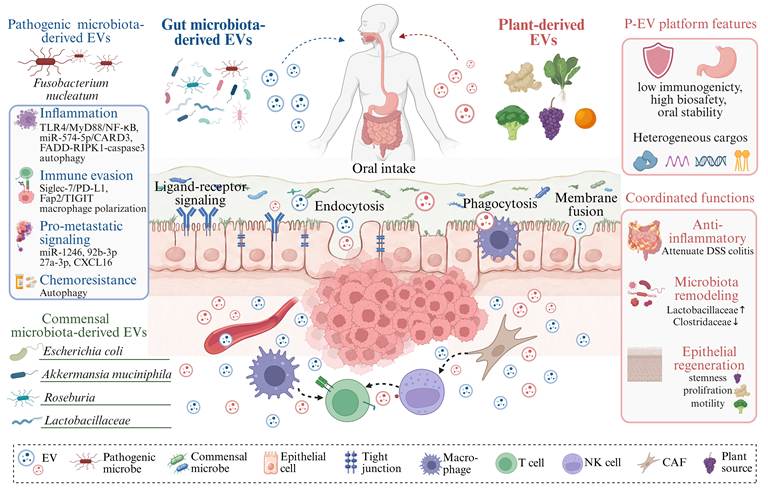
Microbiota-derived EVs initiate tumorigenesis by establishing a chronic inflammatory and barrier-disruptive niche. Fusobacterium nucleatum-derived EVs (FnEVs) activate TLR4/MyD88/NF-κB signaling and induce pro-inflammatory cytokines, while compromising epithelial integrity through miR-574-5p/CARD3-dependent autophagy, tight-junction disruption, and FADD-RIPK1-caspase-3-mediated epithelial injury [93-95]. Beyond this initiating niche, pathogenic EVs further accelerate CRC progression by coordinating immune evasion, intercellular signaling, and stress-adaptive autophagy. FnEVs suppress antitumor immunity through Siglec-7-dependent PD-L1 upregulation, macrophage polarization, and Fap2-mediated TIGIT engagement on NK and T cells [96, 97]. They also disseminate pro-metastatic cues by transferring miR-1246, miR-92b-3p, miR-27a-3p, and CXCL16 [98], while activating autophagic flux to support tumor cell survival, recurrence, and chemoresistance [94, 99, 100]. Notably, EVs derived from both pathogenic and commensal bacteria can globally reshape gene expression profiles in colon cancer cells, highlighting their role as systemic regulators of tumor cell states [101, 102].
Importantly, microbiota-derived EVs exert context-dependent and bidirectional effects. While pathogenic EVs promote tumorigenesis, EVs from commensal and probiotic bacteria exhibit barrier-protective, anti-inflammatory, and antitumor activities. For example, Escherichia coli-derived EVs can induce apoptosis or ROS-mediated mitophagy in CRC cells [103]. EVs from Akkermansia muciniphila and Roseburia intestinalis enhance barrier integrity and suppress inflammation [104-106], while Amuc_2172 (an Akkermansia muciniphila-derived protein) enhances cytotoxic T-cell activation and synergizes with immune checkpoint blockade [106, 107]. Similarly, Lactobacillaceae-derived EVs inhibit CRC progression by modulating PDK1/AKT/BCL-2 signaling, suppressing TLR4/NF-κB/NLRP3 pathways, reversing chemoresistance, and enhancing anti-PD-1 efficacy [108-110]. Collectively, microbiota-derived EVs function as context-dependent molecular switches that integrate inflammatory signaling, immune evasion, autophagy, and intercellular communication networks in CRC. Their bidirectional activity underscores their potential as both biomarkers and therapeutic targets, offering new avenues for precision intervention in CRC.
2.4.2 Plant-Derived EVs
Plant-derived EVs (P-EVs) are membrane-bound nanostructures released by plant cells. They exhibit heterogeneity in size, density, cellular origin, and biochemical composition, reflecting differences in plant tissue and physiological state [111-113]. Despite the presence of a rigid cell wall, accumulating evidence demonstrates that plants actively produce EVs through multiple intracellular pathways, including multivesicular bodies, exocyst-positive organelles, Pen1-positive compartments, vacuoles, and autophagosomes [114-116]. Similar to mammalian EVs, P-EVs carry diverse bioactive cargos including proteins, lipids, RNAs, and metabolites, which underlie their functional versatility. Notably, P-EVs exhibit low immunogenicity, high biosafety, and robust stability after oral administration, underscoring their translational potential [117].
Emerging evidence indicates that P-EVs are bioactive regulators of intestinal homeostasis with substantial anti-inflammatory and regenerative potential. Vesicles derived from edible plants such as ginger [118, 119], grape [120], broccoli [121], red cabbage [122], Portulaca oleracea L [123], and grapefruit [124] have been shown to attenuate dextran sulfate sodium (DSS)-induced colitis, supporting their broad capacity to restrain intestinal inflammation. Beyond direct anti-inflammatory effects, P-EVs actively reshape the gut microbiota by enriching beneficial taxa such as Lactobacillaceae and Bacteroidaceae while reducing pathogenic Clostridaceae [125], thereby contributing to microbial rebalance. They also influence epithelial repair programs. For example, grape-derived EVs promote Lgr5⁺ stem cell expansion via Wnt/β-catenin signaling [120], whereas ginger-derived EVs enhance epithelial survival and proliferation and suppress colitis-associated cancer [126]. In addition, P-EVs can modulate intestinal physiology, as illustrated by broccoli-derived EVs, which improve gut motility and alleviate loperamide-induced constipation [127]. Collectively, these findings position P-EVs as multifunctional modulators of the intestinal ecosystem, with potential relevance for preventing inflammation-driven CRC initiation and improving CRC-associated gut dysfunction.
3. EVs as a Source of Biomarkers
The clinical translation of EVs as liquid biopsy templates relies on their ability to preserve molecular cargo from systemic degradation. However, a substantial translational gap remains. This is largely driven by the low throughput of gold-standard isolation methods and the complexity of biological fluids such as blood, where low-abundance tumor-derived EVs are often masked by a vast background of non-vesicular proteins and commensal vesicles. Consequently, the transition from benchtop discovery to high-throughput clinical assays requires both a deep understanding of the CRC-specific molecular landscape and the integration of advanced analytical platforms.
3.1 Current Landscape of EV-based Biomarkers in CRC
To address the need for early diagnosis and longitudinal monitoring, research has focused on the high molecular diversity of EV cargo. By identifying CRC-specific alterations across different molecular layers, EVs offer a multidimensional resource for biomarker discovery and disease stratification.
3.1.1 RNA Biomarkers
EVs encapsulate multiple RNA species, representing the most extensively studied class of EV-derived biomarkers. Protected by the lipid bilayer, EVs-associated RNAs exhibit enhanced stability compared with freely circulating counterparts, supporting their utility as minimally invasive biomarkers. Accumulating evidence indicates that these RNAs reflect tumor-associated alterations and hold promise for CRC diagnosis, prognosis, and monitoring. Representative studies from the past five years are summarized in Table 1 [12, 128-155], detailing RNA species, sample size, specimen type, and diagnostic performance.
Extracellular vesicle-derived RNAs as diagnostic, prognostic, and predictive biomarkers in colorectal cancer
| Study | Sample Type | Biomarker | Clinical Application | Comparison (Sample Size) | Strategy | AUC / HR (95% CI) | Detection Method | EV Isolation Method |
|---|---|---|---|---|---|---|---|---|
| Cui X et al. 2020 [128] | Serum | miR-1539 | Diagnosis | CRC [n=51] vs healthy [n=49] | Single | 0.673 (0.568-0.779) | RT-qPCR | ExoQuick™ exosome precipitation solution |
| Yang CK et al. 2022 [129] | Plasma | miR-320c | Diagnosis | mCRC [n=18] vs healthy [n=30] | Single | 0.896 | RT-qPCR | ExoEasy Plasma Handbook |
| Qiao D et al. 2022 [130] | Plasma | miR-3937 | Diagnosis | CRC [n=34] vs healthy [n=18] | Single | 0.827 (0.712-0.942) | RT-qPCR | BestBio-Exosome kit |
| Combined+CEA+CA199 | 0.889 (0.801-0.977) | |||||||
| Kim S et al. 2022 [131] | Plasma | miR-92a, miR-141 | Diagnosis | CRC [n=21] vs healthy [n=5] | Single | 0.781-0.895 | DNA barcode-based nucleic acid lateral flow assay | qEV original column |
| Combined | 0.952 | |||||||
| Li T et al. 2023 [132] | Plasma | circTADA2A (hsa_circ_0043278), circGAPVD1(hsa_circ_0003270), circMYO9B (hsa_circ_0004071), and circTJAP1 (hsa_circ_0092310) | Diagnosis | CRC [n=35] vs healthy [n=35] | Single | 0.614-0.750 | Microarray and RT-qPCR | Total Plasma Exosome Isolation Kit |
| Miyazaki K et al. 2023 [133] | Serum | A panel (miR-181b, miR-193b, miR-195, and miR-411) | Diagnosis | T1 CRC with LNM [n=7] vs without LNM [n=51] | Single | 0.69-0.80 | RT-qPCR | Total Exosome Isolation Kit |
| Combined | 0.86 (0.70-1.00) (Train) | |||||||
| Wu Y et al. 2024 [134] | Serum | miR-1470 | Diagnosis | CRC [n=168] vs healthy [n=168] | Single | 0.740 (0.688-0.792) | qRT-PCR | Ultracentrifugation |
| Peng J et al. 2024 [135] | Serum | 3'tRF-ThrCGT, 3'tRF-mtlleGAT | Diagnosis | CRC [n=205] vs healthy [n=201] | Single | 0.656-0.669 | RT-qPCR | Centrifugation |
| Combined | 0.777 (0.732-0.822) | |||||||
| Combined+CEA+CA724 | 0.938 (0.913-0.963) | |||||||
| Mannucci A et al. 2026 [136] | Plasma | 4-miRNA panel (hsa-miR-32-5p, -486-3p, -550a-3-5p, -625-3p) | Diagnosis | EOCRC [n=251] vs non-disease [n=211] | Combined | 0.946 (0.917-0.975) (Train)0.923 (0.879-0.966) (Test) | RT-qPCR | ExoRNeasy Midi kits |
| Chen M et al. 2025 [137] | Plasma | miR-23a, miR-21 | Diagnosis | CRC [n=5] vs non-cancer [n=9] | Single | 0.889-1.000 | CRISPR/Cas13a | ExoQuick ULTRA EV Isolation Kit |
| Dana F et al. 2025 [138] | Plasma | lncRNA CCAT1, lncRNA XIST | Diagnosis | CRC [n=62] vs healthy [n=62] | Single | 0.650-0.860 | RT-qPCR | Centrifugation |
| Xu C et al. 2025 [139] | Plasma | miR-452-5p, miR-487-5p, miR-500a-5p, miR-7976miR-664a-3p, miR-184, miR-4454, miR-218-5p, miR-3200a-3p | Prognosis | Good responders [n=57] vs poor responders [n=37] | Combined | 0.81 | qRT-PCR | ExoRNeasy Midi Kit |
| Handa T et al. 2021 [140] | Serum | miR-4323, miR-4284, miR-1290, miR-1246 | Early diagnosis | Pretreatment and posttreatment of CRA [n=26] | Single | 0.639-0.705 | RT-qPCR | Polymer precipitation |
| Combined | 0.691 (0.528-0.817) | |||||||
| Pretreatment and posttreatment of large CRA [n=13] | Combined | 0.834 (0.628-0.938) | ||||||
| Nakayama S et al. 2025 [141] | Plasma | miR-122-5p, miR-130a-3p, miR-146a-5p, miR-15b-5p, and miR-126 | Early diagnosis | Stage I CRC [n=60] vs healthy [n=60] | Single | 0.730-0.788 | RT-qPCR | QIAzol Lysis Reagent |
| Han L et al. 2021 [142] | Serum | miR-15b, miR-16, miR-21, and miR-31 | Diagnosis and early diagnosis | CRC [n=150] vs healthy [n=123] | Single | 0.58-0.86 | RT-qPCR | ExoQuick kit |
| CRC [n=123] vs CRA [n=117] | Single | 0.55-0.84 | ||||||
| Shi Y et al. 2021 [143] | Serum | miR-126, miR-1290, miR-23a, and miR940 | Diagnosis and early diagnosis | CRC [n=100] vs healthy [n=35] | Single | 0.88-0.94 | RT-qPCR | RiboTM Exosome Isolation Reagent |
| stage I CRC [n=30] vs healthy [n=35] | Single | 0.83-0.90 | ||||||
| Yu M et al. 2021 [144] | Serum | FOXD2-AS1, NRIR, XLOC_009459 | Diagnosis and early diagnosis | CRC [n=203] vs healthy [n=201] | Single | 0.660-0.728 | RT-qPCR | Ultracentrifugation |
| Combined | 0.736 | |||||||
| Early-stage CRC [n=80] vs healthy [n=201] | Single | 0.660-0.743 | ||||||
| Combined | 0.758 | |||||||
| Wang L et al. 2022 [145] | Serum | miR-377-3p, miR-381-3p | Diagnosis and early diagnosis | CRC [n=175] vs healthy [n=172] | Single | 0.792-0.798 | RT-qPCR | Ultracentrifugation |
| Combined | 0.807 (0.7612-0.8520) | |||||||
| Early-stage CRC [n=69] vs healthy [n=172] | Single | 0.798-0.803 | ||||||
| Combined | 0.815 (0.7596-0.8704) | |||||||
| Guo TA et al. 2022 [146] | Plasma | A panel (HIST2H2AA4, H2BFS, UQCRHL, XCL2, AC008269.1, DMC1, RAB6D, KLHDC8B, CA3, APOL4, HIST1H2AI, ANKAR, SGMS1, CYP20A1, HIST1H2BB, STK3, CBWD1) | Diagnosis and early diagnosis | CRC [n=48] vs healthy [n=82] (Train)CRC [n=24] vs healthy [n=40] (Test)CRC [n=14] vs healthy [n=32] (Validation) | Combined | 0.938 (Train); 0.943 (Test); 0.947 (Validation) | RNA-seq | ExoRNeasy Serum/Plasma Kit |
| CRA [n=42] vs healthy [n=80] | Combined | 0.993 (Train); 0.978 (Test) | ||||||
| Shi YJ et al. 2023 [147] | Plasma | miR-185-5p | Diagnosis and early diagnosis | III-IV stage CRC [n=35] vs healthy [n=22] | Single | 0.803 (0.678-0.896) | RT-qPCR | EZBioscience kit |
| I-II stage CRC [n=70] vs healthy [n=50] | 0.887 (0.767-0.959) | |||||||
| CRA [n=65] vs healthy [n=44] | 0.702 (0.607-0.786) | |||||||
| Zhang C et al. 2024 [148] | Plasma | miR-99b-5p, miR-409-3p | Diagnosis and early diagnosis | CRC [n=68] vs healthy [n=27] | Single | 0.604-0.660 | RT-qPCR | ExoRNeasy Midi Kit |
| Combined+CEA | 0.863 (0.774-0.952) | |||||||
| Early CRC [n=28] vs healthy [n=27] | Single | 0.611-0.735 | ||||||
| Combined+CEA | 0.812 (0.679-0.945) | |||||||
| Min L et al. 2024 [149] | Plasma | miR-425-5 p, Let-7f-5p, C19orf43, TOP1, PPDPF, LNC-EV-9572, lnc-MKRN2-42:1, HIST2H2AA4, (MT-ND2) | Diagnosis and early diagnosis | CRC [n=47] vs control [n=53] | 8-RNA signature | 0.87 | RT-qPCR | Ultracentrifugation |
| CRA [n=24] vs control [n=53] | 9-RNA signature | 0.88 | ||||||
| Koo B et al. 2025 [12] | Plasma | miR-23a-3p, miR-92a-3p, miR-125a-3p and miR-150-5p | Diagnosis and early diagnosis | CRC [n=80] vs healthy [n=20] | Single | 0.66-0.87 | RT-qPCR | ZAHVIS platform |
| Combined | 0.986 (0.936-1.0) | |||||||
| Early-stage CRC [n=40] vs healthy [n=20] | Single | 0.61-0.89 | ||||||
| Combined | 0.99 | |||||||
| Combined | 0.900 (Train); 0.900 (Test) | |||||||
| Vychytilova-Faltejskova P et al. 2025 [150] | Serum | SOS1-IT1, RP11-110G2, CSRP1-AS1, DKPK1-AS, SMARCA4-AS, FAR1-IT1, ENSG00000261765, SLC7A9-AS, PAX5-AS, PHB-AS, RP11-190A12, FLOT2-AS, UNC113A-AS, EGR1, RGS2, CXCR4, ITM2B, PTPRCAP, LASP1-AS | Diagnosis and early diagnosis | CRC [n=66] vs healthy [n=66] (Train)CRC [n=105] vs healthy [n=66] (Test) | Combined | 0.854 (Train); 0.852 (Test) | RT-qPCR | qEVsingle/35 nm or qEVoriginal/35 nm columns |
| PL [n=20] vs healthy [n=66] (Train)PL [n=20] vs healthy [n=66] (Test) | 0.977 (Train); 0.983 (Test) | |||||||
| Zhang Z et al. 2025 [151] | Serum | tsRNA-Gly-5-0007 | Diagnosis and early diagnosis | CRC [n=172] vs healthy [n=164] | Single | 0.781 (0.731-0.832) | qRT-PCR | Ultracentrifugation |
| Combined+CEA+CA19-9 | 0.930 (0.877-0.984) | |||||||
| CRC [n=79] vs healthy [n=164] | Single | 0.773 (0.714-0.832) | ||||||
| Combined+CEA+CA199 | 0.901 (0.822-0.980) | |||||||
| Rodríguez-Cobos J et al. 2021 [152] | Plasma | ΔNp73, Δ133p53 | Diagnosis, early diagnosis and prognosis | CRC [n=42] vs healthy [n=29] | Single | 0.650-0.679 | RT-qPCR | Ultracentrifugation |
| PL [n=49] vs healthy [n=29] | Single | 0.723 (ΔNp73) | ||||||
| CRC [n=42] | Single | HR = 0.21 (0.06-0.68) (Δ133p53) | ||||||
| Zheng R et al. 2022 [153] | Plasma | circLPAR1 | Diagnosis and prognosis | CRC [n=112] vs healthy [n=60] | Single | 0.858 (0.786-0.930) | RT-qPCR | ExoQuick Plasma Prep with Thrombin kit |
| Combined+CEA+CA19-9 | 0.875 (0.809-0.941) | |||||||
| CRC [n=112] | Single | HR = 0.46 (0.24-0.87) | ||||||
| Hao YJ et al. 2022 [154] | Plasma | miR-21 | Prognosis | mCRC [n=11] | Single | HR = 0.909 (1.429-68.72) | RT-qPCR | miRCURY Exosome Serum/Plasma kit |
| Yang Y et al. 2023 [155] | Plasma | GOLPH3L, RPL21P1, ANKRD20A17P, CCL2, EPS8L1, CUBN, PODXL, ALG9, TLR5, CLK4, PRICKLE2, HMGB1P24, ME3, RPL13P12, MIB2, SLC5A6, NUDT19, ELMOD3, PGAM5, ERICH6-AS1, BACE1, MYL12BP1 | Prediction of treatment response | mCRC [n = 190]: training cohort [n = 80], testing cohort [n = 62], and external validation cohort [n = 48] | Combined | 0.986 (0.968-1.000) (Train); 0.821 (0.711-0.931) (Test); 0.820 (0.702-0.937) (External validation) | NR | NR |
CRC, colorectal cancer; mCRC, metastatic colorectal cancer; EOCRC, early-onset colorectal cancer; CRA, colorectal adenoma; PL, premalignant lesion; LNM, lymph node metastasis; AUC, area under the curve; HR, hazard ratio; CI, confidence interval; NR, not reported; EV, extracellular vesicle; RNA, ribonucleic acid; miRNA, microRNA; lncRNA, long non-coding RNA; circRNA, circular RNA; tsRNA, transfer RNA-derived small RNA; mRNA, messenger RNA; RT-qPCR, reverse transcription quantitative real-time polymerase chain reaction; PCR, polymerase chain reaction; CEA, carcinoembryonic antigen; CA19-9, carbohydrate antigen 19-9; CA724, carbohydrate antigen 724; ROC, receiver operating characteristic; EV nomenclature follows MISEV2023. Unless endosomal biogenesis was experimentally established, vesicles are referred to as EVs or by appropriate operational terms. The terms “exosome” and “exosomal” are retained only in formal commercial product names and published article titles or when directly reproducing the terminology of the original study.
3.1.1.1 Non-Coding RNAs
miRNAs represent the most extensively investigated EV-derived RNA biomarker in CRC [8, 156]. Numerous studies have demonstrated that specific EV-miRNAs are differentially expressed in CRC patients compared with healthy individuals, often exhibiting superior diagnostic performance to conventional serum markers such as carcinoembryonic antigen (CEA) and CA19-9 [147, 157]. Importantly, several EV-miRNAs retain discriminatory power in colorectal adenomas, early-stage CRC and early-onset CRC, underscoring their potential for early detection. While single-miRNA biomarkers show limited robustness, multi-miRNA panels substantially improve diagnostic accuracy [140]. Beyond diagnosis, EV-miRNAs are closely associated with treatment response, disease progression, and patient survival. Meta-analyses further support their prognostic relevance, with dysregulated circulating EV-miRNAs associating with poorer overall survival (HR = 2.928) and disease-free survival (HR = 2.105) in CRC patients [8].
Beyond miRNAs, other EV-associated non-coding RNAs, including lncRNAs, circRNAs, and tRNA-derived small RNAs (tsRNAs), provide complementary biomarker layers that enhance diagnostic and monitoring capacity. EV-lncRNAs (AUC 0.65-0.96) and circRNAs (AUC 0.61-0.85) have demonstrated moderate-to-high diagnostic performance [132, 158, 159], with increasing evidence supporting multi-marker panels as more robust classifiers than individual transcripts [144]. Notably, EV-derived circLPAR1 exhibit dynamic expression patterns during CRC progression and after surgical resection, supporting their utility in longitudinal disease monitoring.
Emerging evidence also highlights tsRNAs as stable and informative EV cargoes. Specifically, EV-associated tsRNA-Gly-5-0007 demonstrated robust diagnostic efficacy for early-stage CRC (AUC = 0.773), outperforming traditional tumor markers [151]. Moreover, combining tsRNA signatures with established serum markers markedly improves diagnostic accuracy (AUC=0.930-0.938) [135, 151]. Collectively, integrated EV-derived ncRNA signatures provide a multidimensional framework for CRC detection and prognostic stratification.
3.1.1.2 mRNA
Compared with non-coding RNAs, EV-derived mRNAs are generally less abundant and exhibit variable enrichment across transcripts. Nevertheless, EV transcriptomes partially reflect cellular gene expression, supporting their potential as cancer-specific biomarkers [160-162]. In CRC, Cha et al. employed a two-phase approach to identify EV mRNAs associated with diagnostic relevance. Eight candidate mRNAs (MYC, VEGF, CDX2, CD133, CEA, CK19, EpCAM, and CD24) enriched in cancer-derived EVs demonstrated moderate diagnostic performance (AUC 0.54-0.86) in plasma samples, while combinatorial analysis of VEGF and CD133 achieved an AUC of 0.96 with 100% sensitivity and 80% specificity [163]. The clinical utility of EV-mRNAs can be further improved through integrative analysis and statistical modeling. For instance, a six-EV-mRNA (H3F3A, MYL6, FBXO7, TUBA1C, MEF2C, and BANK1) signature derived from combined exoRBase and GEO datasets, constructed using a logistic regression model, demonstrated robust diagnostic performance across training and validation cohorts [164]. In addition, EV-associated transcriptional isoforms expand the biomarker landscape beyond conventional gene-level analysis. Isoforms such as ΔNp73 and Δ133p53 are detectable at premalignant lesion stage (AUC = 0.723 and 0.641, respectively), and EV-derived Δ133p53 show inverse associations with disease-free survival (HR = 0.21) and overall survival (HR = 0.23) [152], highlighting their potential for early detection and prognostic stratification. Collectively, EV-derived mRNAs, particularly when integrated into multi-marker and isoform-resolved frameworks, represent a complementary biomarker layer in CRC.
3.1.2 DNA Biomarkers
EV-derived DNA carries tumor-specific mutations, offering potential as a biomarker for CRC, although research in this area remains limited relative to RNA [165]. In CRC, Fricke et al. identified frameshift mutation patterns in microsatellite stretches of TGFBR2 and other microsatellite instability target genes within CRC cell-derived EVs at the DNA level [166]. Similarly, KRAS and BRAF mutations can be sensitively detected in serum EVs, and EV-KRAS DNA levels increased with tumor progression and decreased after treatment [7, 167]. Moreover, mutant KRAS DNA copy number correlated with tumor size and liver metastasis. Notably, 91% of KRAS-mutated mCRC patients converted to wild type after first-line chemotherapy, with reversion to mutant upon progression in 80% of cases, supporting EV DNA as a tool for monitoring disease dynamics and drug resistance [7]. Current evidence remains limited and systematic validation in large, well-controlled cohorts are still required to establish clinical utility.
3.1.3 Protein Biomarkers
EV-associated proteins have emerged as a promising class of CRC biomarkers, reflecting the phenotypic state of tumor cells more directly than nucleic acids. Recent developments in this area over the past five years are summarized in Table 2 [6, 168-187]. Numerous studies have demonstrated that specific EV proteins are differentially expressed in CRC compared with healthy individuals, often showing diagnostic performance superior to conventional serum markers such as CEA and CA19-9 [169, 175]. Importantly, several EV proteins retain discriminatory power in early-stage CRC, CEA-negative CRC, and colorectal adenomas, highlighting their value for early detection [176, 178, 182].
Extracellular vesicle-derived proteins as diagnostic, prognostic, and predictive biomarkers in colorectal cancer
| Study | Sample Type | Biomarker | Clinical Application | Comparison (Sample Size) | Strategy | AUC / HR (95% CI) | Detection Method | EV Isolation Method |
|---|---|---|---|---|---|---|---|---|
| Nazarova I et al. 2021 [168] | Plasma | CD63 and CD9 | Diagnosis | CRC [n=48] vs healthy [n=50] | Single | 0.79-0.81 | Flow Cytometry | Plasma Two-Phase Polymer System |
| Ganig N et al. 2021 [169] | Plasma | QSOX1 | Diagnosis | CRC [n=48] vs control [n=18] | Single | 0.904 (0.831-0.977) | ELISA | Total Exosome Isolation kit |
| Park J et al. 2021 [170] | Plasma | EGFR, EpCAM, CD24, GPA33, CD133 | Diagnosis | CRC [n=102] vs non-CRC [n=40] | Single | 0.65-0.91 | HiMEX | Immunomagnetic capture |
| Combined | 0.99 | |||||||
| Sun Z et al. 202 [171] | Plasma | FGB and β2-GP1 | Diagnosis | CRC [n=30] vs healthy [n=20] | Single | 0.834-0.871 | ELISA | Exo-Quick™ solution |
| Combined | 0.915 (0.845-0.987) | |||||||
| Rong W et al. 2023 [172] | Plasma | MARCKSL1 | Diagnosis | mCRC [n=38] vs non-mCRC [n=40] | Single | 0.6125 | ELISA | Hieff® Quick exosome isolation kit |
| Combined+CA125+lymphocyte | 0.748 | |||||||
| Zhou GY et al. 2023 [173] | Tissue | NHP2, OLFM4, TOP1, SAMP, TAGL, TRIM28 | Diagnosis | CRC tissue [n=10] vs adjacent normal [n=10] | Single | 0.75-0.97 | LC-MS/MS | Size-exclusion chromatography |
| Feng Y et al. 2025 [174] | Serum | PD-L1, c-Met, PTK7, CD71 | Diagnosis | CRC [n=44] vs healthy [n=18] | Single | 0.802-0.922 | ICP-MS | Ultracentrifugation |
| Combined | 0.975 (0.946-1.000) | |||||||
| Dash S et al. 2022 [175] | Plasma | CD59, TSPAN9, ADAM10 | Diagnosis and early diagnosis | CRC [n=73] vs healthy [n=80] | Single | 0.83-0.95 | Targeted MS | Ultracentrifugation |
| Combined | 0.98 | |||||||
| Stage I/II CRC [n=32] vs healthy [n=80] | Single | 0.86-0.96 | ||||||
| Combined | 0.99 | |||||||
| Li L et al. 2023 [176] | Plasma | SERPINA1, PLG | Diagnosis and early diagnosis | CRC [n=155] vs healthy [n=153] | Single | 0.799-0.934 | ELISA | Differential centrifugation |
| Combined | 0.938 (0.903-0.973) | |||||||
| Combined+CEA+CA19-9 | 0.967 (0.943-0.991) | |||||||
| Early-stage CRC [n=80] vs healthy [n=153] | Single | 0.762-0.919 | ||||||
| Combined | 0.919 (0.866-0.973) | |||||||
| Combined+CEA+CA19-9 | 0.950 (0.910-0.990) | |||||||
| Guo W et al. 2023 [177] | Plasma | ITGAM, ITGB3, CD151, ADAM10 | Diagnosis and early diagnosis | CRC [n=30] vs healthy [n=58] | Single | 0.800-0.856 | Proximity barcoding assay | Differential centrifugation |
| PreCan [n=14] vs healthy [n=28] | 0.783-0.835 | |||||||
| Stage I CRC [n=11] vs healthy [n=28] | 0.784-0.850 | |||||||
| Li P et al. 2023 [178] | Serum | SORL1 | Diagnosis and early diagnosis | CRC [n=79] vs healthy [n=75] | Single | 0.98 | Quantum dot labeling | Microfluidic chip |
| CEA-negative CRC [n=56] vs healthy [n=100] | 0.99 | |||||||
| Early CRC [n=81] vs healthy [n=100] | 0.99 | |||||||
| Huang Z et al. 2024 [179] | Serum | FIBG, PDGF-β, TGF-β | Diagnosis and early diagnosis | CRC [n=135] vs healthy [n=79](Train)CRC [n=84] vs healthy [n=56](Test) | Single | 0.743-0.796 | 4D-DIA quantitative proteomic | Ultracentrifugation |
| Combined+CEA | 0.882 (Train); 0.937 (Test) | |||||||
| AA [n=25] vs healthy [n=79](Train)AA [n=25] vs healthy [n=56](Test) | Combined+CEA | 0.822 (Train); 0.767 (Test) | ||||||
| Zhang J et al. 2024 [180] | Plasma | APOA4, SERPINA3, GPX3, SNCA, YKT6, RAN, ENPP2, ANTXR1, THBS4, SPP2 | Diagnosis and early diagnosis | CRC [n=10] vs healthy [n=10] | Combined | 0.97 | DIA-MS | DSPE-functionalized beads |
| CRC [n=10] vs polyp [n=8] | 0.94 | |||||||
| Polyp [n=8] vs healthy [n=10] | 1 | |||||||
| Yin H et al. 2024 [181] | Serum | PF4, AACT | Diagnosis and early diagnosis | CRC [n=25] vs healthy [n=12] | Single | 0.892-0.984 | 4D-DIA proteomics + ELISA | Size-exclusion chromatography |
| Combined | 0.999 | |||||||
| CRC [n=195] vs healthy [n=96] (Train)CRC [n=161] vs healthy [n=112] (Test)CRC [n=98] vs healthy [n=60](External validation) | Combined+CEA+CA19-9 | 0.960 (Train); 0.963 (Test); 0.895 (External validation) | ||||||
| Early-stage CRC [n=50] vs healthy [n=112] | Combined+CEA+CA19-9 | 0.905 | ||||||
| Wei Y et al. 2025 [182] | Plasma | EpCAM, CD24, EGFR | Diagnosis and early diagnosis | CRC [n=35] vs healthy [n=10] | Single | 0.88-0.97 | Bubble Multimer-Enhanced Fluorescence | Ultracentrifugation |
| Combined | 0.94 | |||||||
| Early-stage CRC [n=8] vs healthy [n=10] | SingleCombined | 0.89-0.960.93 | ||||||
| Wang J et al. 2025 [183] | Plasma | CTTN, HNRNPK, PSMC6 | Diagnosis and early diagnosis | CRC [n=20] vs healthy [n=20] | Single | 0.550-0.750 | Staged MS + ELISA | ExoEasy Maxi Kit |
| CRC [n=123] vs healthy [n=54](Train)CRC [n=53] vs healthy [n=23](Test)CRC [n=75] vs healthy [n=32](Internal validation)CRC [n=142] vs healthy [n=66](External validation) | Combined | 0.992 (Train); 0.990 (Test); 0.986 (Internal validation)0.974 (External validation) | ||||||
| Early-stage (TNM stage I) CRC [n=7] vs CRA [n=27] (Internal validation)Early-stage (TNM stage I) CRC [n=29] vs CRA [n=46] (External validation) | Combined | 1.000 (Internal validation)0.969 (External validation) | ||||||
| Gu C et al. 2023 [184] | Serum | CD147 | Diagnosis and prognosis | CRC [n=120] vs healthy [n=48] | Single | 0.827 (0.764-0.891) | Western blot | Ultracentrifugation |
| Combined+CEA+CA19-9 | 0.852 (0.792-0.912) | |||||||
| Metastasis [n=60] vs non-metastasis [n=60] | Single | 0.644 (0.545-0.743) | ||||||
| CRC [n=120] | Single | HR = 4.306 (1.417-13.083) | ||||||
| Lin K et al. 2022 [6] | Serum | CD14, SerpinA4, CFP, LBP | Prognosis | CRLM [n=320] | Single | CD14: HR = 1.226 (1.082-1.389)LBP: HR = 394.615 (4.059-38360) CFP: HR = 0.992 (0.985-0.998)SerpinA4: HR = 0.924 (0.878-0.973) | ELISA | NR |
| Combined | HR = 3.78 (2.339-6.110) | |||||||
| Raciti G et al. 2025 [185] | Plasma | IL-6, CD81, Cyclin D1 | Prognosis | Recurrence [n=27] vs non-recurrence [n=6] | Single | 0.55-0.63 | dSTORM imaging | Ultracentrifugation |
| Combined | 0.83 | |||||||
| Brocco D et al. 2022 [186] | Plasma | CD133 | Prognosis and prediction of treatment response | mCRC [n=33] | Single | HR = 1.72 (1.24-2.39) | Flow cytometry | Flow cytometry |
| Responders [n=14] vs non-responders [n=16] | Single | 0.728 (0.542-0.913) | ||||||
| Strybel U et al. 2022 [187] | Serum | C8G, SERPINF, CFHR3, THBS4, HGFAC, PGLYRP2, SH3BGRL3, SERPINA3 | Prediction of treatment response | Good responders [n=17] vs poor responders [n=23] | Single | 0.88-0.91 | LC-MS/MS | Size-exclusion chromatography |
CRC, colorectal cancer; mCRC, metastatic colorectal cancer; CRLM, colorectal cancer liver metastasis; AA, advanced adenoma; CRA, colorectal adenoma; PL, premalignant lesion; HR, hazard ratio; AUC, area under the curve; CI, confidence interval; NR, not reported; EV, extracellular vesicle; ELISA, enzyme-linked immunosorbent assay; LC-MS/MS, liquid chromatography coupled with tandem mass spectrometry; DIA-MS, data-independent acquisition mass spectrometry; ICP-MS, inductively coupled plasma mass spectrometry; MS, mass spectrometry; HiMEX, high-throughput integrated magneto-electrochemical device; dSTORM, direct stochastic optical reconstruction microscopy. EV nomenclature follows MISEV2023. Unless endosomal biogenesis was experimentally established, vesicles are referred to as EVs or by appropriate operational terms. The terms “exosome” and “exosomal” are retained only in formal commercial product names and published article titles or when directly reproducing the terminology of the original study.
Given the heterogeneous nature of EV protein expression, multi-protein panels more accurately capture tumor-specific profiles and enhance diagnostic precision. For example, EV-protein panels incorporating tumor-associated surface markers have achieved excellent diagnostic accuracy, with AUC values approaching or exceeding 0.9 across independent cohorts [170, 179, 183]. Beyond diagnosis, EV-associated proteins are closely linked to treatment response, recurrence risk, and survival outcomes. For instance, multi-protein signatures (CD14, SerpinA4, CFP, LBP) have demonstrated the ability to stratify recurrence and mortality in metastatic CRC across discovery, internal, and external validation cohorts [6], while specific EV protein subpopulations (CD133⁺ EVs) correlate with both overall survival and treatment response (AUC = 0.728) [186]. Moreover, combinatorial EV protein clusters (IL-6, CD81, Cyclin D1) further enhance discrimination of CRC recurrence [185], supporting their application in longitudinal monitoring. Collectively, these findings support EV proteins as a complementary biomarker modality for CRC, with applications spanning early detection, prognostic stratification, and disease monitoring.
3.1.4 Lipid Biomarkers
EV-associated metabolites encompass a broad spectrum of molecular classes, among which lipids represent the most extensively characterized components. Beyond their structural roles in EV membranes, lipids contribute to EV biogenesis, cargo sorting, and vesicle release, underscoring their functional relevance. Although comprehensive metabolomic profiling of EVs remains limited, accumulating evidence indicates that EV lipid composition is dynamically altered in CRC and reflects tumor-associated metabolic reprogramming.
Multiple studies have identified disease-specific EV lipid signatures in CRC across both experimental models and patient samples. CRC-derived EVs are enriched in lipid classes such as glycerophospholipids and sphingolipids, with alterations consistently observed in both cell line-derived and clinical EVs [14, 188, 189]. Importantly, specific lipid species have been linked to disease progression; for instance, distinct ceramide profiles are elevated in metastatic CRC, suggesting their potential as indicators of tumor aggressiveness [188]. In parallel, systematic shifts in fatty acid composition and phospholipid species enable discrimination among healthy individuals, patients with adenomas, and CRC cases, supporting their utility in early detection and disease stratification [190]. Similar lipid alterations have also been observed in fecal EVs, reinforcing the concept of disease-specific lipid fingerprints [191]. Collectively, EV lipid profiling holds potential as a noninvasive biomarker for CRC detection and disease monitoring. However, routine clinical application remains distant, and large-scale validation studies with standardized detection methods are required.
3.1.5 Bacterial EV Biomarkers
Bacterial extracellular vesicles (BEVs) have emerged as promising non-invasive biomarkers for CRC. As nanoscale vesicles enriched in proteins, nucleic acids, lipids, and metabolites, BEVs reflect microbial composition and activity, thereby capturing disease-associated dysbiosis beyond conventional microbiome profiling. Increasing evidence indicates that the composition of fecal BEVs differs significantly between CRC patients and healthy individuals, with reduced microbial diversity and distinct shifts in BEV-producing taxa [192]. Notably, Ruminococcus 2-derived BEVs were significantly decreased in distal CRC compared with proximal CRC. In addition, Alistipes-derived EVs have been proposed as biomarkers for CRC diagnosis and stage prediction [192]. Integration of BEV-derived microbial genera with metabolite data further improves diagnostic accuracy, highlighting the advantage of multi-omic approaches. For example, combining leucine and oxalic acid with BEV-producing genera (Collinsella and Solanum melongena) in fecal samples achieved an AUC of 1.0 for CRC detection [191]. However, discrepancies across sample types, such as the limited discriminatory power observed in urinary BEVs, underscore the need for robust methodological standardization [193].
BEV protein markers have also shown high diagnostic and prognostic potential. BEV-derived protein markers (CD147 and A33) have demonstrated high accuracy in distinguishing CRC from healthy donors, with combinatorial panels outperforming conventional biomarkers such as CEA. Moreover, longitudinal analysis of fecal samples demonstrated that CD147/A33-enriched BEVs can track CRC prognosis over time [194]. In addition, BEV-derived microRNAs have shown remarkable sensitivity for CRC detection, with multi-miRNA panels (miR-16-2-3p, miR-375-3p, miR-378a-3p, and miR-7-5p) substantially outperforming traditional serum markers [195]. Collectively, these findings highlight the considerable potential of BEVs as biomarkers for CRC diagnosis, early detection, and prognosis monitoring. However, further studies are required to evaluate their clinical utility, cost-effectiveness, and performance relative to existing diagnostic models.
3.1.6 Positioning EV Biomarkers Relative to Established CRC Screening Tests
Although several EV-based biomarker studies have reported AUC values approaching or exceeding 0.90, these findings should not be interpreted as evidence of superiority over clinically validated CRC screening assays. Established noninvasive approaches have generally undergone larger prospective validation, whereas many EV studies remain retrospective or case-control in design and rely on study-specific analytical thresholds. Representative benchmarks include plasma methylated-SEPT9 testing, next-generation multitarget stool DNA testing, and the cfDNA-based Shield assay, while the recent ENCODER study provides an example of an EV/cell-free miRNA-based approach for early-onset CRC detection (Table 3) [136, 196-198]. Importantly, the reported performance estimates are not directly comparable across studies because the study populations, endpoint definitions, and validation designs differ substantially. EV-based assays may offer additional molecular information by integrating protected nucleic acids, proteins, lipids, and defined EV subpopulations, but they currently lack standardized pre-analytical workflows, universally accepted normalization strategies, locked diagnostic thresholds, and prospective validation in population-based screening cohorts. Accordingly, EV biomarkers should currently be regarded as investigational complements rather than replacements for established CRC screening tests.
Clinical positioning of extracellular vesicle-based biomarkers relative to representative established noninvasive colorectal cancer screening assays
| Study | Sample Type | Biomarker / Principle | Validation Context | CRC Sensitivity | Specificity | Major Advantages | Major Limitations / Clinical Positioning |
|---|---|---|---|---|---|---|---|
| ENCODER EV/cell-free miRNA assay [136] | Plasma | Six circulating cell-free and EV-associated miRNA biomarkers plus machine learning | International multicenter, multiphase EOCRC study with independent external testing | 0.916 (0.842-0.957); stages I-III: 0.973 (0.906-0.993) | 0.875 (0.794-0.927) | High EOCRC discrimination; external testing; combines cell-free and EV-associated miRNA signals | Not an average-risk population screening trial; assay/model-specific; requires prospective screening validation and standardized preanalytics |
| mSEPT9 assays (Epi proColon as a clinical example) [196] | Plasma | Methylated SEPT9 cfDNA; PCR-based detection | Established blood-based screening modality; pooled evidence across mSEPT9 studies | 0.81 (0.77-0.84) | 0.88 (0.79-0.93) | Blood-based sampling; regulatory precedent for blood-based CRC screening; simple PCR workflow | Lower sensitivity for precursor lesions than stool DNA approaches; performance varies by assay and algorithm |
| Next-generation mt-sDNA (BLUE-C) [197] | Stool | Multitarget stool DNA plus hemoglobin | Prospective average-risk screening study (n = 20,176) | 0.939 (0.871-0.977) | 0.906 (0.901-0.910) | High CRC sensitivity; improved precursor detection; large prospective validation | Stool collection; lower specificity than FIT; positive results require colonoscopy |
| Shield cfDNA blood test (ECLIPSE) [198] | Plasma | cfDNA genomic, epigenomic, and fragmentomic features | Large prospective average-risk screening cohort | 0.831 (0.722-0.903) | 0.896 (0.888-0.903) | Blood-based; prospective multicenter validation; clinically deployed assay | Limited sensitivity for advanced precancerous lesions (~13%); positive results require colonoscopy |
CRC, colorectal cancer; cfDNA, cell-free DNA; mSEPT9, methylated SEPT9; mt-sDNA, multitarget stool DNA; FIT, fecal immunochemical test; EV, extracellular vesicle; EOCRC, early-onset colorectal cancer; CI, confidence interval.
3.2 Emerging Analytical Strategies for EV-based Biomarkers in CRC
Despite the molecular promise of EV-associated cargo, the practical utility of these biomarkers is frequently compromised by the limitations of traditional analytical strategies. These limitations primarily arise from three key challenges, including vesicle heterogeneity, low abundance of tumor-derived signals, and the high dimensionality of EV-associated data.
Specifically, single-vesicle analysis resolves inter-vesicular heterogeneity by characterizing individual vesicles. Multi-omics integration captures complementary biological insights across molecular layers, improving biomarker robustness. AI-powered analytics interprets high-dimensional data and enables predictive modeling. Collectively, these advancements have the potential to redefine analytical paradigms in CRC precision oncology, shifting from bulk measurements toward integrated and high-resolution profiling strategies.
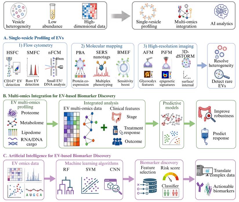
Emerging analytical strategies for EV-based biomarker discovery in colorectal cancer. Extracellular vesicle (EV)-based biomarker development in colorectal cancer (CRC) is challenged by vesicle heterogeneity, the low abundance of tumor-derived signals, and the high dimensionality of EV-associated data. (A) Single-vesicle profiling resolves inter-vesicular heterogeneity by characterizing individual EVs through flow cytometry-based platforms, molecular mapping approaches, and high-resolution imaging techniques, enabling the detection of rare and clinically informative EV subpopulations. (B) Multi-omics integration combines EV proteomic, metabolomic, lipidomic, and nucleic acid profiles with clinical features, thereby improving biomarker robustness and supporting predictive models for diagnosis, treatment response, and outcome stratification. (C) Artificial intelligence and machine learning algorithms further enhance EV-based biomarker discovery by extracting informative features from complex omics datasets, constructing classifiers or risk scores, and translating high-dimensional EV signals into clinically actionable biomarkers. These strategies shift EV biomarker research from bulk-level measurements toward high-resolution, integrated, and data-driven analytical frameworks for CRC precision oncology. Schematic illustrations were created with BioRender.com.
3.2.1 Single-vesicle Profiling of EVs
Single-vesicle analysis enables characterization of individual EVs rather than population-averaged signals. This is particularly important in CRC, where tumor-derived EVs may represent only a minute fraction of total circulating vesicles, especially in early-stage disease. By resolving EV heterogeneity at the single-particle level, these approaches provide the sensitivity needed to identify rare but biologically and clinically informative subpopulations that are obscured by bulk analysis [13].
Flow cytometry-based platforms represent a cornerstone of this field, with high-sensitivity flow cytometry (HSFC), single-molecule flow cytometry (SMFC), and nano-flow cytometry (nFCM) facilitating the precise quantification of surface protein copy numbers and particle distribution. In CRC, HSFC identified a significant enrichment of CD147+ EV in plasma, achieving superior diagnostic performance compared with total EV concentration metrics (AUC: 0.93 vs 0.63) [199]. Comparative studies further suggest that SMFC offers improved sensitivity for detecting small and low-abundance EV subpopulations [200], while nFCM extends analysis to EVs as small as 40 nm and has revealed heterogeneous distributions of EV-associated DNA, including distinct dsDNA-positive subpopulations responsive to genotoxic treatment [201]. Beyond quantitative profiling, emerging molecular mapping and spectroscopic tools provide deeper phenotypic resolution. Proximity barcoding assay (PBA) enables high-resolution mapping of surface protein co-expression and has identified CRC-associated EV subsets with distinct biological relevance and diagnostic potential [177]. Similarly, surface-enhanced raman spectroscopy (SERS) nanotags allow multiplexed phenotyping of CRC-related EV markers [202], whereas bubble multimer-enhanced fluorescence (BMEF) substantially improves analytical sensitivity for stage-discriminating markers [182]. High-resolution imaging techniques further add spatial and structural dimensions to single-vesicle analysis. Atomic force microscopy (AFM) has revealed altered surface glycocalyx features in CRC-derived EVs [203], while photo-induced force microscopy (PiFM) identified CRC-specific epigenetic signatures, including methylation-related features linked to molecular subtypes [204]. Additionally, three-dimensional direct stochastic optical reconstruction microscopy (3D-dSTORM) enabled simultaneous analysis of surface and internal protein distributions, with demonstrated value in distinguishing recurrent from non-recurrent CRC and correlating EV phenotypes with clinicopathological features [185]. Collectively, these advances move single-vesicle analysis beyond bulk quantification toward integrated molecular and structural profiling, thereby providing a powerful framework for the discovery of sensitive and specific EV-based biomarkers in CRC.
3.2.2 Multi-omics Integration for EV-based Biomarker Discovery
Integrative multi-omics profiling of EVs has emerged as a high-dimensional strategy to enhance biomarker discovery in CRC by capturing the coordinated alterations of multiple molecular layers. Compared with single-omics approaches, multi-omics integration enables comprehensive characterization of EV-associated molecular alterations, thereby enhancing the specificity and clinical relevance of identified signatures.
In CRC, integrated analyses of EV proteome, metabolome, and lipidome have effectively discriminated malignant from non-malignant states, with pathway-level insights highlighting metabolic reprogramming, particularly dysregulated fatty acid and amino acid metabolism, as a hallmark of CRC-associated EVs [14]. Similarly, integrated proteomic and lipidomic profiling of serum-derived EVs from rectal cancer patients has demonstrated that EV protein signatures outperform metabolite-based panels in predicting response to neoadjuvant radiotherapy [187]. Notably, this combined analysis further reveals shared pathways related to immune response, complement activation, and cancer-associated signaling that underpin therapeutic sensitivity [187, 205]. Beyond molecular profiling, integration of EV multi-omics datasets with clinical features facilitates the construction of robust disease classification and prediction models. Although still emerging in CRC, evidence from other malignancies, such as pancreatic cancer, demonstrates that machine-learning-driven models incorporating thousands of multi-omics and clinical features significantly outperform single-omics approaches in predicting patient outcomes [205]. Such frameworks underscore the translational potential of multi-dimensional data integration for CRC liquid biopsy.
3.2.3 Artificial Intelligence for EV-based Biomarker Discovery
The complexity and high dimensionality of EV-derived omics data necessitate analytical frameworks beyond conventional statistical approaches. Artificial intelligence (AI), particularly machine learning (ML), provides a robust framework to decipher the non-linear correlations within heterogeneous datasets, thereby unlocking the full diagnostic and prognostic potential of EV-based biomarkers.
Currently, various ML algorithms, including random forest (RF), support vector machine (SVM), and convolutional neural networks (CNN), have been applied for CRC detection. For example, combining 4D-DIA proteomics with an RF model enabled the identification of PF4 and AACT as serum EV biomarkers, which demonstrated strong early-stage CRC diagnostic performance and effective discrimination from benign colorectal conditions [181]. Similarly, an SVM-based model incorporating a five-metabolite EV signature achieved the superior discriminatory power for CRC screening (AUC = 0.985), significantly outperforming alternative statistical approaches [206]. Beyond biomarker selection, AI also strengthens the translational potential of EV-based assays by enabling integrated and scalable diagnostic platforms. For instance, the ZAHV-AI system integrates efficient EV isolation with AI-based analysis to support rapid and cost-effective nucleic acids profiling. Using plasma EV-derived miRNAs (miR-23a-3p, miR-92a-3p, miR-125a-3p, and miR-150-5p) integrated with carcinoembryonic antigen (CEA) yielded an AUC of 0.9861 for overall CRC detection and near-perfect discrimination of stage 0-I disease, surpassing individual markers [12]. Beyond primary CRC detection, ML applications demonstrate remarkable utility in classifying cancers of unknown primary origin [207]. A plasma EV-derived 29-protein signature, optimized via RF algorithms, successfully distinguished melanoma, pancreatic, lung, and colorectal cancers, with its feasibility further confirmed in patient-derived xenograft models [208]. Collectively, these advances position AI as a critical enabler for translating complex EV-derived signals into robust and clinically actionable biomarkers in CRC.
4. Therapeutic Applications of EVs in CRC
Sections 4 and 5 address two complementary levels of EV-based therapeutics. Section 4 focuses on what EV-based interventions are designed to accomplish in CRC, including therapeutic cargo delivery, vaccination, immune microenvironment reprogramming, checkpoint modulation, and disruption of tumor-promoting EV communication. Section 5 focuses on how these therapeutic functions can be technically realized and translated, including cargo-loading strategies, source selection, surface engineering, administration and biodistribution, comparison with established delivery platforms, and scalable manufacturing. Thus, Section 4 is primarily application-oriented, whereas Section 5 addresses the engineering and translational infrastructure required to convert these applications into clinically viable therapeutic platforms.
4.1 EV-Mediated Delivery of Therapeutic Cargos
EVs have emerged as a versatile and biologically compatible nanoplatform for targeted therapy in CRC. Their endogenous membrane architecture confers intrinsic stability, low immunogenicity, and efficient cellular uptake, while enabling immune evasion and prolonged circulation, collectively supporting their use as effective carriers for therapeutic delivery [10].
As natural nucleic acid delivery vehicles, EVs have been engineered to transport miRNA inhibitors [209, 210], miRNA mimics [211], and siRNAs [212, 213] for precise intervention in CRC-associated oncogenic pathways. These engineered EVs have demonstrated therapeutic efficacy by suppressing tumor growth [153], reversing EMT [211], and reducing chemoresistance and metastatic potential [71, 212] in CRC. A representative example is the EV-mediated delivery of siRNA targeting BRAFV600E, which selectively inhibited mutant CRC cells in vitro and in vivo while sparing BRAF wild-type cells [213], highlighting the potential of mutation-specific gene therapy. Beyond nucleic acids, EVs also support the delivery of other therapeutic modalities. They can be engineered for intracellular delivery of bioactive proteins, protecting them from enzymatic degradation and enhancing intracellular bioavailability to improve therapeutic efficacy. For example, EV-mediated delivery of the ribosome-inactivating protein gelonin significantly suppressed tumor growth, resulting in a 14-fold increase in therapeutic efficacy [214]. Similarly, encapsulation of drugs such as oxaliplatin or doxorubicin into EVs improves solubility, tumor accumulation, pharmacokinetics, and systemic tolerability in CRC models [215-217]. In addition, combinatorial cargo strategies further expand the therapeutic potential of EV-based platforms by enabling co-delivery of chemotherapeutic agents with regulatory RNAs [209, 212, 217] or integration of gene-silencing RNAs with metabolic inhibitors [218]. Collectively, these studies establish EVs as programmable therapeutic carriers capable of delivering diverse cargos, including nucleic acids, proteins, small-molecule drugs, and combinatorial therapeutic payloads. Strategies that further improve tumor specificity and delivery precision are discussed in Section 5.3.
4.2 Engineered EV-Based Vaccine Platforms
EV-based vaccines have emerged as a promising cell-free immunotherapeutic strategy in CRC by enabling efficient antigen delivery and immune activation while overcoming the limitations of conventional cell-based vaccines. Unlike traditional approaches, EVs provide a stable and scalable platform for antigen presentation, offering improved safety, manufacturability, and therapeutic versatility.
4.2.1 DC-Derived EV Vaccines
DC-derived EVs (DEVs) represent the most extensively studied EV-based cancer vaccine platform [219]. Enriched in major histocompatibility complexes and costimulatory molecules, DEVs retain intrinsic antigen-presenting capacity, enabling the activation of tumor-specific immune responses without the limitations associated with live DC-based vaccines [220]. Therapeutically, DEV-based vaccines have demonstrated robust antitumor efficacy in preclinical models. Neoantigen-loaded DEV can elicit strong adaptive immune responses, resulting in tumor suppression and durable immune memory [221]. Their efficacy can be further enhanced through engineering strategies. For example, bispecific DEV designed to simultaneously target tumor antigens and modulate immune checkpoints have shown improved therapeutic outcomes in CRC models [222], highlighting the benefit of combining antigen presentation with immune regulation. Importantly, the immunostimulatory activity of DEV has been validated in early-phase clinical studies in melanoma and non-small cell lung cancer, supporting their translational potential [223, 224]. Although clinical evidence in colorectal cancer remains limited, these findings establish DEV as a promising platform for EV-based cancer vaccination.
Engineered EV-based vaccines for colorectal cancer therapy. Engineered extracellular vesicle (EV)-based vaccines provide cell-free platforms for antigen delivery and immune activation in colorectal cancer (CRC). (A) Dendritic cell-derived EVs retain antigen-presenting and costimulatory capacity, which can be enhanced by neoantigen loading or bispecific designs. (B) Tumor-derived EVs carry endogenous tumor antigens but require engineering, such as IFN/IRF1 conditioning, adjuvant loading, anti-PD-1 combination, or TEV-like vesicle design, to improve immunogenicity and reduce immunosuppressive effects. (C) Bacteria-derived EVs, particularly outer membrane vesicles, provide built-in adjuvant activity through pathogen-associated molecular patterns and can be engineered for antigen display or checkpoint modulation. Together, these platforms promote dendritic cell maturation, activate cytotoxic and helper T-cell responses, induce memory T cells, and enhance durable antitumor immunity. Schematic illustrations were created with BioRender.com.
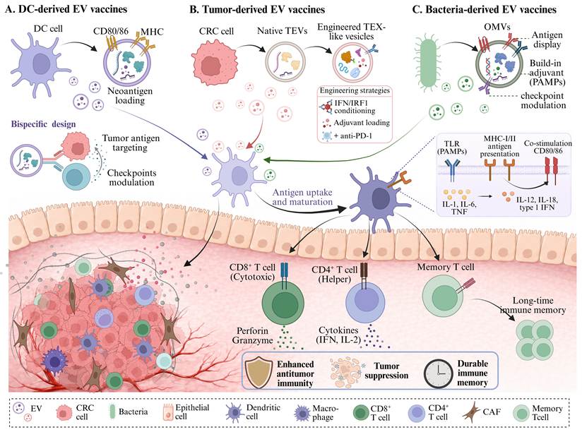
4.2.2 Tumor-Derived EV Vaccines
Tumor-derived EVs (TEVs) provide an alternative vaccine strategy by leveraging their endogenous repertoire of tumor-associated antigens. Preclinical studies have shown that TEV-based vaccination can induce antitumor immune responses and inhibit tumor progression in CRC models [225]. To improve therapeutic efficacy, current strategies focus on enhancing TEV immunogenicity and functionality, including modulation of parental tumor cells, incorporation of immune adjuvants, and combination with immune checkpoint blockade. For instance, interferon- or IRF-1-conditioned TEVs exhibit enhanced antigen-presenting capacity and T cell activation [226], while TEV-based vaccines combined with GM-CSF or anti-PD-1 therapy further augments therapeutic efficacy [227]. However, a major limitation of TEV-based vaccines is their potential to induce immunosuppressive signaling [228]. To address this, engineered TEV-like nanovesicles have been developed to enhance antigen specificity while minimizing immunosuppressive effects [229, 230]. For instance, FAP-targeting TEV-like vesicles can simultaneously induce cytotoxic T cell responses against tumor cells and stromal components, thereby improving therapeutic efficacy [230]. Overall, TEV-based vaccines exploit the intrinsic antigenic landscape of tumor cells while requiring careful engineering to balance immunogenicity and immunosuppression.
4.2.3 Bacteria-Derived EV Vaccines
Bacteria-derived EVs (BEVs), particularly outer membrane vesicles (OMVs), have recently emerged as a highly immunogenic vaccine platform. Unlike mammalian EVs, BEVs intrinsically contain pathogen-associated molecular patterns (PAMPs), which provide a built-in adjuvant effect and support their use as immunogenic vaccine carriers [231]. Preclinical studies showed that systemically administered OMVs preferentially accumulated in tumors, inhibited tumor growth in an IFN-γ-dependent manner, and induced durable immune memory, establishing their feasibility as vaccine-like immunostimulatory nanoplatforms [232]. To improve specificity, bioengineering approaches have been developed to display tumor antigens on OMV surfaces, enabling multi-antigen presentation and enhanced immune activation [233]. This strategy has been further extended to neoantigen delivery, as recombinant OMVs expressing CT26-derived polytope neoantigens showed strong immunogenicity and antitumor activity in vitro and in vivo [234-236]. Moreover, BEV-based vaccines can be combined with immune-modulating strategies to further improve therapeutic efficacy. For example, OMV-based platforms have been engineered to deliver tumor antigens alongside immune checkpoint modulators, resulting in synergistic antitumor effects [237]. These systems also support personalized vaccine design through modular antigen display technologies. Importantly, the clinical use of licensed OMV-containing vaccines such as Bexsero® demonstrates that selected bacterial OMV formulations can be manufactured and administered in humans [238]. However, this experience should not be extrapolated to establish the safety or efficacy of OMV-based cancer vaccines. In CRC, evidence for engineered OMV vaccine platforms remains predominantly preclinical, and key translational issues, including tumor-antigen selection, systemic reactogenicity, biodistribution, dose optimization, and manufacturing consistency, require dedicated clinical evaluation. Thus, BEV/OMV-based cancer vaccines represent a promising but still early-stage therapeutic platform for CRC.
4.3 Engineered EVs for Remodeling the Immunosuppressive TME
Engineered EVs have emerged as programmable platforms to remodel the immunosuppressive TME through coordinated reprogramming of multiple immune compartments. Rather than acting on single pathways, EV-based strategies simultaneously target TAMs, DCs, and T cell subsets, thereby converting an immune-suppressive niche into an immune-reactive state.
Reprogramming TAMs represents a central therapeutic entry point. EV-mediated delivery of STAT6 antisense oligonucleotides effectively induces M2-to-M1 polarization, restores CD8⁺ T cell cytotoxicity, and leads to robust tumor regression in preclinical models [9]. This effect can be further enhanced by engineering EVs with targeting and functional modules. Dual-targeted EVs enable selective delivery to M2 TAMs and improve reprogramming efficiency, while M1 macrophage-derived EVs integrated with therapeutic components such as photosensitizers or nitric oxide-generating systems further amplify antitumor efficacy through combinatorial mechanisms [239, 240]. Beyond macrophage targeting, engineered EVs also reprogram adaptive immunity. Delivery of immunomodulatory cargos such as miR-155 restores DC function and enhances downstream T cell-mediated antitumor responses, thereby alleviating immunosuppression [241]. In parallel, engineered EVs directly correct T cell imbalance within the TME. Delivery of cargos exemplified by miR-206 enhances cytotoxic T cell activity while limiting Treg expansion, reinforcing antitumor immunity [242]. Collectively, engineered EVs remodel the immunosuppressive TME through coordinated, multi-layered immune reprogramming, establishing a therapeutically actionable framework for CRC immunotherapy.
4.4 Targeting EV-Associated Checkpoint Signaling to Enhance Immunotherapy
In addition to cellular reprogramming, EVs critically regulate immune checkpoint signaling and influence the efficacy of immune checkpoint blockade (ICB) therapies. A major mechanism involves modulation of PD-L1 expression and its EV-associated forms, which play key roles in systemic immunosuppression. Pharmacological inhibition of upstream pathways, such as JAK2/STAT3, reduces PD-L1 levels in both tumor cells and EVs, thereby restoring T cell activity [243]. In parallel, EV cargo remodeling can improve responsiveness to ICB [244]; for example, EVs enriched in chemokines promote recruitment of CD8⁺ T cells and NK cells, enhancing sensitivity to PD-1 blockade [245]. Building on these mechanisms, engineered EVs have been developed to directly inhibit checkpoint signaling. EVs loaded with PD-L1 and CTLA-4 siRNA effectively suppress checkpoint signaling, leading to enhanced CD8⁺ T cell activity and reduced Treg infiltration in vivo [246]. Similarly, milk-derived EV-based nanoplatforms such as Super PD-L1exo downregulate PD-L1 expression and promote cytotoxic T cell infiltration [247]. More advanced platforms integrate checkpoint inhibition with cytotoxic modules; for example, KDEL-modified EVs co-delivering PD-L1 siRNA induce immunogenic cell death and amplify antitumor immune responses, achieving synergistic therapeutic effects [248]. Importantly, EV-associated PD-L1 is also a functional handle for improving adoptive T cell therapies. Tumor-derived PD-L1⁺ EVs can facilitate T cell migration to tumor sites and enhance infiltration of effector memory CD8⁺ T cells, thereby improving the tumor-targeting capacity of CAR-T cells [249].
Collectively, EVs occupy a dual position in checkpoint immunotherapy: they act as mediators of immune evasion and therapeutic resistance, yet also provide exploitable platforms for checkpoint-targeted intervention and combinatorial immunotherapy. This duality makes EV-associated checkpoint signaling, particularly PD-L1-centered pathways, a promising axis for optimizing ICB efficacy and overcoming resistance.
4.5 Targeting the Endogenous EV Life Cycle to Disrupt Tumor Communication
Given the central role of EVs in CRC progression and the increased EV output of cancer cells relative to normal cells, inhibition of EV biogenesis, secretion, or uptake has emerged as a rational therapeutic strategy.
One major strategy is to inhibit EV biogenesis and secretion. EV formation occurs within multivesicular bodies (MVBs) and is regulated by both ESCRT-dependent and ESCRT-independent pathways [250]. In the ESCRT-dependent pathway, components such as TSG101 and CHMP5 regulate EV production, and their depletion reduces EV secretion in CRC [251, 252], highlighting the ESCRT machinery as a potential therapeutic target. In contrast, the ESCRT-independent pathway is primarily linked to ceramide metabolism, where pharmacological inhibition of ceramide generation (e.g., GW4869) reduces EV release and associated cargo, with demonstrated antitumor activity in CRC [11]. In addition to MVB formation, EV secretion is regulated by vesicle trafficking machinery, including SNARE proteins, Rab GTPases, and Ca2⁺-dependent processes [62]. In CRC, dysregulation of these pathways (e.g., SEC22B upregulation or KRAS-RAB13 signaling) enhances EV release, whereas their inhibition suppresses EV-mediated effects [27, 28, 253].
A second therapeutic approach is to inhibit EV uptake by recipient cells, thereby disrupting EV-mediated communication. EV internalization occurs via membrane interaction, fusion, and endocytic pathways, which are amenable to pharmacological inhibition [254, 255]. In this context, EV surface molecules represent key determinants of uptake specificity and provide actionable therapeutic targets [175, 256, 257]. In RAS-transformed cells, oncogenic RAS has been shown to promote CRAF-dependent macropinocytosis and enhanced EV uptake [256], while CD166 (ALCAM) has been implicated in the binding and uptake of cancer-derived EVs [257]. Beyond targeting individual molecules, modulation of EV surface properties offers an additional strategy to regulate cellular interactions. Surface glycosylation, for instance, influences EV uptake [258], and artificial glycosylation can be exploited to improve therapeutic delivery [254]. Similarly, engineering the EV biomolecular corona provides a means to modulate EV behavior, as exemplified by albumin enrichment, which prolongs circulation and reduces macrophage uptake, while removal of endogenous components can enhance functional activity [255]. Notably, EV uptake exhibits heterogeneity across CRC cell populations, as illustrated by IFITM1-dependent differences in uptake capacity [259], suggesting that cell-intrinsic variability should be considered in the design of EV-targeted therapies.
5. Engineering and Translational Optimization of EV Therapeutics in CRC
While EV-based therapeutic strategies have demonstrated substantial preclinical efficacy, their successful clinical translation critically depends on rational engineering design and scalable manufacturing approaches.
5.1 Engineering Strategies for Cargo Loading
Whereas Section 4.1 focuses on the therapeutic cargos delivered by EVs and their biological effects in CRC, this section focuses specifically on the engineering methods used to incorporate these cargos into EVs. Advanced bioengineering techniques have refined the methods for loading therapeutic cargo into EVs, broadly categorized into pre-loading (endogenous loading), post-loading (exogenous loading), each tailored to different applications [16]. In brief, pre-loading exploits EV biogenesis to achieve endogenous cargo encapsulation and is simple and suitable for nucleic acids and proteins, but is limited by transfection efficiency and may perturb cellular homeostasis. In contrast, post-loading introduces cargos after EV isolation, offering greater control; methods such as electroporation, ultrasound, and freeze-thaw enhance loading efficiency (with ultrasound often showing superior performance), but may compromise membrane integrity and cargo stability [260]. To overcome the limitations of conventional loading techniques, next-generation technologies have been developed. Microfluidic systems have been shown to significantly enhance loading efficiency under controlled flow conditions, for example increasing doxorubicin encapsulation from 7.86% to 31.98%. In parallel, acoustofluidic platforms enable one-step, reproducible drug loading with improved scalability [261, 262]. These technologies may be particularly attractive for CRC therapy, where controlled dosing, scalability, and regulatory compliance are essential for clinical translation [16, 261, 262].
5.2 Optimizing EV Sources for Therapeutic Application
The choice of donor source fundamentally shapes EV composition, therapeutic function, and translational feasibility. In CRC therapy, selection of an appropriate donor source must account for scalability, immunogenicity, intrinsic bioactivity, and compatibility with intended therapeutic payloads.
5.2.1 Mammalian Cell-Derived EVs
EVs derived from stem cells, tumor cells, and immune cells exhibit distinct biological properties, informing their application-specific suitability. MSC-derived EVs are widely used due to scalable culture conditions, high proliferation, and strong immunomodulatory activity. For instance, MSC-EVs have been engineered to deliver functional miRNAs or chemotherapeutic agents, demonstrating effective cargo delivery and enhanced intratumoral accumulation in CRC models [81, 210, 263]. Tumor-derived EVs, such as from CT26 cells, exhibit preferential uptake by parental tumor cells and superior tumor penetration, although safety concerns necessitate cautious selection [264]. HEK293T-derived EVs are frequently used due to their high production efficiency and ease of genetic modification, enabling delivery of therapeutic RNAs and chemotherapeutic agents [217, 228]. Immune cell-derived EVs exhibit intrinsic immunostimulatory properties, making them suitable for delivering immune modulators and enhancing antitumor immunity [260].
5.2.2 Non-Mammalian Cell-Derived EVs
Alternative EV sources, including bacteria, milk, and plants, have emerged as complementary platforms to enhance therapeutic performance. Bacterial OMVs exhibit strong immunogenicity and enable efficient antigen delivery, supporting their use in immunotherapy-oriented strategies [233]. Milk-derived EVs demonstrate high stability in the gastrointestinal tract and are well suited for oral delivery, offering a practical route for non-invasive administration [265]. Plant-derived EVs offer advantages such as low toxicity, high yield, and cost-effective production. A recent study developed a hybrid therapeutic platform combining plant-derived EVs with a dendrimer-based micelle nanoparticle loaded with an miR-146 mimic. This system achieved a fivefold increase in miRNA uptake compared with either component alone and demonstrated effective cargo delivery and antitumor activity in mouse models [266].
5.2.3 Bio-mimetic EV Platforms
Bio-mimetic EVs, including artificially synthesized EV-like nanoparticles, hybrid EVs, EV membrane-camouflaged nanoparticles and physical-origin nanovesicles, have recently been developed to overcome the key limitations of natural EVs [17]. These platforms may address some limitations of natural EVs by increasing production yield and enabling design flexibility, although compositional control and manufacturing standardization remain important translational challenges [267, 268]. Different bio-mimetic strategies provide complementary advantages. Artificial EV-like nanoparticles enable precise compositional control and reproducible fabrication; hybrid EVs combine the biological functionality of natural EVs with the engineering flexibility of synthetic nanocarriers; EV membrane-camouflaged nanoparticles integrate immune evasion and targeting capability with high loading efficiency; and EV-mimetic nanovesicles achieve substantially increased production yield while retaining essential biomimetic properties. In a CT26 colon adenocarcinoma mouse model, doxorubicin-loaded nanovesicles inhibited tumor growth as effectively as natural EVs, and achieved 20-fold greater efficacy than free doxorubicin, without obvious systemic side effects [268], suggesting that nanovesicles are a promising alternative to natural EVs for chemotherapeutic delivery.
5.3 Strategies to Enhance EV Targeting Specificity
Distinct from Section 4.5, which focuses on therapeutic disruption of endogenous tumor-associated EV biogenesis, secretion, and uptake, this section addresses the engineering of administered therapeutic EVs to enhance tissue- and cell-specific delivery. The therapeutic efficacy of EV-based platforms depends not only on cargo potency but also on their ability to reach tumor cells or disease-relevant microenvironments with sufficient specificity. However, like many nanocarriers, systemically administered EVs are frequently sequestered by the liver, which limits delivery efficiency to extrahepatic tumors such as CRC.
To overcome this limitation, surface engineering strategies have been developed to enhance targeting specificity. A key approach involves functionalizing EV membranes with targeting ligands such as peptides [18], antibodies, or aptamers [263, 269] to enable receptor-specific delivery to tumor cells. Peptide-mediated targeting has been widely explored due to its small size and ease of modification. For example, DOX-loaded EVs modified with the EGFR-targeting peptide GE11 demonstrated enhanced uptake in EGFR-positive tumor cells [18]. Aptamer-based targeting strategies further expand the versatility of EV engineering. For example, AS1411 (anti-nucleolin) or MUC1 aptamer-functionalized EVs enable prolonged circulation and preferential tumor accumulation, thereby enhancing the delivery and intra-tumoral retention of chemotherapeutic agents such as doxorubicin in CRC models [269]. In CRC, an anti-PD-L1-functionalized exosome platform carrying apatinib has been investigated to combine tumor-targeted delivery with immune reprogramming [15]. Dual-targeted EV platforms combining tumor-targeting ligands with immune-modulatory components have demonstrated the capacity to simultaneously enhance tumor specificity and remodel the tumor microenvironment, thereby improving overall therapeutic outcomes [15]. These strategies indicate that EV surface engineering should be viewed not merely as a delivery enhancement technique, but as a key determinant of therapeutic selectivity, biodistribution, and clinical translatability.
5.4 Administration Routes and Biodistribution in CRC
The route of administration is a major determinant of EV biodistribution and should be matched to the anatomical and metastatic characteristics of CRC. Intravenous administration provides systemic exposure and is therefore attractive for disseminated or metastatic disease; however, intravenously administered EVs are rapidly exposed to phagocytic clearance and frequently accumulate in the liver and spleen. In CRC, this hepatic distribution may reduce delivery to extrahepatic primary lesions, whereas in colorectal liver metastasis it could potentially be exploited for liver-directed therapy if tumor-selective uptake can be distinguished from nonspecific uptake by resident phagocytic cells.
Intraperitoneal administration provides an alternative strategy for peritoneal dissemination by increasing local exposure while limiting systemic distribution. In a preclinical colorectal carcinomatosis model, intraperitoneally administered MSC-derived EVs loaded with the photosensitizer mTHPC showed markedly greater tumor selectivity than either the free drug or a liposomal formulation [270]. Nevertheless, penetration into larger lesions, dose reproducibility, and clinical scalability remain unresolved. Oral administration has been explored particularly with milk- and plant-derived EVs because some preparations can retain vesicular integrity or protect therapeutic cargo under gastrointestinal conditions and access the intestinal mucosa [271]. However, gastrointestinal stability is source- and formulation-dependent, and mucus penetration, epithelial uptake, gastrointestinal variability, and quantitative dose delivery remain important barriers. Rectal or intracolonic administration offers a potential route for localized colonic exposure without upper gastrointestinal transit. Recent proof-of-concept studies in inflammatory colonic disease have demonstrated that locally administered EV formulations can enhance mucosal retention and sustained delivery, although CRC-specific evidence remains limited [272]. Thus, local retention, mucosal coverage, and formulation design require further investigation.
Thus, administration route should be integrated with EV source, cargo properties, targeting strategy, primary tumor location, and metastatic pattern rather than treated as a purely procedural variable.
5.5 Positioning EVs Relative to Established Therapeutic Delivery Platforms
EVs should be considered alongside established therapeutic delivery systems rather than in isolation (Table 4) [273-277]. Lipid nanoparticles (LNPs) provide efficient nucleic-acid encapsulation and comparatively mature, scalable manufacturing, although systemic formulations remain strongly influenced by hepatic biodistribution and extrahepatic targeting remains challenging [273]. Adeno-associated virus (AAV) vectors can support efficient and durable transgene expression but are constrained by limited cargo capacity, pre-existing or treatment-induced immunity, and difficulties with repeated administration [275]. Liposomes and polymeric nanocarriers offer chemically defined and highly tunable formulations, but tumor penetration, targeting efficiency, formulation reproducibility, and scale-up remain context-dependent challenges [276, 277]. EVs differ from these platforms in their biomimetic membrane composition and capacity to transport diverse molecular cargos, but they face substantial limitations in product heterogeneity, cargo loading, dose definition, batch consistency, systemic clearance, and large-scale manufacturing [273, 274]. Thus, EVs should not be viewed as intrinsically superior to established delivery systems. In CRC, platform selection should instead be guided by the therapeutic payload, required duration of activity, target site, need for repeated administration, and manufacturing feasibility.
Translational comparison of extracellular vesicles with established therapeutic delivery platforms
| Platform | Typical Cargo / Functional Features | Targeting and Biodistribution | Major Advantages | Major Limitations | Clinical / Translational Status |
|---|---|---|---|---|---|
| Extracellular vesicles (EVs) [273, 274] | Nucleic acids, proteins, small molecules; native membrane proteins and lipids can contribute to cell interactions | Source-dependent biodistribution and tropism; substantial mononuclear phagocyte system uptake; targeting can be enhanced by surface engineering | High biocompatibility; natural intercellular cargo transfer; accommodates diverse cargo classes; membrane can support biological targeting | Source and product heterogeneity; variable yield and cargo loading; rapid systemic clearance; batch consistency, potency assays, and GMP manufacturing remain challenging | Preclinical and early clinical development; active bioengineering, manufacturing, and regulatory standardization efforts |
| Lipid nanoparticles (LNPs) [273] | Primarily mRNA, siRNA, and other nucleic-acid therapeutics; chemically defined lipid formulations | Systemic formulations often show liver-biased distribution; ionizable lipid composition and ligand engineering can modify tissue targeting | Efficient nucleic-acid encapsulation and cytosolic delivery; scalable and reproducible manufacturing; multiple approved products | Extrahepatic targeting remains difficult; endosomal escape is incomplete; formulation-dependent inflammatory or tolerability issues may occur | Clinically mature nucleic-acid delivery platform with approved siRNA and mRNA products; extensive manufacturing and regulatory experience |
| Adeno-associated virus (AAV) [275] | DNA transgenes with durable expression; packaging capacity is limited (~4.7 kb) | Serotype- and capsid-dependent tissue tropism; capsid engineering can alter organ and cell targeting | High transduction efficiency in selected tissues; sustained transgene expression; several approved gene therapies | Limited cargo capacity; pre-existing and treatment-induced immunity; redosing is difficult; dose-related toxicity and off-target transduction remain concerns | Clinically validated gene-transfer platform with multiple approved products; generally optimized for durable, often single-administration gene delivery |
| Liposomes [276] | Hydrophilic or lipophilic small molecules; selected nucleic acids and combination formulations | Passive tumor accumulation is variable; PEGylation, ligands, and stimuli-responsive designs can modify circulation and targeting | Clinically established; flexible drug encapsulation; can improve pharmacokinetics and reduce toxicity of conventional drugs | Variable tumor penetration and EPR dependence; stability and premature drug release can limit performance; reticuloendothelial clearance and repeated-dose reactions may occur | Mature nanomedicine platform with multiple approved formulations, including established oncology products |
| Polymeric nanoparticles / micelles [277] | Small molecules, proteins, and nucleic acids; tunable polymers enable controlled release and surface functionalization | Size, charge, polymer composition, and ligands can be engineered for passive or active targeting | Highly tunable physicochemical properties; controlled drug release; modular surface chemistry | Complex formulation optimization; scale-up and batch reproducibility; purification, storage, and regulatory standardization remain barriers | Broad preclinical and clinical investigation, but overall clinical translation is more limited than for lipid-based nanocarriers |
EV, extracellular vesicle; LNP, lipid nanoparticle; AAV, adeno-associated virus; siRNA, small interfering RNA; mRNA, messenger RNA; EPR, enhanced permeability and retention; GMP, good manufacturing practice.
5.6 Manufacturing and Purification Strategies for Clinical Translation
Despite advances in EV engineering, their therapeutic potential in CRC is ultimately constrained by challenges in scalable production, standardization, and purity. Intrinsic heterogeneity in EV size, composition, and morphology further complicates clinical-grade manufacturing.
To address these limitations, emerging bioengineering strategies have focused on improving both yield and quality. Microfluidic systems enable precise control of culture conditions, enhancing EV yield and quality. For example, shear stress-regulated microfluidic culture (0.5-5 dyn/cm²) markedly increases EV production from MSCs while preserving vesicle integrity. Beyond production, microfluidic platforms have also emerged as efficient tools for EV isolation, integrating size-based separation and affinity capture to overcome the low recovery and poor scalability of conventional methods such as ultracentrifugation. For instance, a porous microfluidic chip coupled with anti-CD9 capture significantly improves EV enrichment efficiency compared with traditional approaches [178]. In parallel, affinity-based bead-assisted strategies provide a simple and rapid alternative for EV enrichment. DSPE-functionalized beads enable direct capture of EVs from biological fluids within minutes without the need for specialized equipment, achieving efficient and broadly applicable EV enrichment [129]. Collectively, these strategies establish a more efficient and scalable framework for EV production and purification, thereby strengthening the translational potential of EV-based therapeutics in CRC.
Engineered EVs remodel the immunosuppressive tumor microenvironment in colorectal cancer. Engineered extracellular vesicles (EVs) provide programmable platforms for converting an immunosuppressive tumor microenvironment (TME) into an immune-reactive state in colorectal cancer (CRC). By delivering modular cargos, including nucleic acids, proteins, small molecules, and surface-engineered functional components, EV-based strategies can coordinately reprogram multiple immune compartments. These approaches promote M2-to-M1 tumor-associated macrophage (TAM) polarization, enhance dendritic cell (DC) maturation and antigen presentation, restore cytotoxic CD8⁺ T-cell activity, and limit regulatory T-cell expansion. In parallel, EV-based platforms can target checkpoint-associated signaling by suppressing PD-L1/PD-1 or CTLA-4 pathways, while chemokine-enriched EVs facilitate recruitment of CD8⁺ T cells and NK cells. Tumor-derived PD-L1⁺ EVs may also be exploited to improve CAR-T cell trafficking and tumor infiltration. Schematic illustrations were created with BioRender.com.
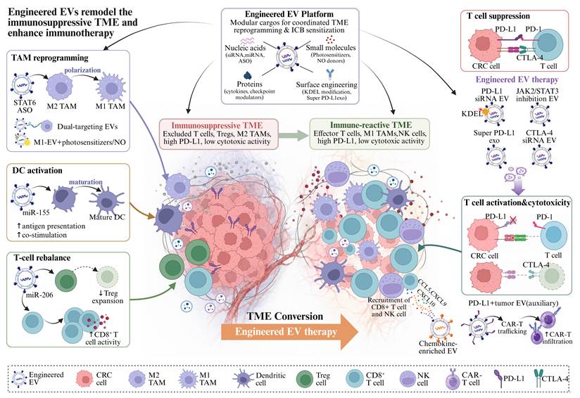
Strategies to improve the therapeutic potential of EVs in colorectal cancer. The clinical translation of extracellular vesicle (EV)-based therapeutics in colorectal cancer (CRC) depends on coordinated optimization of engineering design and manufacturing workflows. (A) Cargo loading strategies include pre-loading through donor-cell engineering, post-loading into isolated EVs, and next-generation platforms such as microfluidic and acoustofluidic systems, which improve loading efficiency and reproducibility. (B) EV source optimization involves selecting appropriate mammalian, non-mammalian, or bio-mimetic platforms according to scalability, bioactivity, immunogenicity, and manufacturability. (C) Targeting specificity can be enhanced through surface engineering with peptides, antibodies, or aptamers, as well as multifunctional or dual-targeting designs that improve tumor accumulation and reduce off-target delivery, particularly liver sequestration. (D) Manufacturing and purification strategies, including shear stress-regulated microfluidic culture, on-chip microfluidic isolation, and bead-assisted affinity enrichment, further improve EV yield, purity, and scalability. Schematic illustrations were created with BioRender.com.
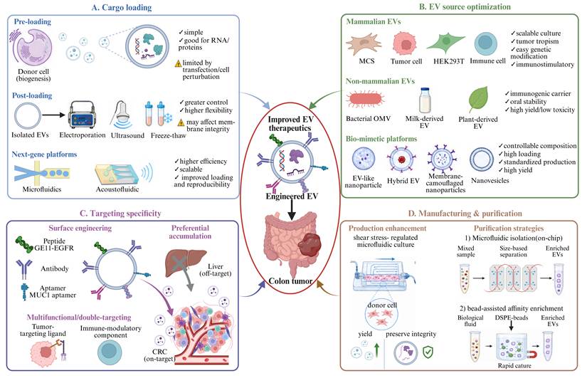
6. Challenges and Future Perspectives
Despite substantial progress, the clinical translation of EV-based strategies in CRC remains constrained by several interrelated challenges. Intrinsic EV heterogeneity arising from biogenesis, cargo composition, cellular origin, and species source creates context-dependent functions that complicate mechanistic studies, biomarker discovery, and therapeutic design. Different EV subpopulations may exert opposing pro- or anti-tumor effects, making functional interpretation challenging. Population-level analyses yield averaged metrics essential for scalable biomarker discovery and production yet obscure rare and functionally distinct EV subpopulations. High-resolution analytical technologies, including single-vesicle profiling combined with multi-omics integration, computational modeling, and AI-assisted analytics, enable linkage of EV heterogeneity with clinical phenotypes. However, low abundance of tumor-derived EVs, molecular variability, cargo instability, and insufficient cohort validation remain critical barriers.
Beyond heterogeneity, the design and optimization of EV-based therapeutics face multiple interrelated challenges, including EV source selection, engineering strategies, and in vivo barriers. EV source critically influences immunogenicity, yield, safety, and functional cargo compatibility. Autologous EVs offer low immunogenicity but limited scalability, whereas allogeneic or engineered sources provide higher yield yet introduce donor variability or tumorigenicity risks. Engineering strategies, including cargo loading, surface modification, targeted delivery, and immune modulation, aim to enhance therapeutic efficacy while preserving native biological activity. However, engineered EVs face batch-to-batch variability, unstable modifications, suboptimal in vivo stability, poor targeting specificity, and unassessed long-term safety. Additional limitations include rapid clearance by the mononuclear phagocyte system, inefficient intracellular trafficking, and incomplete cargo release.
Manufacturing and standardization remain major hurdles, as efficient, scalable, and reproducible EV isolation, characterization, and production are lacking. Current isolation methods (e.g., ultracentrifugation, precipitation) face trade-offs between yield, purity, and scalability, while emerging microfluidic and affinity-based approaches require further validation for clinical-grade production. Microfluidic technologies offer several advantages over conventional EV isolation methods. They enable precise fluid control, reduced reagent consumption, and automated workflows, leading to improved yield, purity, and consistency while minimizing sample processing time. By integrating size-based filtration, immunoaffinity capture, or inertial focusing, microfluidic platforms can achieve high recovery efficiency and enhanced selectivity for rare functional EV subpopulations. Additionally, closed-loop designs with in-line quality control sensors facilitate real-time monitoring of EV size, concentration, and purity, supporting scalable and reproducible production. However, current microfluidic systems face challenges in throughput, cost-effective fabrication, and sterility assurance at clinical scales, and their widespread adoption for GMP-compliant EV manufacturing remains limited, requiring further validation and standardization.
Clinical translation remains constrained by inadequate preclinical models that incompletely recapitulate the human tumor microenvironment, limited clinical trial data, and insufficient characterization of optimal dosage, administration routes, predictive biomarkers, long-term efficacy, and adverse effects across CRC subtypes, including metastatic, chemotherapy-resistant, and MSI/MSS tumors. The absence of large-scale, randomized controlled trials further hinders clinical adoption.
Importantly, these translational challenges are unlikely to be uniform across CRC clinical contexts. In dMMR/MSI-H CRC, where immune-checkpoint blockade can produce substantial and durable responses, EV profiling may provide a dynamic complement to tissue-based biomarkers for monitoring immune escape and acquired therapeutic resistance. By contrast, pMMR/MSS CRC remains substantially less responsive to checkpoint blockade and is biologically heterogeneous; here, EV-defined immune, stromal, or metabolic states may help identify biologically selected subgroups that are more likely to benefit from rational combination immunotherapy. In metastatic CRC, particularly in patients with liver metastases, EV-based approaches face a distinct biodistribution problem: the prominent hepatic sequestration of systemically administered EVs may represent both an off-target clearance mechanism and a potential opportunity for liver-directed delivery, provided that reticuloendothelial uptake can be distinguished from metastasis-selective targeting. In chemotherapy-resistant disease, longitudinal EV RNA, protein, and DNA signatures may enable dynamic tracking of treatment-induced adaptation and emerging resistance. However, prospective comparison with established approaches, including ctDNA, conventional serum markers, and imaging, will be essential before these applications can enter routine clinical practice.
From a translational perspective, future priorities should also be distinguished by clinical readiness. In the near term, the most feasible goals include standardization of pre-analytical workflows, reproducible EV isolation and characterization, prospective validation of focused EV biomarker panels, integration of EV readouts with established clinical assays, optimization of administration routes, and development of relatively simple targeting modifications compatible with GMP manufacturing. These approaches build directly on technologies that are already experimentally mature and can therefore be evaluated in prospective clinical cohorts. In the longer term, population-scale single-EV multi-omics, AI-guided patient-specific EV signatures, personalized EV vaccine platforms, multifunctional EVs carrying multiple therapeutic cargos, and closed-loop manufacturing with real-time quality control could enable more individualized intervention. However, these strategies will require substantially greater advances in analytical standardization, manufacturing reproducibility, safety assessment, and prospective clinical validation.
7. Conclusions
In conclusion, EVs serve as key mediators of intercellular communication and have witnessed substantial research and technological advances. These include precise identification through single-vesicle analysis and surface-enhanced Raman spectroscopy, functional characterization via multi-omics integration and single-vesicle sequencing, and efficient isolation using microfluidic platforms. These innovations have deepened our understanding of EV biology, revealing their roles in tumor progression, metastasis, and immune modulation. Moreover, they have catalyzed the development of EV-based applications in liquid biopsy diagnostics and therapeutic platforms. Despite these advances, challenges remain, including standardization of isolation and characterization methods, scalable production, optimization of in vivo stability and targeting specificity, and rigorous validation in clinical settings. Addressing these challenges will be essential to translate EV research into precision diagnostic and therapeutic strategies, thereby realizing the full theranostic potential of EVs in CRC and other malignancies.
Abbreviations
CRC: colorectal cancer; EVs: extracellular vesicles; ILVs: intraluminal vesicles; MVBs: multivesicular bodies; MISEV: Minimal Information for Studies of Extracellular Vesicles; PD-L1: programmed death-ligand 1; TRAIL: TNF-related apoptosis-inducing ligand; miRNAs: microRNAs; siRNAs: small interfering RNAs; rRNAs: ribosomal RNAs; lncRNAs: long noncoding RNAs; circRNAs: circular RNAs; mRNAs: messenger RNAs; RBP: RNA-binding protein; dsDNA: double-stranded DNA; ssDNA: single-stranded DNA; mtDNA: mitochondrial DNA; gDNA: genomic DNA; cGAS: cyclic GMP-AMP synthase; STING: stimulator of interferon genes; AIM2: absent in melanoma 2; TME: tumor microenvironment; CAFs: cancer-associated fibroblasts; ECM: extracellular matrix; EMT: epithelial-mesenchymal transition; PMN: premetastatic niche; MSCs: mesenchymal stromal/stem cells; MSC-EVs: mesenchymal stromal/stem cell-derived extracellular vesicles; FnEVs: Fusobacterium nucleatum-derived extracellular vesicles; NK cells: natural killer cells; P-EVs: plant-derived extracellular vesicles; HR: hazard ratio; AUC: area under the receiver operating characteristic curve; tsRNAs: tRNA-derived small RNAs; ncRNAs: non-coding RNAs; GEO: Gene Expression Omnibus; mCRC: metastatic colorectal cancer; BEVs: bacterial extracellular vesicles; DCs: dendritic cells; DEVs: dendritic cell-derived extracellular vesicles; TEVs: tumor-derived extracellular vesicles; IFN: interferon; IRF-1: interferon regulatory factor 1; GM-CSF: granulocyte-macrophage colony-stimulating factor; OMVs: outer membrane vesicles; PAMPs: pathogen-associated molecular patterns; FDA: Food and Drug Administration; TAMs: tumor-associated macrophages; STAT6: signal transducer and activator of transcription 6; ASO: antisense oligonucleotide; Tregs: regulatory T cells; ICB: immune checkpoint blockade; PD-1: programmed cell death protein 1; CTLA-4: cytotoxic T-lymphocyte-associated protein 4; CAR-T cells: chimeric antigen receptor T cells; ESCRT: endosomal sorting complex required for transport; BM-MSCs: bone marrow-derived mesenchymal stromal/stem cells; UC-MSCs: umbilical cord-derived mesenchymal stromal/stem cells; 4D-DIA: four-dimensional data-independent acquisition; AAV: adeno-associated virus; AFM: atomic force microscopy; AI: artificial intelligence; BMEF: bubble multimer-enhanced fluorescence; cfDNA: cell-free DNA; CNN: convolutional neural network; dMMR: deficient mismatch repair; DSS: dextran sulfate sodium; DSPE: 1,2-distearoyl-sn-glycero-3-phosphoethanolamine; ER: endoplasmic reticulum; FAP: fibroblast activation protein; GMP: good manufacturing practice; HSFC: high-sensitivity flow cytometry; IFN-γ: interferon gamma; LNPs: lipid nanoparticles; ML: machine learning; MSI: microsatellite instability; MSI-H: microsatellite instability-high; MSS: microsatellite-stable; nFCM: nano-flow cytometry; PBA: proximity barcoding assay; pMMR: proficient mismatch repair; PiFM: photo-induced force microscopy; RF: random forest; ROS: reactive oxygen species; SERS: surface-enhanced Raman spectroscopy; SMFC: single-molecule flow cytometry; SVM: support vector machine; 3D-dSTORM: three-dimensional direct stochastic optical reconstruction microscopy; TA-MSC-EVs: tumor-associated mesenchymal stem cell-derived extracellular vesicles; ctDNA: circulating tumor DNA; VAP-A: vesicle-associated membrane protein-associated protein A; CERT: ceramide transfer protein.
Acknowledgements
Figures were created under the academic license of BioRender.com.
Funding
This work was supported by grants from Natural Science Foundation of Sichuan Province (No. 2025ZNSFSC1866 and 2025ZNSFSC1896), the China Postdoctoral Science Foundation (No. 2025M772233), the National Natural Science Foundation of China (No.82573169), the Sichuan University Interdisciplinary Innovation Fund, and the Postdoctoral Research Foundation of West China Hospital (No. 2025HXBH119).
Author contributions
Yining Sun: Writing, original draft preparation, drawing, and figure-making. Xian Zhang: Writing, original draft preparation, drawing, and figure-making. Feifan Chen: Critical revision. Binyang Luo: Critical revision. Zhu Wang: Conception, critical revision. Jin-Lin Yang: Conception, critical revision.
Competing Interests
The authors have declared that no competing interest exists.
References
1. Sung H, Ferlay J, Siegel RL, Laversanne M, Soerjomataram I, Jemal A. et al. Global Cancer Statistics 2020: GLOBOCAN Estimates of Incidence and Mortality Worldwide for 36 Cancers in 185 Countries. CA Cancer J Clin. 2021;71:209-49
2. Gerstberger S, Jiang Q, Ganesh K. Metastasis. Cell. 2023;186:1564-79
3. Biller LH, Schrag D. Diagnosis and Treatment of Metastatic Colorectal Cancer: A Review. JAMA. 2021;325:669-85
4. Rahmati S, Moeinafshar A, Rezaei N. The multifaceted role of extracellular vesicles (EVs) in colorectal cancer: metastasis, immune suppression, therapy resistance, and autophagy crosstalk. J Transl Med. 2024;22:452
5. Li B, Wu W, Xu W, Qian H, Ji C. Advances of extracellular vesicles isolation and detection frontier technology: from heterogeneity analysis to clinical application. J Nanobiotechnology. 2025;23:678
6. Lin K, Baenke F, Lai X, Schneider M, Helm D, Polster H. et al. Comprehensive proteomic profiling of serum extracellular vesicles in patients with colorectal liver metastases identifies a signature for non-invasive risk stratification and early-response evaluation. Mol Cancer. 2022;21:91
7. Lucchetti D, Zurlo IV, Colella F, Ricciardi-Tenore C, Di Salvatore M, Tortora G. et al. Mutational status of plasma exosomal KRAS predicts outcome in patients with metastatic colorectal cancer. Sci Rep. 2021;11:22686
8. Gheytanchi E, Tajik F, Razmi M, Babashah S, Cho WCS, Tanha K. et al. Circulating exosomal microRNAs as potential prognostic biomarkers in gastrointestinal cancers: a systematic review and meta-analysis. Cancer Cell Int. 2023;23:10
9. Kamerkar S, Leng C, Burenkova O, Jang SC, McCoy C, Zhang K. et al. Exosome-mediated genetic reprogramming of tumor-associated macrophages by exoASO-STAT6 leads to potent monotherapy antitumor activity. Sci Adv. 2022;8:eabj7002
10. Wang L, Wang D, Ye Z, Xu J. Engineering Extracellular Vesicles as Delivery Systems in Therapeutic Applications. Adv Sci (Weinh). 2023;10:e2300552
11. Gu YY, Yu J, Zhang JF, Wang C. Suppressing the secretion of exosomal miR-19b by gw4869 could regulate oxaliplatin sensitivity in colorectal cancer. Neoplasma. 2019;66:39-45
12. Koo B, Kim YI, Lee M, Lim SB, Shin Y. Enhanced Early Detection of Colorectal Cancer via Blood Biomarker Combinations Identified Through Extracellular Vesicle Isolation and Artificial Intelligence Analysis. J Extracell Vesicles. 2025;14:e70088
13. Ferguson S, Yang KS, Zelga P, Liss AS, Carlson JCT, Del Castillo CF. et al. Single-EV analysis (sEVA) of mutated proteins allows detection of stage 1 pancreatic cancer. Sci Adv. 2022;8:eabm3453
14. Eylem CC, Yilmaz M, Derkus B, Nemutlu E, Camci CB, Yilmaz E. et al. Untargeted multi-omic analysis of colorectal cancer-specific exosomes reveals joint pathways of colorectal cancer in both clinical samples and cell culture. Cancer Lett. 2020;469:186-94
15. Liu M, Ma X, Zhou R, Yang X, Zhou Y, Ma B. et al. Bioengineered Anti-PD-L1 Functionalized Nanoplatform for Targeted Delivery and Tumor Immune Reprogramming Against Colorectal Cancer. Biomater Res. 2025;29:0284
16. Xu G, Jin J, Fu Z, Wang G, Lei X, Xu J. et al. Extracellular vesicle-based drug overview: research landscape, quality control and nonclinical evaluation strategies. Signal Transduct Target Ther. 2025;10:255
17. Zhang X, Zhang H, Gu J, Zhang J, Shi H, Qian H. et al. Engineered Extracellular Vesicles for Cancer Therapy. Adv Mater. 2021;33:e2005709
18. Yang Y, Wang F, Li Y, Chen R, Wang X, Chen J. et al. Engineered extracellular vesicles with polypeptide for targeted delivery of doxorubicin against EGFR-positive tumors. Oncol Rep. 2024;52:154
19. Cocucci E, Meldolesi J. Ectosomes and exosomes: shedding the confusion between extracellular vesicles. Trends Cell Biol. 2015;25:364-72
20. Welsh JA, Goberdhan DCI, O'Driscoll L, Buzas EI, Blenkiron C, Bussolati B. et al. Minimal information for studies of extracellular vesicles (MISEV2023): From basic to advanced approaches. J Extracell Vesicles. 2024;13:e12404
21. Scavo MP, Rizzi F, Depalo N, Fanizza E, Ingrosso C, Curri ML. et al. A Possible Role of FZD10 Delivering Exosomes Derived from Colon Cancers Cell Lines in Inducing Activation of Epithelial-Mesenchymal Transition in Normal Colon Epithelial Cell Line. Int J Mol Sci. 2020;21:6705
22. Setroikromo R, Zhang B, Reis CR, Mistry RH, Quax WJ. Death Receptor 5 Displayed on Extracellular Vesicles Decreases TRAIL Sensitivity of Colon Cancer Cells. Front Cell Dev Biol. 2020;8:318
23. Lee CH, Bae JH, Choe EJ, Park JM, Park SS, Cho HJ. et al. Macitentan improves antitumor immune responses by inhibiting the secretion of tumor-derived extracellular vesicle PD-L1. Theranostics. 2022;12:1971-87
24. Ji Q, Zhou L, Sui H, Yang L, Wu X, Song Q. et al. Primary tumors release ITGBL1-rich extracellular vesicles to promote distal metastatic tumor growth through fibroblast-niche formation. Nat Commun. 2020;11:1211
25. Higginbotham JN, Demory Beckler M, Gephart JD, Franklin JL, Bogatcheva G, Kremers GJ. et al. Amphiregulin exosomes increase cancer cell invasion. Curr Biol. 2011;21:779-86
26. Komarova EY, Suezov RV, Nikotina AD, Aksenov ND, Garaeva LA, Shtam TA. et al. Hsp70-containing extracellular vesicles are capable of activating of adaptive immunity in models of mouse melanoma and colon carcinoma. Sci Rep. 2021;11:21314
27. Wei D, Zhan W, Gao Y, Huang L, Gong R, Wang W. et al. RAB31 marks and controls an ESCRT-independent exosome pathway. Cell Res. 2021;31:157-77
28. Hinger SA, Abner JJ, Franklin JL, Jeppesen DK, Coffey RJ, Patton JG. Rab13 regulates sEV secretion in mutant KRAS colorectal cancer cells. Sci Rep. 2020;10:15804
29. Demory Beckler M, Higginbotham JN, Franklin JL, Ham AJ, Halvey PJ, Imasuen IE. et al. Proteomic analysis of exosomes from mutant KRAS colon cancer cells identifies intercellular transfer of mutant KRAS. Mol Cell Proteomics. 2013;12:343-55
30. Long Y, Dan Y, Jiang Y, Ma J, Zhou T, Fang L. et al. Colorectal Cancer Cell-Derived Extracellular Vesicles Promote Angiogenesis Through JAK/STAT3/VEGFA Signaling. Biology (Basel). 2024;13:873
31. Tóth E, Turiák L, Visnovitz T, Cserép C, Mázló A, Sódar BW. et al. Formation of a protein corona on the surface of extracellular vesicles in blood plasma. J Extracell Vesicles. 2021;10:e12140
32. Lozano-Andrés E, Enciso-Martinez A, Gijsbers A, Ridolfi A, Van Niel G, Libregts S. et al. Physical association of low density lipoprotein particles and extracellular vesicles unveiled by single particle analysis. J Extracell Vesicles. 2023;12:e12376
33. Varga Z, Fehér B, Kitka D, Wacha A, Bóta A, Berényi S. et al. Size Measurement of Extracellular Vesicles and Synthetic Liposomes: The Impact of the Hydration Shell and the Protein Corona. Colloids Surf B Biointerfaces. 2020;192:111053
34. Busatto S, Yang Y, Walker SA, Davidovich I, Lin WH, Lewis-Tuffin L. et al. Brain metastases-derived extracellular vesicles induce binding and aggregation of low-density lipoprotein. J Nanobiotechnology. 2020;18:162
35. Giulimondi F, Digiacomo L, Pozzi D, Palchetti S, Vulpis E, Capriotti AL. et al. Interplay of protein corona and immune cells controls blood residency of liposomes. Nat Commun. 2019;10:3686
36. Shaw JR, Caprio N, Truong N, Weldemariam M, Tran A, Pilli N. et al. Inflammatory disease progression shapes nanoparticle biomolecular corona-mediated immune activation profiles. Nat Commun. 2025;16:924
37. Försönits AI, Tóth E, Jezsoviczky S, Bárkai T, Khamari D, Galinsoga A. et al. Improved Accessibility of Extracellular Vesicle Surface Molecules Upon Partial Removal of the Protein Corona by High Ionic Strength. J Extracell Vesicles. 2025;14:e70124
38. Wang D, Wang X, Song Y, Si M, Sun Y, Liu X. et al. Exosomal miR-146a-5p and miR-155-5p promote CXCL12/CXCR7-induced metastasis of colorectal cancer by crosstalk with cancer-associated fibroblasts. Cell Death Dis. 2022;13:380
39. Zhao S, Mi Y, Zheng B, Wei P, Gu Y, Zhang Z. et al. Highly-metastatic colorectal cancer cell released miR-181a-5p-rich extracellular vesicles promote liver metastasis by activating hepatic stellate cells and remodelling the tumour microenvironment. J Extracell Vesicles. 2022;11:e12186
40. Liu YD, Zhuang XP, Cai DL, Cao C, Gu QS, Liu XN. et al. Let-7a regulates EV secretion and mitochondrial oxidative phosphorylation by targeting SNAP23 in colorectal cancer. J Exp Clin Cancer Res. 2021;40:31
41. Dou R, Liu K, Yang C, Zheng J, Shi D, Lin X. et al. EMT-cancer cells-derived exosomal miR-27b-3p promotes circulating tumour cells-mediated metastasis by modulating vascular permeability in colorectal cancer. Clin Transl Med. 2021;11:e595
42. Rabienezhad Ganji N, Urzì O, Tinnirello V, Costanzo E, Polito G, Palumbo Piccionello A. et al. Proof-of-Concept Study on the Use of Tangerine-Derived Nanovesicles as siRNA Delivery Vehicles toward Colorectal Cancer Cell Line SW480. Int J Mol Sci. 2023;25:546
43. Tan JN, Yu JH, Hou D, Xie YQ, Lai DM, Zheng F. et al. Extracellular Vesicle-Packaged circTAX1BP1 from Cancer-Associated Fibroblasts Regulates RNA m6A Modification through Lactylation of VIRMA in Colorectal Cancer Cells. Adv Sci (Weinh). 2025;12:e14008
44. Xian D, Niu L, Zeng J, Wang L. LncRNA KCNQ1OT1 Secreted by Tumor Cell-Derived Exosomes Mediates Immune Escape in Colorectal Cancer by Regulating PD-L1 Ubiquitination via MiR-30a-5p/USP22. Front Cell Dev Biol. 2021;9:653808
45. Yang LJ, Zhang J, Chen QP, Pan EG, Huang ZJ, Yin XF. et al. Metastatic colorectal cancer cell preferentially secrets ribosomes, eukaryotic initiation factors and tRNA ligases via extracellular vesicles and aberrantly activate macrophages. Int J Biol Macromol. 2025;311:143938
46. Fuji T, Umeda Y, Nyuya A, Taniguchi F, Kawai T, Yasui K. et al. Detection of circulating microRNAs with Ago2 complexes to monitor the tumor dynamics of colorectal cancer patients during chemotherapy. Int J Cancer. 2019;144:2169-80
47. Zhao S, Mi Y, Guan B, Zheng B, Wei P, Gu Y. et al. Tumor-derived exosomal miR-934 induces macrophage M2 polarization to promote liver metastasis of colorectal cancer. J Hematol Oncol. 2020;13:156
48. Barman B, Sung BH, Krystofiak E, Ping J, Ramirez M, Millis B. et al. VAP-A and its binding partner CERT drive biogenesis of RNA-containing extracellular vesicles at ER membrane contact sites. Dev Cell. 2022;57:974-94.e8
49. Xiao H, Ye X, Vishwakarma V, Preet R, Dixon DA. CRC-derived exosomes containing the RNA binding protein HuR promote lung cell proliferation by stabilizing c-Myc mRNA. Cancer Biol Ther. 2022;23:139-49
50. Monoe Y, Jingushi K, Taniguchi K, Hirosuna K, Arima J, Inomata Y. et al. Cancer-Specific RNA Modifications in Tumour-Derived Extracellular Vesicles Promote Tumour Growth. J Extracell Vesicles. 2025;14:e70083
51. Choi J, Cho HY, Jeon J, Kim KA, Han YD, Ahn JB. et al. Detection of circulating KRAS mutant DNA in extracellular vesicles using droplet digital PCR in patients with colon cancer. Front Oncol. 2022;12:1067210
52. Thakur K, Singh MS, Feldstein-Davydova S, Hannes V, Hershkovitz D, Tsuriel S. Extracellular Vesicle-Derived DNA vs. CfDNA as a Biomarker for the Detection of Colon Cancer. Genes (Basel). 2021;12:1171
53. Kim KA, Kim S, Wortzel I, Lee S, Han YD, Kim TM. et al. Genome-wide methylation profiling reveals extracellular vesicle DNA as an ex vivo surrogate of cancer cell-derived DNA. Sci Rep. 2024;14:24110
54. Zhao F, Zheng T, Gong W, Wu J, Xie H, Li W. et al. Extracellular vesicles package dsDNA to aggravate Crohn's disease by activating the STING pathway. Cell Death Dis. 2021;12:815
55. Lian Q, Xu J, Yan S, Huang M, Ding H, Sun X. et al. Chemotherapy-induced intestinal inflammatory responses are mediated by exosome secretion of double-strand DNA via AIM2 inflammasome activation. Cell Res. 2017;27:784-800
56. Dellar ER, Hill C, Carter DRF, Baena-Lopez LA. Oxidative stress-induced changes in the transcriptomic profile of extracellular vesicles. J Extracell Biol. 2024;3:e150
57. Winston T, Song Y, Shi H, Yang J, Alsudais M, Kontaridis MI. et al. Lineage-Specific Mesenchymal Stromal Cells Derived from Human iPSCs Showed Distinct Patterns in Transcriptomic Profile and Extracellular Vesicle Production. Adv Sci (Weinh). 2024;11:e2308975
58. Wu B, Liu DA, Guan L, Myint PK, Chin L, Dang H. et al. Stiff matrix induces exosome secretion to promote tumour growth. Nat Cell Biol. 2023;25:415-24
59. Xiong L, Wei Y, Jia Q, Chen J, Chen T, Yuan J. et al. The application of extracellular vesicles in colorectal cancer metastasis and drug resistance: recent advances and trends. J Nanobiotechnology. 2023;21:143
60. Sun H, Meng Q, Shi C, Yang H, Li X, Wu S. et al. Hypoxia-Inducible Exosomes Facilitate Liver-Tropic Premetastatic Niche in Colorectal Cancer. Hepatology. 2021;74:2633-51
61. Patras L, Fens M, Vader P, Barendrecht A, Sesarman A, Banciu M. et al. Normoxic Tumour Extracellular Vesicles Modulate the Response of Hypoxic Cancer and Stromal Cells to Doxorubicin In Vitro. Int J Mol Sci. 2020;21:5951
62. Bebelman MP, Janssen E, Pegtel DM, Crudden C. The forces driving cancer extracellular vesicle secretion. Neoplasia. 2021;23:149-57
63. Lee YJ, Shin KJ, Chae YC. Regulation of cargo selection in exosome biogenesis and its biomedical applications in cancer. Exp Mol Med. 2024;56:877-89
64. Zhou M, Liu H, Hui J, Chen Q, Zhao Y, Wang H. et al. Extracellular Vesicle-Packaged miR-99a Reprograms Fibroblasts to Create an Inflammatory Niche That Drives Colorectal Cancer Metastasis. Cancer Res. 2026;86:131-45
65. Abdouh M, Floris M, Gao ZH, Arena V, Arena M, Arena GO. Colorectal cancer-derived extracellular vesicles induce transformation of fibroblasts into colon carcinoma cells. J Exp Clin Cancer Res. 2019;38:257
66. Hasegawa K, Kuwata K, Yoshitake J, Shimomura S, Uchida K, Shibata T. Extracellular vesicles derived from inflamed murine colorectal tissue induce fibroblast proliferation via epidermal growth factor receptor. FEBS J. 2021;288:1906-17
67. Kalra H, Gangoda L, Fonseka P, Chitti SV, Liem M, Keerthikumar S. et al. Extracellular vesicles containing oncogenic mutant β-catenin activate Wnt signalling pathway in the recipient cells. J Extracell Vesicles. 2019;8:1690217
68. Huang Z, Yang M, Li Y, Yang F, Feng Y. Exosomes Derived from Hypoxic Colorectal Cancer Cells Transfer Wnt4 to Normoxic Cells to Elicit a Prometastatic Phenotype. Int J Biol Sci. 2018;14:2094-102
69. Zhang C, Wang XY, Zhang P, He TC, Han JH, Zhang R. et al. Cancer-derived exosomal HSPC111 promotes colorectal cancer liver metastasis by reprogramming lipid metabolism in cancer-associated fibroblasts. Cell Death Dis. 2022;13:57
70. Dokhanchi M, Pakravan K, Zareian S, Hussen BM, Farid M, Razmara E. et al. Colorectal cancer cell-derived extracellular vesicles transfer miR-221-3p to promote endothelial cell angiogenesis via targeting suppressor of cytokine signaling 3. Life Sci. 2021;285:119937
71. Zeng Z, Li Y, Pan Y, Lan X, Song F, Sun J. et al. Cancer-derived exosomal miR-25-3p promotes pre-metastatic niche formation by inducing vascular permeability and angiogenesis. Nat Commun. 2018;9:5395
72. Cao J, Qin S, Li B, Zhang Z, Miao P, Yan H. et al. Extracellular vesicle-induced lipid dysregulation drives liver premetastatic niche formation in colorectal cancer. Gut. 2025;74:2012-23
73. Hoshino A, Costa-Silva B, Shen TL, Rodrigues G, Hashimoto A, Tesic Mark M. et al. Tumour exosome integrins determine organotropic metastasis. Nature. 2015;527:329-35
74. Cortés-Hernández LE, Eslami SZ, Attina A, Batista S, Cayrefourcq L, Vialeret J. et al. Proteomic profiling and functional analysis of extracellular vesicles from metastasis-competent circulating tumor cells in colon cancer. J Exp Clin Cancer Res. 2025;44:102
75. Wang Z, Kim SY, Tu W, Kim J, Xu A, Yang YM. et al. Extracellular vesicles in fatty liver promote a metastatic tumor microenvironment. Cell Metab. 2023;35:1209-26.e13
76. Hu JL, Wang W, Lan XL, Zeng ZC, Liang YS, Yan YR. et al. CAFs secreted exosomes promote metastasis and chemotherapy resistance by enhancing cell stemness and epithelial-mesenchymal transition in colorectal cancer. Mol Cancer. 2019;18:91
77. Ren J, Ding L, Zhang D, Shi G, Xu Q, Shen S. et al. Carcinoma-associated fibroblasts promote the stemness and chemoresistance of colorectal cancer by transferring exosomal lncRNA H19. Theranostics. 2018;8:3932-48
78. Ghofrani-Shahpar M, Pakravan K, Razmara E, Amooie F, Mahmoudian M, Heshmati M. et al. Cancer-associated fibroblasts drive colorectal cancer cell progression through exosomal miR-20a-5p-mediated targeting of PTEN and stimulating interleukin-6 production. BMC Cancer. 2024;24:400
79. Yang P, Zhang D, Wang T, Ji J, Jin C, Peng C. et al. CAF-derived exosomal WEE2-AS1 facilitates colorectal cancer progression via promoting degradation of MOB1A to inhibit the Hippo pathway. Cell Death Dis. 2022;13:796
80. Shi Y, Zhu H, Jiang H, Yue H, Yuan F, Wang F. Cancer-associated fibroblasts-derived exosomes from chemoresistant patients regulate cisplatin resistance and angiogenesis by delivering VEGFA in colorectal cancer. Anticancer Drugs. 2023;34:422-30
81. Li T, Wan Y, Su Z, Li J, Han M, Zhou C. Mesenchymal Stem Cell-Derived Exosomal microRNA-3940-5p Inhibits Colorectal Cancer Metastasis by Targeting Integrin α6. Dig Dis Sci. 2021;66:1916-27
82. Xu Y, Shen L, Li F, Yang J, Wan X, Ouyang M. microRNA-16-5p-containing exosomes derived from bone marrow-derived mesenchymal stem cells inhibit proliferation, migration, and invasion, while promoting apoptosis of colorectal cancer cells by downregulating ITGA2. J Cell Physiol. 2019;234:21380-94
83. Wang Y, Lin C. Exosomes miR-22-3p Derived from Mesenchymal Stem Cells Suppress Colorectal Cancer Cell Proliferation and Invasion by Regulating RAP2B and PI3K/AKT Pathway. J Oncol. 2021;2021:3874478
84. Du Q, Ye X, Lu SR, Li H, Liu HY, Zhai Q. et al. Exosomal miR-30a and miR-222 derived from colon cancer mesenchymal stem cells promote the tumorigenicity of colon cancer through targeting MIA3. J Gastrointest Oncol. 2021;12:52-68
85. Li H, Li F. Exosomes from BM-MSCs increase the population of CSCs via transfer of miR-142-3p. Br J Cancer. 2018;119:744-55
86. Guo J, Wang X, Guo Q, Zhu S, Li P, Zhang S. et al. M2 Macrophage Derived Extracellular Vesicle-Mediated Transfer of MiR-186-5p Promotes Colon Cancer Progression by Targeting DLC1. Int J Biol Sci. 2022;18:1663-76
87. Zhou W, Li S, Zhang X, Li C, Zhang J. miR-143-3p shuttled by M2 macrophage-derived extracellular vesicles induces progression of colorectal cancer through a ZC3H12A/C/EBPβ axis-dependent mechanism. Int Immunopharmacol. 2023;119:110137
88. Tian J, Zhang L, La X, Fan X, Li Z. MiR-769-5p of macrophage exosomes induced by GRP78 promotes stemness and chemoresistance in colorectal cancer. Cell Death Dis. 2025;16:156
89. Liu F, Ai F, Tang A, Yang Z, Li Z, Liu S. Macrophage-Derived Exosomes Promoted the Development and Stemness of Inflammatory Bowel Disease-Related Colorectal Cancer via nuclear paraspeckle assembly transcript 1-Mediated miRNA-34a-5p/phosphoprotein enriched in astrocytes 15 Axis. Inflamm Bowel Dis. 2025;31:524-38
90. Zhou M, Guan B, Liu Y, Gu Q, Chen W, Xie B. et al. Fibrinogen-like 2 in tumor-associated macrophage-derived extracellular vesicles shapes an immunosuppressive microenvironment in colorectal liver metastases by promoting tumor stemness and neutrophil extracellular traps formation. Cancer Lett. 2025;618:217642
91. Chen W, Zhou M, Guan B, Xie B, Liu Y, He J. et al. Tumour-associated macrophage-derived DOCK7-enriched extracellular vesicles drive tumour metastasis in colorectal cancer via the RAC1/ABCA1 axis. Clin Transl Med. 2024;14:e1591
92. Dai Z, Coker OO, Nakatsu G, Wu WKK, Zhao L, Chen Z. et al. Multi-cohort analysis of colorectal cancer metagenome identified altered bacteria across populations and universal bacterial markers. Microbiome. 2018;6:70
93. Liu L, Liang L, Yang C, Zhou Y, Chen Y. Extracellular vesicles of Fusobacterium nucleatum compromise intestinal barrier through targeting RIPK1-mediated cell death pathway. Gut Microbes. 2021;13:1-20
94. Chen Y, Chen Y, Zhang J, Cao P, Su W, Deng Y. et al. Fusobacterium nucleatum Promotes Metastasis in Colorectal Cancer by Activating Autophagy Signaling via the Upregulation of CARD3 Expression. Theranostics. 2020;10:323-39
95. Wei S, Zhang J, Wu X, Chen M, Huang H, Zeng S. et al. Fusobacterium nucleatum Extracellular Vesicles Promote Experimental Colitis by Modulating Autophagy via the miR-574-5p/CARD3 Axis. Inflamm Bowel Dis. 2023;29:9-26
96. Gur C, Ibrahim Y, Isaacson B, Yamin R, Abed J, Gamliel M. et al. Binding of the Fap2 protein of Fusobacterium nucleatum to human inhibitory receptor TIGIT protects tumors from immune cell attack. Immunity. 2015;42:344-55
97. Lamprinaki D, Garcia-Vello P, Marchetti R, Hellmich C, McCord KA, Bowles KM. et al. Siglec-7 Mediates Immunomodulation by Colorectal Cancer-Associated Fusobacterium nucleatum ssp. animalis. Front Immunol. 2021;12:744184
98. Guo S, Chen J, Chen F, Zeng Q, Liu WL, Zhang G. Exosomes derived from Fusobacterium nucleatum-infected colorectal cancer cells facilitate tumour metastasis by selectively carrying miR-1246/92b-3p/27a-3p and CXCL16. Gut. 2022;71:e1-e3
99. Yu T, Guo F, Yu Y, Sun T, Ma D, Han J. et al. Fusobacterium nucleatum Promotes Chemoresistance to Colorectal Cancer by Modulating Autophagy. Cell. 2017;170:548-63.e16
100. Lévy J, Cacheux W, Bara MA, L'Hermitte A, Lepage P, Fraudeau M. et al. Intestinal inhibition of Atg7 prevents tumour initiation through a microbiome-influenced immune response and suppresses tumour growth. Nat Cell Biol. 2015;17:1062-73
101. Wu X, Xu J, Yang X, Wang D, Xu X. Integrating Transcriptomics and Metabolomics to Explore the Novel Pathway of Fusobacterium nucleatum Invading Colon Cancer Cells. Pathogens. 2023;12:201
102. Vdovikova S, Gilfillan S, Wang S, Dongre M, Wai SN, Hurtado A. Modulation of gene transcription and epigenetics of colon carcinoma cells by bacterial membrane vesicles. Sci Rep. 2018;8:7434
103. Jiang Y, Ma J, Long Y, Dan Y, Fang L, Wang Z. Extracellular Membrane Vesicles of Escherichia coli Induce Apoptosis of CT26 Colon Carcinoma Cells. Microorganisms. 2024;12:1446
104. Han HS, Hwang S, Choi SY, Hitayezu E, Humphrey MA, Enkhbayar A. et al. Roseburia intestinalis-derived extracellular vesicles ameliorate colitis by modulating intestinal barrier, microbiome, and inflammatory responses. J Extracell Vesicles. 2024;13:e12487
105. Ashrafian F, Behrouzi A, Shahriary A, Ahmadi Badi S, Davari M, Khatami S. et al. Comparative study of effect of Akkermansia muciniphila and its extracellular vesicles on toll-like receptors and tight junction. Gastroenterol Hepatol Bed Bench. 2019;12:163-8
106. Wang X, Lin S, Wang L, Cao Z, Zhang M, Zhang Y. et al. Versatility of bacterial outer membrane vesicles in regulating intestinal homeostasis. Sci Adv. 2023;9:eade5079
107. Jiang Y, Xu Y, Zheng C, Ye L, Jiang P, Malik S. et al. Acetyltransferase from Akkermansia muciniphila blunts colorectal tumourigenesis by reprogramming tumour microenvironment. Gut. 2023;72:1308-18
108. Shi Y, Meng L, Zhang C, Zhang F, Fang Y. Extracellular vesicles of Lacticaseibacillus paracasei PC-H1 induce colorectal cancer cells apoptosis via PDK1/AKT/Bcl-2 signaling pathway. Microbiol Res. 2022;255:126921
109. Lu S, Xu J, Zhao Z, Guo Y, Zhang H, Jurutka PW. et al. Dietary Lactobacillus rhamnosus GG extracellular vesicles enhance antiprogrammed cell death 1 (anti-PD-1) immunotherapy efficacy against colorectal cancer. Food Funct. 2023;14:10314-28
110. Tong L, Zhang X, Hao H, Liu Q, Zhou Z, Liang X. et al. Lactobacillus rhamnosus GG Derived Extracellular Vesicles Modulate Gut Microbiota and Attenuate Inflammatory in DSS-Induced Colitis Mice. Nutrients. 2021;13:3319
111. Mathieu M, Martin-Jaular L, Lavieu G, Théry C. Specificities of secretion and uptake of exosomes and other extracellular vesicles for cell-to-cell communication. Nat Cell Biol. 2019;21:9-17
112. Wang Q, Zhuang X, Mu J, Deng ZB, Jiang H, Zhang L. et al. Delivery of therapeutic agents by nanoparticles made of grapefruit-derived lipids. Nat Commun. 2013;4:1867
113. Woith E, Melzig MF. Extracellular Vesicles from Fresh and Dried Plants-Simultaneous Purification and Visualization Using Gel Electrophoresis. Int J Mol Sci. 2019;20:357
114. Nemati M, Singh B, Mir RA, Nemati M, Babaei A, Ahmadi M. et al. Plant-derived extracellular vesicles: a novel nanomedicine approach with advantages and challenges. Cell Commun Signal. 2022;20:69
115. Wang J, Ding Y, Wang J, Hillmer S, Miao Y, Lo SW. et al. EXPO, an exocyst-positive organelle distinct from multivesicular endosomes and autophagosomes, mediates cytosol to cell wall exocytosis in Arabidopsis and tobacco cells. Plant Cell. 2010;22:4009-30
116. Kwon C, Neu C, Pajonk S, Yun HS, Lipka U, Humphry M. et al. Co-option of a default secretory pathway for plant immune responses. Nature. 2008;451:835-40
117. You JY, Kang SJ, Rhee WJ. Isolation of cabbage exosome-like nanovesicles and investigation of their biological activities in human cells. Bioact Mater. 2021;6:4321-32
118. Chen X, Zhou Y, Yu J. Exosome-like Nanoparticles from Ginger Rhizomes Inhibited NLRP3 Inflammasome Activation. Mol Pharm. 2019;16:2690-9
119. Teng Y, Ren Y, Sayed M, Hu X, Lei C, Kumar A. et al. Plant-Derived Exosomal MicroRNAs Shape the Gut Microbiota. Cell Host Microbe. 2018;24:637-52.e8
120. Ju S, Mu J, Dokland T, Zhuang X, Wang Q, Jiang H. et al. Grape exosome-like nanoparticles induce intestinal stem cells and protect mice from DSS-induced colitis. Mol Ther. 2013;21:1345-57
121. Deng Z, Rong Y, Teng Y, Mu J, Zhuang X, Tseng M. et al. Broccoli-Derived Nanoparticle Inhibits Mouse Colitis by Activating Dendritic Cell AMP-Activated Protein Kinase. Mol Ther. 2017;25:1641-54
122. Kang SJ, Lee JH, Rhee WJ. Engineered plant-derived extracellular vesicles for targeted regulation and treatment of colitis-associated inflammation. Theranostics. 2024;14:5643-61
123. Zhu MZ, Xu HM, Liang YJ, Xu J, Yue NN, Zhang Y. et al. Edible exosome-like nanoparticles from portulaca oleracea L mitigate DSS-induced colitis via facilitating double-positive CD4(+)CD8(+)T cells expansion. J Nanobiotechnology. 2023;21:309
124. Wang B, Zhuang X, Deng ZB, Jiang H, Mu J, Wang Q. et al. Targeted drug delivery to intestinal macrophages by bioactive nanovesicles released from grapefruit. Mol Ther. 2014;22:522-34
125. Qiu FS, Wang JF, Guo MY, Li XJ, Shi CY, Wu F. et al. Rgl-exomiR-7972, a novel plant exosomal microRNA derived from fresh Rehmanniae Radix, ameliorated lipopolysaccharide-induced acute lung injury and gut dysbiosis. Biomed Pharmacother. 2023;165:115007
126. Zhang M, Viennois E, Prasad M, Zhang Y, Wang L, Zhang Z. et al. Edible ginger-derived nanoparticles: A novel therapeutic approach for the prevention and treatment of inflammatory bowel disease and colitis-associated cancer. Biomaterials. 2016;101:321-40
127. Duan T, Wang X, Dong X, Wang C, Wang L, Yang X. et al. Broccoli-Derived Exosome-like Nanoparticles Alleviate Loperamide-Induced Constipation, in Correlation with Regulation on Gut Microbiota and Tryptophan Metabolism. J Agric Food Chem. 2023;71:16568-80
128. Cui X, Lv Z, Ding H, Xing C, Yuan Y. MiR-1539 and Its Potential Role as a Novel Biomarker for Colorectal Cancer. Front Oncol. 2020;10:531244
129. Yang CK, Hsu HC, Liu YH, Tsai WS, Ma CP, Chen YT. et al. EV-miRome-wide profiling uncovers miR-320c for detecting metastatic colorectal cancer and monitoring the therapeutic response. Cell Oncol (Dordr). 2022;45:621-38
130. Qiao D, Gu C, Wang W, Yan W, Jiang C, Hu J. et al. Tumor-Originated Exosomal hsa-miR-3937 as a Minimally Invasive Early Biomarker for Liquid Biopsy of Colorectal Cancer. J Oncol. 2022;2022:6990955
131. Kim S, Han J, Park JS, Kim JH, Lee ES, Cha BS. et al. DNA barcode-based detection of exosomal microRNAs using nucleic acid lateral flow assays for the diagnosis of colorectal cancer. Talanta. 2022;242:123306
132. Li T, Zhou T, Wu J, Lv H, Zhou H, Du M. et al. Plasma exosome-derived circGAPVD1 as a potential diagnostic marker for colorectal cancer. Transl Oncol. 2023;31:101652
133. Miyazaki K, Wada Y, Okuno K, Murano T, Morine Y, Ikemoto T. et al. An exosome-based liquid biopsy signature for pre-operative identification of lymph node metastasis in patients with pathological high-risk T1 colorectal cancer. Mol Cancer. 2023;22:2
134. Wu Y, Zhang J, Lin F, Zhao Y, Zheng B, Zhou N. et al. Exosomal miR-1470 is a diagnostic biomarker and promotes cell proliferation and metastasis in colorectal cancer. Cancer Med. 2024;13:e7117
135. Peng J, Bu F, Duan L, Song A, Wang G, Zhang Z. Serum extracellular vesicles 3'tRF-ThrCGTand 3'tRF-mtlleGAT combined with tumor markers can serve as minimally invasive diagnostic predictors for colorectal cancer. Front Oncol. 2024;14:1474095
136. Mannucci A, Balaguer F, Yamada Y, Nagasaka T, Toiyama Y, Okugawa Y. et al. An Exosome-Based Liquid Biopsy for the Detection of Early-Onset Colorectal Cancer: The ENCODER Multicenter Study. Gastroenterology. 2026;170:330-43
137. Chen M, Choi HK, Goldston LL, Hou Y, Jiang C, Lee KB. Advanced Cancer Liquid Biopsy Platform for miRNA Detection in Extracellular Vesicles Using CRISPR/Cas13a and Gold Nanoarrays. ACS Nano. 2025;19:31438-56
138. Dana F, Mahjoub S, Shokati Eshkiki Z, Namazi A, Tabaeian SP, Akbari A. Clinical relevance of plasma-derived exosomal long non-coding RNAs (lncRNAs) CCAT1 and XIST in colorectal cancer patients. Mol Biol Res Commun. 2025;14:157-66
139. Xu C, Mannucci A, Esposito F, Oliveres H, Alonso-Orduña V, Yubero A. et al. An Exosome-Based Liquid Biopsy Predicts Depth of Response and Survival Outcomes to Cetuximab and Panitumumab in Metastatic Colorectal Cancer: The EXONERATE Study. Clin Cancer Res. 2025;31:1002-15
140. Handa T, Kuroha M, Nagai H, Shimoyama Y, Naito T, Moroi R. et al. Liquid Biopsy for Colorectal Adenoma: Is the Exosomal miRNA Derived From Organoid a Potential Diagnostic Biomarker? Clin Transl Gastroenterol. 2021;12:e00356
141. Nakayama S, Umeda M, Kobayashi K, Nakano Y, Hori K, Umemura T. et al. MicroRNAs in circulating extracellular vesicles as biomarkers of early colorectal cancer captured using high mannose N-glycan-specific lectin from Oscillatoria Agardhii. Front Oncol. 2025;15:1619460
142. Han L, Shi WJ, Xie YB, Zhang ZG. Diagnostic value of four serum exosome microRNAs panel for the detection of colorectal cancer. World J Gastrointest Oncol. 2021;13:970-9
143. Shi Y, Zhuang Y, Zhang J, Chen M, Wu S. Four circulating exosomal miRNAs as novel potential biomarkers for the early diagnosis of human colorectal cancer. Tissue Cell. 2021;70:101499
144. Yu M, Song XG, Zhao YJ, Dong XH, Niu LM, Zhang ZJ. et al. Circulating Serum Exosomal Long Non-Coding RNAs FOXD2-AS1, NRIR, and XLOC_009459 as Diagnostic Biomarkers for Colorectal Cancer. Front Oncol. 2021;11:618967
145. Wang L, Song X, Yu M, Niu L, Zhao Y, Tang Y. et al. Serum exosomal miR-377-3p and miR-381-3p as diagnostic biomarkers in colorectal cancer. Future Oncol. 2022;18:793-805
146. Guo TA, Lai HY, Li C, Li Y, Li YC, Jin YT. et al. Plasma Extracellular Vesicle Long RNAs Have Potential as Biomarkers in Early Detection of Colorectal Cancer. Front Oncol. 2022;12:829230
147. Shi YJ, Fang YX, Tian TG, Chen WP, Sun Q, Guo FQ. et al. Discovery of extracellular vesicle-delivered miR-185-5p in the plasma of patients as an indicator for advanced adenoma and colorectal cancer. J Transl Med. 2023;21:421
148. Zhang C, Zhang L, Huang Q, Jiang S, Peng T, Wang S. et al. Diagnostic and screening potential of plasma exosome miR-99b-5p and its combination with other miRNAs for colorectal cancer. Oncol Lett. 2024;28:461
149. Min L, Bu F, Meng J, Liu X, Guo Q, Zhao L. et al. Circulating small extracellular vesicle RNA profiling for the detection of T1a stage colorectal cancer and precancerous advanced adenoma. Elife. 2024;12:RP88675
150. Vychytilova-Faltejskova P, Pavlikova M, Pifkova L, Jugas R, Machackova T, Radova L. et al. Comprehensive profiling of lncRNAs and mRNAs enriched in small extracellular vesicles for early noninvasive detection of colorectal cancer: diagnostic panel assembly and extensive validation. Mol Oncol. 2025;19:3445-62
151. Zhang Z, Li G, Li L, Jiang W, Wang S, Zhou N. et al. Exosomal tsRNA-Gly-5-0007 may be used as a diagnostic marker for colorectal cancer. Sci Rep. 2025;15:26751
152. Rodríguez-Cobos J, Viñal D, Poves C, Fernández-Aceñero MJ, Peinado H, Pastor-Morate D. et al. ΔNp73, TAp73 and Δ133p53 Extracellular Vesicle Cargo as Early Diagnosis Markers in Colorectal Cancer. Cancers (Basel). 2021;13:2240
153. Zheng R, Zhang K, Tan S, Gao F, Zhang Y, Xu W. et al. Exosomal circLPAR1 functions in colorectal cancer diagnosis and tumorigenesis through suppressing BRD4 via METTL3-eIF3h interaction. Mol Cancer. 2022;21:49
154. Hao YJ, Yang CY, Chen MH, Chang LW, Lin CP, Lo LC. et al. Potential Values of Circulating microRNA-21 to Predict Early Recurrence in Patients with Colorectal Cancer after Treatments. J Clin Med. 2022;11:2400
155. Yang Y, Zhang J, Zhang W, Wang Y, Zhai Y, Li Y. et al. A liquid biopsy signature of circulating extracellular vesicles-derived RNAs predicts response to first line chemotherapy in patients with metastatic colorectal cancer. Mol Cancer. 2023;22:199
156. Santos DAR, Gaiteiro C, Santos M, Santos L, Dinis-Ribeiro M, Lima L. MicroRNA Biomarkers as Promising Tools for Early Colorectal Cancer Screening-A Comprehensive Review. Int J Mol Sci. 2023;24:11023
157. Karimi N, Ali Hosseinpour Feizi M, Safaralizadeh R, Hashemzadeh S, Baradaran B, Shokouhi B. et al. Serum overexpression of miR-301a and miR-23a in patients with colorectal cancer. J Chin Med Assoc. 2019;82:215-20
158. Xie Y, Li J, Li P, Li N, Zhang Y, Binang H. et al. RNA-Seq Profiling of Serum Exosomal Circular RNAs Reveals Circ-PNN as a Potential Biomarker for Human Colorectal Cancer. Front Oncol. 2020;10:982
159. Wang L, Duan W, Yan S, Xie Y, Wang C. Circulating long non-coding RNA colon cancer-associated transcript 2 protected by exosome as a potential biomarker for colorectal cancer. Biomed Pharmacother. 2019;113:108758
160. Laish I, Levi Z, Mahajna H, Albshesh A, Horesh N, Katz E. et al. Characterization of blood-derived exosomal hTERT mRNA as a biomarker for colon cancer and Lynch syndrome. Front Oncol. 2022;12:962473
161. Melo SA, Luecke LB, Kahlert C, Fernandez AF, Gammon ST, Kaye J. et al. Glypican-1 identifies cancer exosomes and detects early pancreatic cancer. Nature. 2015;523:177-82
162. Prieto-Vila M, Yoshioka Y, Ochiya T. Biological Functions Driven by mRNAs Carried by Extracellular Vesicles in Cancer. Front Cell Dev Biol. 2021;9:620498
163. Cha BS, Park KS, Park JS. Signature mRNA markers in extracellular vesicles for the accurate diagnosis of colorectal cancer. J Biol Eng. 2020;14:4
164. Lei T, Zhang Y, Wang X, Liu W, Feng W, Song W. A Diagnostic Model Using Exosomal Genes for Colorectal Cancer. Front Genet. 2022;13:863747
165. Kuriyama S, Yamada T, Miyasaka T, Uehara K, Ohta R, Matsuda A. et al. Detection of KRAS Mutations Using Extracellular Vesicle DNA in Colorectal Cancer Patients. Cancer Sci. 2025;116:1661-70
166. Fricke F, Lee J, Michalak M, Warnken U, Hausser I, Suarez-Carmona M. et al. TGFBR2-dependent alterations of exosomal cargo and functions in DNA mismatch repair-deficient HCT116 colorectal cancer cells. Cell Commun Signal. 2017;15:14
167. Hao YX, Li YM, Ye M, Guo YY, Li QW, Peng XM. et al. KRAS and BRAF mutations in serum exosomes from patients with colorectal cancer in a Chinese population. Oncol Lett. 2017;13:3608-16
168. Nazarova I, Slyusarenko M, Sidina E, Nikiforova N, Semiglazov V, Semiglazova T. et al. Evaluation of Colon-Specific Plasma Nanovesicles as New Markers of Colorectal Cancer. Cancers (Basel). 2021;13:3905
169. Ganig N, Baenke F, Thepkaysone ML, Lin K, Rao VS, Wong FC. et al. Proteomic Analyses of Fibroblast- and Serum-Derived Exosomes Identify QSOX1 as a Marker for Non-invasive Detection of Colorectal Cancer. Cancers (Basel). 2021;13:1351
170. Park J, Park JS, Huang CH, Jo A, Cook K, Wang R. et al. An integrated magneto-electrochemical device for the rapid profiling of tumour extracellular vesicles from blood plasma. Nat Biomed Eng. 2021;5:678-89
171. Sun Z, Ji S, Wu J, Tian J, Quan W, Shang A. et al. Proteomics-Based Identification of Candidate Exosomal Glycoprotein Biomarkers and Their Value for Diagnosing Colorectal Cancer. Front Oncol. 2021;11:725211
172. Rong W, Shao S, Pu Y, Ji Q, Zhu H. Circulating extracellular vesicle-derived MARCKSL1 is a potential diagnostic non-invasive biomarker in metastatic colorectal cancer patients. Sci Rep. 2023;13:9957
173. Zhou GY, Zhao DY, Yin TF, Wang QQ, Zhou YC, Yao SK. Proteomics-based identification of proteins in tumor-derived exosomes as candidate biomarkers for colorectal cancer. World J Gastrointest Oncol. 2023;15:1227-40
174. Feng Y, Luo C, Yang Y, Zheng X, Du Z, Yang Y. et al. Engineering Lanthanide Ion-Labeled Aptamers for ICP-MS Profiling of Serum-Derived Extracellular Vesicles from Colorectal Cancer. JACS Au. 2025;5:4740-9
175. Dash S, Wu CC, Wu CC, Chiang SF, Lu YT, Yeh CY. et al. Extracellular Vesicle Membrane Protein Profiling and Targeted Mass Spectrometry Unveil CD59 and Tetraspanin 9 as Novel Plasma Biomarkers for Detection of Colorectal Cancer. Cancers (Basel). 2022;15:177
176. Li L, Song X, Chen G, Zhang Z, Zheng B, Zhang Q. et al. Plasma exosomal protein PLG and SERPINA1 in colorectal cancer diagnosis and coagulation abnormalities. J Cancer Res Clin Oncol. 2023;149:8507-19
177. Guo W, Cai Y, Liu X, Ji Y, Zhang C, Wang L. et al. Single-Exosome Profiling Identifies ITGB3+ and ITGAM+ Exosome Subpopulations as Promising Early Diagnostic Biomarkers and Therapeutic Targets for Colorectal Cancer. Research (Wash D C). 2023;6:0041
178. Li P, Chen J, Chen Y, Song S, Huang X, Yang Y. et al. Construction of Exosome SORL1 Detection Platform Based on 3D Porous Microfluidic Chip and its Application in Early Diagnosis of Colorectal Cancer. Small. 2023;19:e2207381
179. Huang Z, Deng C, Ma C, He G, Tao J, Zhang L. et al. Identification and validation of the surface proteins FIBG, PDGF-β, and TGF-β on serum extracellular vesicles for non-invasive detection of colorectal cancer: experimental study. Int J Surg. 2024;110:4672-87
180. Zhang J, Gao Z, Xiao W, Jin N, Zeng J, Wang F. et al. A simplified and efficient extracellular vesicle-based proteomics strategy for early diagnosis of colorectal cancer. Chem Sci. 2024;15:18419-30
181. Yin H, Xie J, Xing S, Lu X, Yu Y, Ren Y. et al. Machine learning-based analysis identifies and validates serum exosomal proteomic signatures for the diagnosis of colorectal cancer. Cell Rep Med. 2024;5:101689
182. Wei Y, Wei B, Mo X, Xu X, Huang J, Lu H. et al. Bubble Multimer-Enhanced Fluorescence Enables Self-Contained Profiling of Colorectal Cancer Extracellular Vesicles. Nano Lett. 2025;25:16411-20
183. Wang J, Gu CZ, Wang PX, Xian JR, Wang H, Shang AQ. et al. Integrative proteomic profiling of tumor and plasma extracellular vesicles identifies a diagnostic biomarker panel for colorectal cancer. Cell Rep Med. 2025;6:102090
184. Gu C, Shang A, Liu G, Zhu J, Zhang W, Jin L. et al. Identification of CD147-positive extracellular vesicles as novel non-invasive biomarkers for the diagnosis and prognosis of colorectal cancer. Clin Chim Acta. 2023;548:117510
185. Raciti G, Cavallaro G, Giuffrida R, Grange C, Leggio L, Catania M. et al. Single-Vesicle Molecular Profiling by dSTORM Imaging in a Liquid Biopsy Assay Predicts Early Relapse in Colorectal Cancer. Biomolecules. 2025;15:1307
186. Brocco D, Simeone P, Buca D, Marino PD, De Tursi M, Grassadonia A. et al. Blood Circulating CD133+ Extracellular Vesicles Predict Clinical Outcomes in Patients with Metastatic Colorectal Cancer. Cancers (Basel). 2022;14:1357
187. Strybel U, Marczak L, Zeman M, Polanski K, Mielańczyk Ł, Klymenko O. et al. Molecular Composition of Serum Exosomes Could Discriminate Rectal Cancer Patients with Different Responses to Neoadjuvant Radiotherapy. Cancers (Basel). 2022;14:993
188. Elmallah MIY, Ortega-Deballon P, Hermite L, Pais-De-Barros JP, Gobbo J, Garrido C. Lipidomic profiling of exosomes from colorectal cancer cells and patients reveals potential biomarkers. Mol Oncol. 2022;16:2710-8
189. Lydic TA, Townsend S, Adda CG, Collins C, Mathivanan S, Reid GE. Rapid and comprehensive 'shotgun' lipidome profiling of colorectal cancer cell derived exosomes. Methods. 2015;87:83-95
190. Bestard-Escalas J, Reigada R, Reyes J, de la Torre P, Liebisch G, Barceló-Coblijn G. Fatty Acid Unsaturation Degree of Plasma Exosomes in Colorectal Cancer Patients: A Promising Biomarker. Int J Mol Sci. 2021;22:5060
191. Kim DJ, Yang J, Seo H, Lee WH, Ho Lee D, Kym S. et al. Colorectal cancer diagnostic model utilizing metagenomic and metabolomic data of stool microbial extracellular vesicles. Sci Rep. 2020;10:2860
192. Park J, Kim NE, Yoon H, Shin CM, Kim N, Lee DH. et al. Fecal Microbiota and Gut Microbe-Derived Extracellular Vesicles in Colorectal Cancer. Front Oncol. 2021;11:650026
193. Yoon H, Kim NE, Park J, Shin CM, Kim N, Lee DH. et al. Analysis of the gut microbiome using extracellular vesicles in the urine of patients with colorectal cancer. Korean J Intern Med. 2023;38:27-38
194. Zhang Z, Liu X, Yang X, Jiang Y, Li A, Cong J. et al. Identification of faecal extracellular vesicles as novel biomarkers for the non-invasive diagnosis and prognosis of colorectal cancer. J Extracell Vesicles. 2023;12:e12300
195. Zhang Z, Liu X, Peng C, Du R, Hong X, Xu J. et al. Machine Learning-Aided Identification of Fecal Extracellular Vesicle microRNA Signatures for Noninvasive Detection of Colorectal Cancer. ACS Nano. 2025;19:10013-25
196. Zhang Y, Song K, Zhou Y, Chen Y, Cheng X, Dai M. et al. Accuracy and long-term effectiveness of established screening modalities and strategies in colorectal cancer screening: An umbrella review. Int J Cancer. 2025;157:126-38
197. Imperiale TF, Porter K, Zella J, Gagrat ZD, Olson MC, Statz S. et al. Next-Generation Multitarget Stool DNA Test for Colorectal Cancer Screening. N Engl J Med. 2024;390:984-93
198. Chung DC, Gray DM 2nd, Singh H, Issaka RB, Raymond VM, Eagle C. et al. A Cell-free DNA Blood-Based Test for Colorectal Cancer Screening. N Engl J Med. 2024;390:973-83
199. Tian Y, Ma L, Gong M, Su G, Zhu S, Zhang W. et al. Protein Profiling and Sizing of Extracellular Vesicles from Colorectal Cancer Patients via Flow Cytometry. ACS Nano. 2018;12:671-80
200. Kim J, Xu S, Jung SR, Nguyen A, Cheng Y, Zhao M. et al. Comparison of EV characterization by commercial high-sensitivity flow cytometers and a custom single-molecule flow cytometer. J Extracell Vesicles. 2024;13:e12498
201. Liu H, Tian Y, Xue C, Niu Q, Chen C, Yan X. Analysis of extracellular vesicle DNA at the single-vesicle level by nano-flow cytometry. J Extracell Vesicles. 2022;11:e12206
202. Zhang W, Jiang L, Diefenbach RJ, Campbell DH, Walsh BJ, Packer NH. et al. Enabling Sensitive Phenotypic Profiling of Cancer-Derived Small Extracellular Vesicles Using Surface-Enhanced Raman Spectroscopy Nanotags. ACS Sens. 2020;5:764-71
203. Paul D, Roy A, Nandy A, Datta B, Borar P, Pal SK. et al. Identification of Biomarker Hyaluronan on Colon Cancer Extracellular Vesicles Using Correlative AFM and Spectroscopy. J Phys Chem Lett. 2020;11:5569-76
204. Kang M, Lee ES, Lee ES, Yu H, Lee TG, Suh YD. et al. Nanoscale Epigenetic Profiling of Colorectal Cancer Cell-Derived Exosomes via Single-Vesicle Nanoscopy. Small Methods. 2025;9:e00919
205. Wojakowska A, Marczak L, Zeman M, Chekan M, Zembala-Nożyńska E, Polanski K. et al. Proteomic and metabolomic signatures of rectal tumor discriminate patients with different responses to preoperative radiotherapy. Front Oncol. 2024;14:1323961
206. Yagin FH, Alkhateeb A, Colak C, Azzeh M, Yagin B, Rueda L. A Fecal-Microbial-Extracellular-Vesicles-Based Metabolomics Machine Learning Framework and Biomarker Discovery for Predicting Colorectal Cancer Patients. Metabolites. 2023;13:589
207. Barlin M, Erdmann-Gilmore P, Mudd JL, Zhang Q, Seymour RW, Guo Z. et al. Proteins in Tumor-Derived Plasma Extracellular Vesicles Indicate Tumor Origin. Mol Cell Proteomics. 2023;22:100476
208. Hoshino A, Kim HS, Bojmar L, Gyan KE, Cioffi M, Hernandez J. et al. Extracellular Vesicle and Particle Biomarkers Define Multiple Human Cancers. Cell. 2020;182:1044-61.e18
209. Liang G, Zhu Y, Ali DJ, Tian T, Xu H, Si K. et al. Engineered exosomes for targeted co-delivery of miR-21 inhibitor and chemotherapeutics to reverse drug resistance in colon cancer. J Nanobiotechnology. 2020;18:10
210. Han S, Li G, Jia M, Zhao Y, He C, Huang M. et al. Delivery of Anti-miRNA-221 for Colorectal Carcinoma Therapy Using Modified Cord Blood Mesenchymal Stem Cells-Derived Exosomes. Front Mol Biosci. 2021;8:743013
211. Rezaei R, Baghaei K, Amani D, Piccin A, Hashemi SM, Asadzadeh Aghdaei H. et al. Exosome-mediated delivery of functionally active miRNA-375-3p mimic regulate epithelial mesenchymal transition (EMT) of colon cancer cells. Life Sci. 2021;269:119035
212. Huang C, Zhou Y, Feng X, Wang J, Li Y, Yao X. Delivery of Engineered Primary Tumor-Derived Exosomes Effectively Suppressed the Colorectal Cancer Chemoresistance and Liver Metastasis. ACS Nano. 2023;17:10313-26
213. Wang D, Wang L, Zhang W, Xu K, Chen L, Guo Z. et al. Extracellular vesicle-mediated gene therapy targets BRAF(V600E)-mutant colorectal cancer by inhibiting the MEK1/2-ERK1/2 pathway. J Nanobiotechnology. 2025;23:129
214. Cheng G, Li W, Ha L, Han X, Hao S, Wan Y. et al. Self-Assembly of Extracellular Vesicle-like Metal-Organic Framework Nanoparticles for Protection and Intracellular Delivery of Biofunctional Proteins. J Am Chem Soc. 2018;140:7282-91
215. Han Y, Zheng W, Zhang Y, Tong C, Song P, Qi Z. et al. Oxaliplatin-loaded natural killer cell-derived exosomes for a safe and efficient chemoimmunotherapy of colorectal cancer. J Pharm Sci. 2025;114:103783
216. Li Y, Gao Y, Gong C, Wang Z, Xia Q, Gu F. et al. A33 antibody-functionalized exosomes for targeted delivery of doxorubicin against colorectal cancer. Nanomedicine. 2018;14:1973-85
217. Hui B, Lu C, Wang J, Xu Y, Yang Y, Ji H. et al. Engineered exosomes for co-delivery of PGM5-AS1 and oxaliplatin to reverse drug resistance in colon cancer. J Cell Physiol. 2022;237:911-33
218. Xiang Y, Liu Q, Liu K, Chen L, Chen F, Li T. et al. An exosome-based nanoplatform for siRNA delivery combined with starvation therapy promotes tumor cell death through autophagy, overcoming refractory KRAS-mutated tumors and restoring cetuximab chemosensitivity. Mater Today Bio. 2025;32:101732
219. Xiong X, Ke X, Wang L, Lin Y, Wang S, Yao Z. et al. Neoantigen-based cancer vaccination using chimeric RNA-loaded dendritic cell-derived extracellular vesicles. J Extracell Vesicles. 2022;11:e12243
220. Shpigelman J, Rao K. Dendritic Cell-Derived Extracellular Vesicles as Therapeutic Cancer Vaccines: Mechanisms and Optimization Strategies. Immunology. 2025 [Epub ahead of print]
221. Li J, Li J, Peng Y, Du Y, Yang Z, Qi X. Dendritic cell derived exosomes loaded neoantigens for personalized cancer immunotherapies. J Control Release. 2023;353:423-33
222. Xu F, Jiang D, Xu J, Dai H, Fan Q, Fei Z. et al. Engineering of dendritic cell bispecific extracellular vesicles for tumor-targeting immunotherapy. Cell Rep. 2023;42:113138
223. Liu J, Lu J, Miao F, Wu L, Ding L, Ma T. et al. Dendritic cell-derived biomimetic vesicles as cancer vaccine platforms for melanoma immunotherapy. J Control Release. 2026;389:114405
224. Than UTT, Le HT, Hoang DH, Nguyen XH, Pham CT, Bui KTV. et al. Induction of Antitumor Immunity by Exosomes Isolated from Cryopreserved Cord Blood Monocyte-Derived Dendritic Cells. Int J Mol Sci. 2020;21:1834
225. Ganji A, Farahani I, Shojapour M, Ghazavi A, Mosayebi G. In vivo therapeutic effects of colorectal cancer cell-derived exosomes. Iran J Basic Med Sci. 2020;23:1439-44
226. Yang MQ, Du Q, Varley PR, Goswami J, Liang Z, Wang R. et al. Interferon regulatory factor 1 priming of tumour-derived exosomes enhances the antitumour immune response. Br J Cancer. 2018;118:62-71
227. Dai S, Wei D, Wu Z, Zhou X, Wei X, Huang H. et al. Phase I clinical trial of autologous ascites-derived exosomes combined with GM-CSF for colorectal cancer. Mol Ther. 2008;16:782-90
228. Zhang M, Hu S, Liu L, Dang P, Liu Y, Sun Z. et al. Engineered exosomes from different sources for cancer-targeted therapy. Signal Transduct Target Ther. 2023;8:124
229. Zhang Y, Zuo B, Yu Z, Zhao K, Zhang Y, He K. et al. Complete remission of tumors in mice with neoantigen-painted exosomes and anti-PD-1 therapy. Mol Ther. 2023;31:3579-93
230. Hu S, Ma J, Su C, Chen Y, Shu Y, Qi Z. et al. Engineered exosome-like nanovesicles suppress tumor growth by reprogramming tumor microenvironment and promoting tumor ferroptosis. Acta Biomater. 2021;135:567-81
231. Gabaran SG, Hassani S, Mahmoodi M, Salehi M, Rezaie J. Bacterial extracellular vesicles in colorectal cancer: mechanisms, biomarker potential, and therapeutic engineering. Cell Commun Signal. 2026;24:99
232. Kim OY, Park HT, Dinh NTH, Choi SJ, Lee J, Kim JH. et al. Bacterial outer membrane vesicles suppress tumor by interferon-γ-mediated antitumor response. Nat Commun. 2017;8:626
233. Cheng K, Zhao R, Li Y, Qi Y, Wang Y, Zhang Y. et al. Bioengineered bacteria-derived outer membrane vesicles as a versatile antigen display platform for tumor vaccination via Plug-and-Display technology. Nat Commun. 2021;12:2041
234. Sharif E, Mobasheri T, Mohit E. Bioengineered ClearColi™-derived outer membrane vesicles displaying CT26 neoepitopes as potent vaccine adjuvants against colon carcinoma in a preventive mouse model. Vaccine. 2025;53:127088
235. Sharif E, Nezafat N, Ahmadi FM, Mohit E. In Silico Design of CT26 Polytope and its Surface Display by ClearColi™-Derived Outer Membrane Vesicles as a Cancer Vaccine Candidate Against Colon Carcinoma. Appl Biochem Biotechnol. 2024;196:8820-47
236. Sharif E, Nezafat N, Mohit E. Recombinant ClearColi™-derived outer membrane vesicles as an effective carrier for development of neoepitope-based vaccine candidate against colon carcinoma. Int Immunopharmacol. 2024;143:113283
237. Liu C, Yang C, Jiang X, Lu X, Huang X, Wei J. et al. Bacterial Membrane Vesicles as Heteroantigen Reservoirs for Potentiated Colorectal Cancer Immunotherapy. ACS Nano. 2025;19:23760-77
238. Kuhdari P, Stefanati A, Lupi S, Valente N, Gabutti G. Meningococcal B vaccination: real-world experience and future perspectives. Pathog Glob Health. 2016;110:148-56
239. Zhang RY, Cheng K, Huang ZY, Zhang XS, Li Y, Sun X. et al. M1 macrophage-derived exosome for reprograming M2 macrophages and combining endogenous NO gas therapy with enhanced photodynamic synergistic therapy in colorectal cancer. J Colloid Interface Sci. 2024;654:612-25
240. Chen Y, Gong L, Cao Y, Liu Z, Wang Y, Cheng H. et al. Reprogramming tumor-associated macrophages by a dually targeted milk exosome system as a potent monotherapy for cancer. J Control Release. 2024;366:395-409
241. Asadirad A, Baghaei K, Hashemi SM, Dehnavi S, Ghanbarian H, Mortaz E. et al. Dendritic cell immunotherapy with miR-155 enriched tumor-derived exosome suppressed cancer growth and induced antitumor immune responses in murine model of colorectal cancer induced by CT26 cell line. Int Immunopharmacol. 2022;104:108493
242. Tian J, Liu M, Yang X, Wang T, Zhou R, Liu J. et al. Tumor-derived exosome-based microRNA-206 delivery system as dual modulators of the immune microenvironment and gut microbiota for colorectal cancer therapy. Int J Biol Macromol. 2025;321:146489
243. Wen J, Chang X, Bai B, Gao Q, Zhao Y. Orexin A Suppresses the Expression of Exosomal PD-L1 in Colon Cancer and Promotes T Cell Activity by Inhibiting JAK2/STAT3 Signaling Pathway. Dig Dis Sci. 2022;67:2173-81
244. Zhao X, Yuan C, Wangmo D, Subramanian S. Tumor-Secreted Extracellular Vesicles Regulate T-Cell Costimulation and Can Be Manipulated To Induce Tumor-Specific T-Cell Responses. Gastroenterology. 2021;161:560-74.e11
245. Li R, He J, Liu C, Jiang Z, Qin J, Liang K. et al. PBRM1 deficiency enhances PD1 immunotherapeutic sensitivity via chromosomal accessibility in colorectal cancer. Theranostics. 2025;15:3316-31
246. Li J, Chen Y, Liao M, Yu S, Yuan B, Jia Z. et al. Exosomes-delivered PD-L1 siRNA and CTLA-4 siRNA protect against growth and tumor immune escape in colorectal cancer. Genomics. 2023;115:110646
247. Liu D, Wang J, You W, Ma F, Sun Q, She J. et al. A d-peptide-based oral nanotherapeutic modulates the PD-1/PD-L1 interaction for tumor immunotherapy. Front Immunol. 2023;14:1228581
248. Wang J, Zhang Z, Zhuo Y, Zhang Z, Chen R, Liang L. et al. Endoplasmic reticulum-targeted delivery of celastrol and PD-L1 siRNA for reinforcing immunogenic cell death and potentiating cancer immunotherapy. Acta Pharm Sin B. 2024;14:3643-60
249. Cho J, Tae N, Song Y, Kim CW, Lee SJ, Ahn JH. et al. The expression of PD-L1 on tumor-derived exosomes enhances infiltration and anti-tumor activity of αCD3 × αPD-L1 bispecific antibody-armed T cells. Cancer Immunol Immunother. 2024;73:196
250. Lee YJ, Shin KJ, Jang HJ, Ryu JS, Lee CY, Yoon JH. et al. GPR143 controls ESCRT-dependent exosome biogenesis and promotes cancer metastasis. Dev Cell. 2023;58:320-34.e8
251. Marie PP, Fan SJ, Mason J, Wells A, Mendes CC, Wainwright SM. et al. Accessory ESCRT-III proteins are conserved and selective regulators of Rab11a-exosome formation. J Extracell Vesicles. 2023;12:e12311
252. Haque PS, Paris SL, Souza RF, Onyiah JC, Coughlan C, Orlicky DJ. et al. Obese adipose tissue-derived extracellular vesicles enriched with glycolytic cargo promote colorectal cancer tumorigenesis. Cancer Lett. 2025;635:218104
253. Choi JH, Park SY, Lee WJ, Lee CJ, Kim JH, Jang TY. et al. SEC22B inhibition attenuates colorectal cancer aggressiveness and autophagic flux under unfavorable environment. Biochem Biophys Res Commun. 2023;665:10-8
254. Ripoll L, Zickler AM, Vader P, El Andaloussi S, Verweij FJ, van Niel G. Biology and therapeutic potential of extracellular vesicle targeting and uptake. Nat Rev Mol Cell Biol. 2026;27:358-76
255. Liam-Or R, Faruqu FN, Walters A, Han S, Xu L, Wang JT. et al. Cellular uptake and in vivo distribution of mesenchymal-stem-cell-derived extracellular vesicles are protein corona dependent. Nat Nanotechnol. 2024;19:846-55
256. Choi D, Montermini L, Meehan B, Lazaris A, Metrakos P, Rak J. Oncogenic RAS drives the CRAF-dependent extracellular vesicle uptake mechanism coupled with metastasis. J Extracell Vesicles. 2021;10:e12091
257. Cardeñes B, Clares I, Bezos T, Toribio V, López-Martín S, Rocha A. et al. ALCAM/CD166 Is Involved in the Binding and Uptake of Cancer-Derived Extracellular Vesicles. Int J Mol Sci. 2022;23:5753
258. Nishida-Aoki N, Tominaga N, Kosaka N, Ochiya T. Altered biodistribution of deglycosylated extracellular vesicles through enhanced cellular uptake. J Extracell Vesicles. 2020;9:1713527
259. Kelemen A, Carmi I, Oszvald Á, Lőrincz P, Petővári G, Tölgyes T. et al. IFITM1 expression determines extracellular vesicle uptake in colorectal cancer. Cell Mol Life Sci. 2021;78:7009-24
260. Wang X, Chen W, Zeng W, Feng K, Zheng Y, Wang P. et al. Extracellular vesicles as biomarkers and drug delivery systems for tumor. Acta Pharm Sin B. 2025;15:3460-86
261. Wang Z, Rich J, Hao N, Gu Y, Chen C, Yang S. et al. Acoustofluidics for simultaneous nanoparticle-based drug loading and exosome encapsulation. Microsyst Nanoeng. 2022;8:45
262. Thakur A, Sidu RK, Zou H, Alam MK, Yang M, Lee Y. Inhibition of Glioma Cells' Proliferation by Doxorubicin-Loaded Exosomes via Microfluidics. Int J Nanomedicine. 2020;15:8331-43
263. Bagheri E, Abnous K, Farzad SA, Taghdisi SM, Ramezani M, Alibolandi M. Targeted doxorubicin-loaded mesenchymal stem cells-derived exosomes as a versatile platform for fighting against colorectal cancer. Life Sci. 2020;261:118369
264. Nguyen VD, Kim HY, Choi YH, Park JO, Choi E. Tumor-derived extracellular vesicles for the active targeting and effective treatment of colorectal tumors in vivo. Drug Deliv. 2022;29:2621-31
265. Samuel M, Fonseka P, Sanwlani R, Gangoda L, Chee SH, Keerthikumar S. et al. Oral administration of bovine milk-derived extracellular vesicles induces senescence in the primary tumor but accelerates cancer metastasis. Nat Commun. 2021;12:3950
266. Corvigno S, Liu Y, Bayraktar E, Stur E, Bayram NN, Ahumada AL. et al. Enhanced plant-derived vesicles for nucleotide delivery for cancer therapy. NPJ Precis Oncol. 2024;8:86
267. Li A, Zhao Y, Li Y, Jiang L, Gu Y, Liu J. Cell-derived biomimetic nanocarriers for targeted cancer therapy: cell membranes and extracellular vesicles. Drug Deliv. 2021;28:1237-55
268. Jang SC, Kim OY, Yoon CM, Choi DS, Roh TY, Park J. et al. Bioinspired exosome-mimetic nanovesicles for targeted delivery of chemotherapeutics to malignant tumors. ACS Nano. 2013;7:7698-710
269. Hosseini NF, Amini R, Ramezani M, Saidijam M, Hashemi SM, Najafi R. AS1411 aptamer-functionalized exosomes in the targeted delivery of doxorubicin in fighting colorectal cancer. Biomed Pharmacother. 2022;155:113690
270. Pinto A, Marangon I, Méreaux J, Nicolás-Boluda A, Lavieu G, Wilhelm C. et al. Immune Reprogramming Precision Photodynamic Therapy of Peritoneal Metastasis by Scalable Stem-Cell-Derived Extracellular Vesicles. ACS Nano. 2021;15:3251-63
271. Guan X, Zhu M, Zhu H, Wang Q, Chen J, Chen Y. et al. Oral natural extracellular vesicles for biomedical applications: Advances and clinical perspectives. J Adv Res. 2026;83:1105-25
272. Mairal A, Tariq U, Mehrotra S, Matheshwaran S, Marsal J, Kumar A. Probiotic-Derived Vesicles Rectally Delivered via Thermo-Gelling Copolymer Promote Mucosal Healing and Reduce Inflammation in Ulcerative Colitis. Adv Sci (Weinh). 2026: e76585.
273. Bader J, Brigger F, Leroux JC. Extracellular vesicles versus lipid nanoparticles for the delivery of nucleic acids. Adv Drug Deliv Rev. 2024;215:115461
274. Ma Y, Dong S, Grippin AJ, Teng L, Lee AS, Kim BYS. et al. Engineering therapeutical extracellular vesicles for clinical translation. Trends Biotechnol. 2025;43:61-82
275. Dhungel BP, Winburn I, Pereira CDF, Huang K, Chhabra A, Rasko JEJ. Understanding AAV vector immunogenicity: from particle to patient. Theranostics. 2024;14:1260-88
276. Chen J, Hu S, Sun M, Shi J, Zhang H, Yu H. et al. Recent advances and clinical translation of liposomal delivery systems in cancer therapy. Eur J Pharm Sci. 2024;193:106688
277. Mahmud MM, Pandey N, Winkles JA, Woodworth GF, Kim AJ. Toward the scale-up production of polymeric nanotherapeutics for cancer clinical trials. Nano Today. 2024;56:102314
Author contact
Corresponding author: Department of Gastroenterology and Hepatology, West China Hospital of Sichuan University, Guoxue Lane 37, the No. 1 residential building, Chengdu, Sichuan Province 610041, China. Email addresses: yangjinlincn (JinLin Yang); wangzhucn (Zhu Wang).