Impact Factor ISSN: 1449-2288




Int J Biol Sci 2027; 23(1):321-340. doi:10.7150/ijbs.134435 This issue Cite
Review
Vascularization of Endometrial Organoids: From Basic Biology to Clinical Translation in Reproductive Medicine
Dan Mo1#, Zhonghong Zeng1#, Dezhi Mao2#, Jiayi Ma1, Haiyan Huang1, Jizhou Li1, Xi Cheng1, Yandong Chen3, Yueqi Leng1, Mengqing Gu1, Yang Yu1,3  , Xunsi Qin1 , Rong Li1
1. State Key Laboratory of Female Fertility Promotion, Center for Reproductive Medicine, Department of Obstetrics and Gynecology, Peking University Third Hospital; National Clinical Research Center for Obstetrics and Gynecology, Peking University Third Hospital, Beijing 100191, China.
2. Department of Orthopedics, Peking University Third Hospital, Beijing 100191, China.
3. Stem Cell Research Center, Peking University Third Hospital, Beijing 100191, China.
#Equal contribution.
Received 2026-3-14; Accepted 2026-8-25; Published 2027-1-1
Abstract
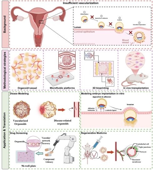
The endometrium undergoes repeated regeneration, and menstrual cycling and embryo implantation depend on regulated angiogenesis. Endometrial organoids are in vitro models that reproduce selected features of endometrial biology. However, conventional systems consist largely of epithelial cells and lack the vascular networks needed to model tissue-level functions. Bioengineering approaches are beginning to address this limitation by incorporating vascular components. This review summarizes the biological rationale, engineering strategies, and potential applications of vascularized endometrial organoids. We discuss the endometrial vasculature and approaches to developing models for disease research, drug screening, and regenerative medicine. These models may improve understanding of endometrial physiology and pathology and support research into personalized diagnostic and therapeutic approaches in reproductive medicine.
Keywords: endometrial organoids, vascularization, microfluidics, reproductive medicine, disease modeling, regenerative medicine
Introduction
The endometrium, the inner mucosal layer of the mammalian uterus, undergoes cycles of regeneration, differentiation, and shedding. Ovarian hormones, primarily estrogen and progesterone, regulate this process [1]. Its regenerative capacity and receptivity to embryo implantation are central to reproduction [2]. Endometrial proliferation is primarily driven by estrogen receptor (ER)-mediated nuclear transcription, together with paracrine/autocrine signaling networks. These include Wnt/β-catenin [3], Notch [4], PI3K/AKT [5] and MAPK/ERK [6] cascades, with additional regulation by transforming growth factor-β (TGF-β)/bone morphogenetic protein (BMP) [7] and Hippo pathways [8]. Spatiotemporal interactions among these pathways balance proliferation and differentiation. Their dysregulation is associated with endometrial hyperplastic disorders.
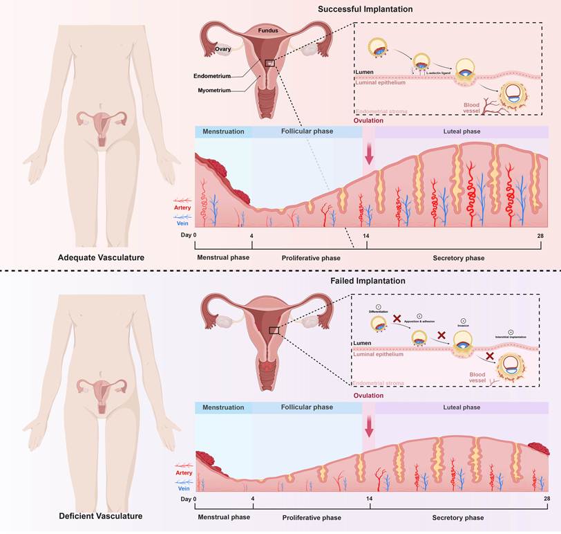
The integrity, maintenance, and repair of the endometrial functional layer depend on vascular function [9]. The vascular network undergoes morphological and functional remodeling throughout the menstrual cycle [10]. These changes support endometrial repair, preparation for implantation, and the occurrence of menstruation. During the proliferative phase, estrogen promotes the proliferation of epithelial cells and stromal cells, accompanied by angiogenesis. The newly formed capillaries provide nutrients and oxygen to the dilated tissues [11]. During the secretory phase, rising progesterone levels slow angiogenesis [12]. Existing vessels, particularly the spiral arteries, undergo remodeling, becoming more tortuous, dilated, and permeable in preparation for embryo implantation [12]. Vascular changes during the implantation window contribute to the establishment of the maternal-fetal interface. In the absence of pregnancy, corpus luteum regression causes hormone levels to decline. Hormone withdrawal contributes to vasoconstriction, tissue ischemia and hypoxia, vascular breakdown in the functional layer, and tissue shedding during menstruation. Repair and renewed angiogenesis then support endometrial regeneration. The perivascular microenvironment also contributes to this process, as decidualization begins around blood vessels [1]. Regulated vessel formation, remodeling, and regression therefore support reproductive health. Disruption of these processes can contribute to implantation failure, miscarriage, and endometrial disease (Figure 1).
Endometrial vascular dynamics across the menstrual cycle and their role in implantation success.
Conventional endometrial research models, including two-dimensional (2D) cell culture and animal models, have limitations in reproducing human three-dimensional (3D) tissue architecture, intercellular interactions, and the in vivo physiological microenvironment [13,14]. Animal models are also constrained by species-specific differences and by ethical considerations, costs, and difficulties with real-time observation [15]. These limitations restrict investigation of human endometrial biology and disease mechanisms.
Endometrial organoids are self-organizing 3D cultures derived from endometrial tissue stem cells or induced pluripotent stem cells that reproduce selected structural and functional characteristics of the native tissue. Since their establishment in 2017, endometrial organoid methods have continued to develop [16,17]. These organoids express endometrium-specific markers and can respond to hormonal stimulation [16]. They can also reproduce some molecular events related to the menstrual cycle and the implantation window [18,19], providing models for the development, physiology, pathology, and drug response of the human endometrium. Organoids usually represent one major cellular component. In contrast, assembloids combine different organoids or cell populations to simulate the interactions among their components [20]. In this review, endometrial epithelial organoids thus refer to self-organizing structures containing only epithelium. The model that combines endometrial epithelial organoids with stromal cells is called an epithelial-stromal endometrial assembly. The model that integrates endothelial cells simultaneously is called a vascularized endometrial assembly. Platform-specific terms, such as endometrium-on-a-chip and vascularized organoid-on-a-chip, were retained when used in the original studies. The microfluidic “endometrium-on-a-chip (EoC)” systems support the construction of patient-specific vascularized models under dynamic perfusion conditions [21,22], while 3D bioprinting has supported centimeter-scale vascularized endometrial cancer organoids for drug testing [23]. Assembloids integrating epithelial, stromal, and vascular components have expanded the research on embryo implantation and uterine regeneration to multicellular endometrial models [24-26].
Most existing endometrial organoid models only contain epithelial components and lack vascular networks [27,28]. This limits their ability to reproduce the physiological and pathological processes involving the vascular system [11]. Embryo implantation involves vascular remodeling and signal transduction at the maternal-fetal interface [29]. The establishment and growth of endometriotic lesions require neovascularization [30], and the progression of endometrial cancer is related to tumor angiogenesis [31]. When vascular networks are lacking, organoids rely on the diffusion of nutrients and oxygen, which may lead to central necrosis and limit long-term culture, expansion, size, and structural complexity [32]. Therefore, integrating functional vascular systems may improve the physiological relevance of endometrial organoid models and expand their application scope, supporting their eventual clinical translation [21].
This review examines advances, challenges, and future directions in vascularized endometrial organoid research. We discuss construction strategies and functional validation methods, then consider applications in embryo implantation, endometrial diseases modeling, drug screening, and regenerative medicine. We also identify technical and biological limitations that should guide further model development and evaluation for reproductive medicine.
Methodological strategies for engineering vascularized endometrial organoids
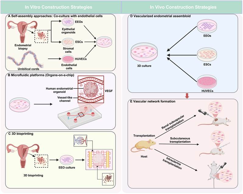
Vascularization strategies aim to generate branched, perfusable networks that respond to tissue demands. Standard endometrial organoid models generally lack functional vasculature, limiting the study of vascular-dependent processes such as embryo implantation. Developing vascularized models requires approaches from developmental biology, tissue engineering, vascular biology, and microfluidics. The main strategies described below provide different routes to improving endometrial model structure and function (Figure 2). Their clinical translation requires further validation and appropriate ethical oversight.
Methods for vascularization of endometrial organoids. (A) Self-assembly by co-culture with endothelial cells (Purple: endometrial epithelial organoids (EEOs); Orange: stromal cells; Red: HUVECs). (B) Microfluidic platforms (Organs-on-a-chip) for perfusable vasculature. (C) 3D bioprinting for spatial patterning of vascular networks. (D) Vascularized endometrial assembloid. (E) Vascular network formation in mice.
Self-assembly approaches: Co-culture with endothelial cells
Co-culture combines organoids with exogenous vascular components in a heterogeneous cell system (Figure 2A). Organoids are cultured with primary or immortalized human endothelial cells, such as human umbilical vein endothelial cells (HUVECs), within a 3D matrix gel such as Matrigel [33,34]. Endothelial cells isolated from endometrial microvasculature provide greater tissue specificity, but limited availability, expansion capacity, and high culture costs favor the use of more accessible endothelial sources. Supporting cells (including pericytes, fibroblasts, or mesenchymal stem cells (MSCs)) are often added to improve network stability and function [35]. The microenvironment around the blood vessels of the endometrium contains different cell populations. CD146+ pericytes support microvessel stability, while CD34+ outer membrane cells (En-ADVs) occupy the outer wall of blood vessels and have regenerative and angiogenic properties [36,37]. Single-cell RNA sequencing (scRNA-seq) also identified a CD9+SUSD2+ subpopulation with unique transcriptional and functional characteristics [38]. Supporting cells secrete basement membrane components and surround new blood vessels, providing mechanical and biochemical signals for maturation and stability. The proportion of cells varies depending on the model and construction strategy. Gu et al. mixed epithelial cells, stromal cells, and HUVECs in a ratio of 1:1:1 based on the volume of cell sediment [39]. Dai et al. employed two formulations: a vascularized endometrial assembloid containing 100 endometrial epithelial organoids (EEOs), 5 × 10⁴ endometrial stromal cells (ESCs), and 1.5 × 10⁵ HUVECs per 30 μL of hydrogel; an avascular dual-lineage control containing 100 EEOs and 2 × 10⁵ ESCs per 30 μL of hydrogel, but no HUVECs [21]. In these systems, endothelial cells can self-assemble around organoids to form capillary-like networks [34,40]. Co-culture supports the interaction among cell populations and the formation of spontaneous networks. However, there are differences in the results among different batches. The matrix composition, cell ratio, inoculation sequence, and culture medium all need to be carefully optimized. Self-assembly also has relatively limited control over the network geometry and spatial distribution, which increases the difficulty of developing standardized disease models.
Rodríguez-Eguren et al. [41] identified the integration of vascular and epithelial-stromal components as an important consideration in endometrial regeneration treatment in their systematic review. Endometrial assemblies containing epithelial, stromal, and endothelial cells can reproduce multicellular tissue structures beyond pure epithelial organoids [21,24,25].
Co-culture systems can be used to study the communication between endothelial and epithelial/stromal cells, as well as the synergistic effects of estrogen and progesterone on angiogenesis. These studies may help clarify the disease mechanism and provide a basis for treatment development.
The formation of vascular networks should be supplemented by functional maturity assessment. Barrier integrity can be evaluated by trans-endothelial electrical resistance (TEER) or the permeability of fluorescent tracers such as fluorescein isothiocyanate-dextran (FITC-dextran) [41,42]. Responses to stimuli such as histamine or vascular endothelial growth factor (VEGF) can be evaluated through endothelial cell shape, junctional protein distribution, and permeability changes. VE-cadherin and claudin-5 expression, together with pericyte coverage evaluated using markers such as NG2 and α-SMA, can provide additional evidence for vascular maturation [43]. These measurements help determine which vascular functions the model reproduces and whether it is suitable for studying the physiology or pathology of the endometrium.
Bioengineering-driven strategies
Microfluidic platforms (Organs-on-a-chip) for perfusable vasculature
Organ-on-a-chip technology utilizes microfluidic control to create perfusable vascular channels and expose cells to fluid shear stress, providing a dynamic culture environment (Figure 2B) [26]. A typical design includes parallel microchannels separated by porous membranes. The endothelial-lined vascular channels are perfused, typically using a syringe or peristaltic pump at a flow rate of 0.1 to 10 μL/min to generate shear stress similar to blood flow [42,44]. Adjacent tissue channels contain endometrial organoids or epithelial and stromal cells. This arrangement allows the exchange of soluble factors (including hormones and cytokines) and cell migration (including immune cell movement) to occur among the components [45,46].
Dai et al. [21] developed a vascularized endometrial assembloid by integrating EEOs, stromal cells, and endothelial cells in a microfluidic chip with optimized hydrogel and dynamic perfusion culture. Animal studies subsequently evaluated endometrial repair and fertility restoration. Lee et al.[22] established a patient-derived vascularized EoC model using a polydimethylsiloxane (PDMS) chip with parallel microchannels separated by porous membranes. Patient-derived stromal cells, HUVECs, and EEOs were sequentially seeded into their respective channels. Under perfusion and hormonal stimulation, the model formed an epithelium-stroma-vasculature trilayer structure reproducing selected features of the endometrial microenvironment. The endometrial receptivity scoring system (ERS²) was developed on this platform. It quantifies receptivity markers, integrin αvβ3/osteopontin (OPN), by immunofluorescence and angiogenic parameters by confocal microscopy. ERS² supports in vitro assessment of patient-specific endometrial function and monitoring of vascular responses to sequential platelet-rich plasma (PRP) therapy. Its clinical utility requires further validation.
Microfluidic platforms can support real-time, high-resolution studies of immune cell migration under flow, including uterine natural killer cell (uNK) extravasation from the vascular lumen into endometrial tissue [47]. This approach may clarify immune regulation during embryo implantation. Chips also allow dynamic control of hormone concentrations to approximate menstrual cycle fluctuations. Biosensors can provide additional measurements of barrier integrity, metabolites, and oxygen levels within the same system.
3D bioprinting for spatial patterning of vascular networks
3D bioprinting deposits organoids, endothelial cells, supporting cells, and bioinks to produce vascularized tissue constructs with predefined architectures (Figure 2C) [48,49]. Extrusion-based and light-based methods have different strengths and limitations. Direct co-printing mixes endothelial and tissue cells in a single bioink, supporting self-assembly resembling angiogenesis [50], but often yields irregular networks with delayed lumen maturation [51]. Sacrificial mold printing uses removable templates to create defined, immediately perfusable channels [52]. This approach requires more complex fabrication and, for transplantation, subsequent integration with host vasculature [53]. The choice of method therefore depends on the required balance between cellular self-organization and geometric control. Printing can define the relative positions of vascular and glandular structures, which may be useful for developing constructs to repair extensive endometrial injury. Gong et al. [23] used projection micro-stereolithography (PμSL) 3D printing, a light-based technique, with BIO-1 biocompatible resin to fabricate a microvascular network with 80 μm inner diameters, 20 μm wall thicknesses, and 400 μm spacing. The platform supported centimeter-scale endometrial cancer organoid culture and carboplatin sensitivity testing, illustrating its use for large tissue models in pharmacological research.
In vivo transplantation
In vivo transplantation supports vascularization of endometrial organoids and assessment of host-graft integration (Figure 2D, 2E). Non-vascularized or pre-vascularized constructs are implanted into immunodeficient mice beneath the renal capsule, subcutaneously, or in the uterine cavity. Host endothelial cells can enter the graft and form vessels with vascular precursor cells in the construct, connecting it to the host circulation [54]. Transplantation therefore permits direct assessment of vascular integration and tissue function in vivo. Vascular density at the implantation site influences graft vascularization. For example, pre-vascularized organoids transplanted beneath the vascular-rich renal capsule can form stable, branched networks containing arteries and veins [55]. Site selection may thus affect vascular maturation and function.
Ma et al. [56] provided early proof-of-concept evidence that transplanted EEOs could differentiate into functional glands and promote endometrial structural regeneration in recipient rats. Dai et al. [21] evaluated vascularized endometrial assembloids after subcutaneous implantation. Constructs containing epithelial, stromal, and endothelial cells formed vascular networks with human and mouse-derived endothelial cells within 14 days post-implantation and were perfused by the host circulation. These findings support early vascular integration in vivo. Sun-Young Hwang et al. [57] also investigated direct transplantation of human endometrial organoids into injured uterine tissue. The organoids survived and integrated with the host tissue, accompanied by endometrial proliferation, increased angiogenesis marker expression, and improved pregnancy outcomes. These studies provide preclinical methods and outcome measures for evaluating organoid-based intrauterine transplantation.
A comparative framework for selecting vascularization strategies
The choice of strategy depends on the experimental problem and the required level of structural and functional control. Table 1 compares the four main methods in terms of technical and biological parameters. Microfluidic platforms provide controllable shear stress and real-time monitoring, but they require dedicated equipment. In vivo transplantation can evaluate functional anastomosis but the controllability of the experiment is relatively low. These trade-offs should guide the selection of models in mechanism research, preclinical drug screening, and regenerative studies.
Comparative analysis of vascularization strategies for endometrial organoids.
| Feature | Self-Assembly (Co-culture) | Microfluidic Platforms (Organs-on-a-Chip) | 3D Bioprinting | In Vivo Transplantation | References |
|---|---|---|---|---|---|
| Core Principle | Spontaneous endothelial organization in ECM | Controlled perfusion in microchannels | Precise deposition of cells/materials | Integration with host vasculature | [33,42,44,48,49,54] |
| Vascular Patency | Moderate (lumen formation variable) | High (actively perfused) | High (immediate if templated) | High (anastomosis with host) | [21,22,40,52,55] |
| Shear Stress Integration | Not applicable (static) | High (physiological flow) | Low (static post-printing) | Physiological (host blood flow) | [42,50,51,54,124] |
| Spatial Control | Low (random network) | High (defined geometry) | Very High (programmable) | Low (host-dependent) | [23,34,45,52,55] |
| Scalability / Throughput | High (well-plate compatible) | Moderate (chip-based, limited) | Low (slow fabrication) | Very Low (labor-intensive) | [21-23,39] |
| Biological Complexity | Moderate (multiple cell types) | High (multi-tissue, flow) | High (defined multicellular geometry) | Very High (full systemic) | [21,23,25,54,56] |
| Real-time Monitoring | Limited (end-point assays) | High (integrated biosensors) | Limited (end-point assays) | Low (invasive imaging) | [16,21-23] |
| Technical Difficulty | Low | High | High | High | [21-23,33] |
| Cost | Low | Medium | Medium | High | [22,23,34,54] |
| Key Applications | Basic cell-cell interaction studies | Drug screening, personalized medicine | Constructing defined anatomical tissues | Validating regenerative potential | [21-23,25,56] |
| Key Limitations | Batch variability, no flow | Specialized equipment, membrane barriers | Complex fabrication, maturation | Ethical concerns, low throughput | [15,21,42,51] |
Abbreviations: 3D, three-dimensional; ECM, extracellular matrix.
Beyond VEGF: Non-canonical angiogenic pathways in the endometrial context
In addition to the classic VEGF/VEGFR axis, multiple pathways are also involved in endometrial angiogenesis. The angiopoietin (Ang)/Tie2 system regulates vascular stability and remodeling, and the Ang-1/Ang-2 balance may be involved in vascular changes related to the menstrual cycle [58-61]. Semaphorin class 3 members (such as SEMA3A) and ephrin/Eph receptor signals also regulate angiogenesis and vascular modeling. Imbalances in these signaling pathways are associated with endometrial cancer and implantation defects [62,63]. Hypoxia-inducible factor-2α (HIF-2α) provides another transcriptional pathway that links metabolic regulation to vascular function during the secretory and menstrual phases [64]. These pathways may interact with VEGF during cyclical vascular remodeling. Incorporating them into vascularized endometrial models is helpful for identifying mechanisms and candidate therapeutic targets.
Validation and functional characterization
To support cross-study comparison and standardization, studies should report quantitative parameters of vascular maturity: (i) Vascular density, expressed as the total vascular length per unit area (mm/mm²) or the number of vessels per field of view; (ii) Pericyte coverage, expressed as the percentage of endothelial circumference or area covered by pericyte markers (such as platelet-derived growth factor receptor β (PDGFRβ), neuron-glial antigen 2 (NG2), and alpha-smooth muscle actin (α-SMA)), relative to CD31⁺ or vWF⁺ endothelium; (iii) Barrier integrity, assessed using TEER (Ω·cm²) or macromolecular permeability (such as FITC-dextran clearance rate); (iv) Lumen diameter (μm), measured by confocal Z-axis tomography or electron microscopy. These indicators have been used in the research of vascular organoids and organ-on-a-chip [21,43,55,65], providing a basis for comparing the maturity under different construction methods and culture conditions.
Morphological assessment: From macroscopic connectivity to ultrastructure
Morphological verification utilizes complementary imaging methods to assess network integrity, maturity, and similarity to natural vascular structures. Fluorescence and bright-field microscopy can identify endothelial tube-like structures [21,25]. Endothelial marker staining (such as CD31 and vWF) combined with confocal 3D reconstruction and transmission electron microscopy (TEM) can evaluate connectivity and hollow lumen formation [21]. TEM can also show the connection between the basement membrane and endothelium, providing ultrastructural evidence related to barrier formation [43]. These structural observation results should be interpreted in combination with the measurement results of functional barriers.
Functional validation: From perfusion capacity to physiological utility
Morphological assessment establishes structural features, while functional testing determines whether the network performs the vascular functions required for a specific application. Both are necessary for evaluating the physiological relevance of vascularized endometrial organoids.
Perfusion, nutrient delivery, and barrier integrity
Perfusion provides direct evidence of network function. Introducing fluorescent tracers (such as dextrans or FITC-albumin) into the vascular lumens on microfluidic chips or through in vivo intravenous injection can display the flow and extravasation conditions. These experiments evaluate patency and permeability [43]. Vascularization should also be evaluated for its impact on tissue activity. Comparative studies with avascular organoids have reported that vascularization can reduce central necrosis, which is consistent with improved nutrient and oxygen delivery, and is accompanied by changes in glucose consumption and lactic acid production [23]. These findings support the assessment of activity and metabolism, but should not replace direct functional assays. TEER and macromolecular permeability experiments can characterize the integrity of the endothelial barrier and selective transport [42].
Hormonal responsiveness: an endometrium-specific hallmark
Responsiveness to ovarian hormones is a defining feature of endometrial models. Vascularized endometrial assembloids can reproduce selected hormone-dependent changes associated with the menstrual cycle. Turco et al. showed that EEOs responded to sequential estrogen and progesterone stimulation in vitro and expressed corresponding molecular markers [16]. Dynamic perfusion culture in microfluidic chips has also supported secretory-phase transformation in vascularized models [21]. Reported changes included upregulation of prolactin (PRL), progestagen-associated endometrial protein (PAEP), and secreted phosphoprotein 1 (SPP1). Protein and mRNA measurements were accompanied by epithelial morphological changes, including cytoplasmic secretory vacuoles, increased mucin 1 (MUC1) expression, and stronger periodic acid-Schiff (PAS) staining. Together, these observations are consistent with an early secretory phenotype.
Hormone-driven vascular remodeling
Hormonal responses have also been reported in statically cultured vascularized endometrial assembloids, suggesting that these responses are not restricted to perfusion culture. In addition to epithelial and stromal responses, vascular networks may undergo hormone-dependent remodeling. Under sequential estrogen (E2) and progesterone (P4) stimulation, endothelial changes can be assessed against vascular responses observed in vivo [12]. Relevant endpoints include sprouting during the proliferative phase and expression of permeability-regulating proteins, such as vascular endothelial cadherin, during the secretory window. Evaluating interactions between hormones and vascular function may help investigate the vascular contributions to disorders associated with hormonal imbalance, including abnormal uterine bleeding and endometriosis [11].
Transcriptomic and proteomic signatures: Assessing maturation and similarity to in vivo endometrium
Omics analyses can characterize molecular maturation and mechanisms in vascularized organoids. Transcriptome analysis (including scRNA-seq) can identify subtypes of arterial, venous, and capillary endothelium. García-Alonso et al. [66] constructed a spatiotemporal map of the human endometrium during the menstrual cycle and identified groups of arterial and venous endothelial cells with different transcriptional characteristics. Venous cells are enriched with genes related to white blood cell migration, while arterial cells have different expression profiles, suggesting functional specialization. The perivascular cell population includes endometrial-restricted six-transmembrane epithelial antigen of prostate 4-positive perivascular cells (PV-STEAP4) and myometrial myosin heavy chain 11-positive perivascular cells (PV MYH11). These expression profiles provide reference data for evaluating engineered endothelial and perivascular cell populations and their participation in diseases such as endometriosis. Proteomics, including secretome analysis, can complement transcriptomics by characterizing paracrine signaling. Dong et al. [67] reported that endometrial organoids regulate trophoblast function through specific miRNA modules. In vascularized models, correlation analysis can characterize the vascular secretion profile: that is, the cytokines, chemokines, and growth factors released by vascular cells. This helps to clarify the contribution of blood vessels to maternal-fetal signal transduction during implantation and their role in the disease-related microenvironment [21,25].
Extracellular vesicles (EVs) may also mediate paracrine regulation of endometrial angiogenesis. EVs from endometrial organoids or endothelial cells contain miRNAs, proteins, and lipids that can affect recipient cells [67]. For instance, EVs from adenomyosis organoids contain unique miRNA profiles associated with angiogenesis and embryo implantation [68]. Direct evidence that endothelial-derived or organoid-derived EVs regulate endometrial vascular networks remains limited. However, studies in other reproductive and cancer settings have shown effects on endothelial proliferation, migration, and tube formation through pro-angiogenic miRNAs, such as miR-126 and miR-210, and proteins, such as VEGF and Angs [69]. In vascularized endometrial models, EVs may mediate signaling between vascular and epithelial/stromal compartments and influence maturation, barrier integrity, and hormonal remodeling. Combining EV profiling with vascular functional assays could test these mechanisms and assess candidate biomarkers for endometrial disorders.
Construction and validation methods establish which physiological and pathological questions a vascularized endometrial model can address. Patency, barrier integrity, hormonal responses, and molecular profiles should be assessed together. Perfusable, hormone-responsive networks may support investigation of implantation-associated vascular remodeling and diseases involving vascular dysfunction. Model suitability depends on validation of the specific function under study, rather than vascular morphology alone.
Applications of vascularized endometrial organoids in modeling physiology and pathophysiology
Vascularized endometrial models extend in vitro research to interactions involving the vascular compartment. This section considers applications in embryo implantation and menstruation, followed by disease models in which vascular dysfunction contributes to pathology.
Modeling endometrial physiology
Reflecting endometrial receptivity and mimicking menstruation
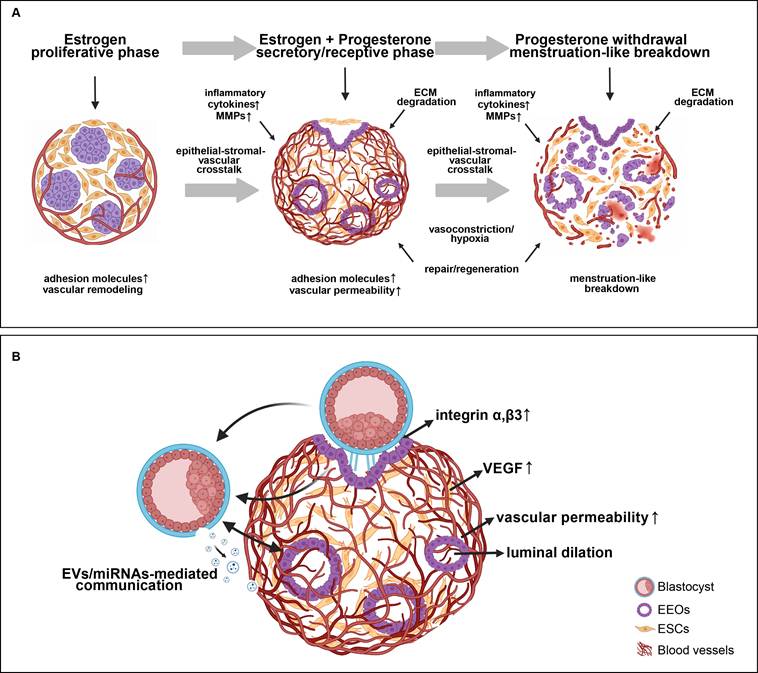
Endometrial receptivity is a transient, hormonally defined state in the mid-secretory phase that permits embryo attachment and early implantation [70]. It involves coordinated epithelial-stromal-vascular signaling, morphological changes, adhesion molecule expression, and vascular remodeling [71-73]. Vascularized endometrial organoids may support investigation of receptivity and the initiation of menstruation, both of which involve vascular function. Sequential hormonal stimulation allows assessment of vascular changes associated with proliferative and secretory conditions. Progesterone withdrawal, a trigger for menstruation, could be used to test whether these models reproduce inflammatory responses and tissue breakdown. Relevant endpoints include pro-inflammatory cytokine and matrix metalloproteinase (MMP) expression by stromal and endothelial cells, extracellular matrix degradation, and features of tissue shedding [1,11]. Functional endothelial networks may also permit investigation of vasoconstriction and hypoxia-related signaling during repair and regeneration. The extent to which these models reproduce cyclical breakdown and regeneration requires direct validation. Such studies could clarify vascular contributions to endometrial homeostasis and disorders such as abnormal uterine bleeding [74] (Figure 3A).
Vascularized endometrial organoids for modeling endometrial physiology. (A) Hormone-regulated endometrial receptivity and menstruation-like remodeling. (B) Embryo implantation and maternal-fetal crosstalk.
Modeling embryo implantation and intercellular crosstalk
Vascularized organoid models have been used to investigate vascular remodeling during embryo implantation in vitro. Co-culture studies have reported localized changes in vascular permeability and luminal dilation accompanying blastoid attachment under hormonally induced receptive conditions [25]. These platforms could also test how embryo-derived signals, including human chorionic gonadotropin (hCG), affect endothelial angiogenic responses, junctional organization, and barrier function. These specific endothelial responses require experimental validation and should not be assumed to be established features of current models.
Several co-culture systems have extended the study of implantation in vitro. Song J et al. [75] established a 3D post-implantation co-culture system supporting human embryo development up to day 14 post-fertilization. Endometrial co-culture was associated with enhanced trophoblast differentiation and earlier emergence of extravillous trophoblasts (EVTs), suggesting that the maternal microenvironment influences trophoblast lineage specification. Molè MA et al. [76] developed cell-engineered receptive endometrial scaffold technology (CREST), reproducing glandular architecture, hormone-responsive epithelium, and stromal decidualization. This system supported embryo attachment, invasion, and early post-implantation development in vitro. Microfluidic “artificial uterus” chips incorporating patient-derived EEOs and stromal cells have also been used to examine implantation and deficiencies in patients with recurrent implantation failure (RIF) [22].
These models also support investigation of intercellular communication in the endometrial microenvironment. Systems containing epithelial, stromal, and endothelial compartments have identified signaling networks, including epithelial VEGFA signaling to endothelial and stromal cells [21,25]. Human endometrial assembloids have been used to study the effects of decidual senescence on implantation [77]. Vascular responses during implantation can be assessed through TEER or impedance measurements alongside FITC-dextran permeability assays [78]. These platforms may also support studies of extracellular vesicle-mediated maternal-embryonic communication [67] (Figure 3B).
Modeling vascular-related endometrial diseases
Patient-derived vascularized organoids combine tissue architecture and cellular heterogeneity with a vascular compartment. These features may support studies of vascular contributions to endometrial disease and the evaluation of candidate therapies. Their usefulness depends on how well each model reproduces the disease phenotype of interest.
Thin endometrium (TE) and intrauterine adhesion (IUA)/Asherman's syndrome (insufficient angiogenesis)
TE and IUA/Asherman's syndrome impair fertility through structural and functional endometrial deficits [79,80]. TE is characterized by insufficient endometrial thickness (< 7 mm) during the implantation window [81], whereas IUA/Asherman's syndrome involves fibrotic adhesions after injury [82]. Impaired angiogenesis and inadequate blood supply contribute to reduced receptivity and implantation failure in both conditions [83-86]. Their vascular phenotypes also differ, as summarized in Table 2.
Shared and distinct vascular phenotypes of TE and IUA/Asherman's syndrome.
| Feature | TE | IUA/Asherman's syndrome | References |
|---|---|---|---|
| Core vascular defect | Quantitative: reduced vessel density | Mixed: reduced density + fibrosis-associated vascular distortion | [84,144,145] |
| Endothelial dysfunction | Impaired tube formation, reduced VEGF response | Impaired tube formation + EndMT | [144,145] |
| Extracellular matrix | Relatively preserved | Dense fibrotic matrix with aberrant collagen deposition | [146,147] |
| Pericyte/perivascular cell involvement | Dysfunctional CD9⁺SUSD2⁺ perivascular cells with reduced proliferation and aberrant ECM remodeling | Pericyte loss associated with vascular instability | [38,145] |
| Key mechanistic drivers | Estrogen deficiency, reduced angiogenesis signaling | Injury-induced inflammation, TGF-β-driven fibrosis, impaired stem cell niches | [145,148] |
| Investigated regenerative interventions | PRP and experimental pro-angiogenic approaches | Cell- and scaffold-assisted repair | [149-151] |
Abbreviations: TE, thin endometrium; IUA, intrauterine adhesion; EndMT, endothelial-mesenchymal transition; TGF-beta, transforming growth factor beta; PRP, platelet-rich plasma.
Patient-specific vascularized organoids could be used to assess endothelial deficits associated with these conditions, including reduced tube formation and migration [87]. Peng et al. [88] suggested that oxygen-glucose deprivation-induced cytoskeletal abnormalities may impair vascular stability by disrupting endothelial-pericyte interactions. Delivery strategies for En-ADVs, including spheroid-loaded microneedle patches, are discussed in the regenerative medicine section [89]. Gu et al. [39] developed a microfluidic-engineered vascularized endometrial micro-organoid platform by co-encapsulating ESCs, epithelial organoids, and endothelial cells in hydrogel microspheres. The platform reproduced pathological remodeling and promoted endometrial regeneration and fertility recovery in a murine IUA model. Combining patient-derived vascularized models with scRNA-seq may help identify endothelial subpopulations associated with vascular dysfunction [82,90]. Hwang et al. [57] also found that endometrial organoids can act as reservoirs of functional mitochondria, suggesting an opportunity to examine links between energy metabolism and angiogenesis in vascularized models.
Combining endometrial progenitor/stem cells (En-PSCs) with bioactive molecules such as hydroxysafflor yellow A (HSYA) has enhanced angiogenic and reparative effects in preclinical models [91]. However, transplanted human cells have shown limited survival and regeneration in some xenotransplantation settings, suggesting that paracrine mechanisms may contribute alongside direct engraftment [36]. Endometrial regeneration also requires remodeling of the fibrotic extracellular matrix. Xu et al. reported that matrix metalloproteinase-9 (MMP-9)-mediated collagen degradation was required for functional remodeling in the studied system [92]. Vascularization and matrix remodeling should therefore be considered together when developing regenerative approaches.
Chronic endometritis (CE) (inflammation-induced vascular dysfunction)
CE is a persistent inflammatory condition characterized by plasma cell infiltration into the endometrial stroma and is associated with infertility and RIF [93,94]. Inflammation and angiogenesis can reinforce one another. Reported vascular abnormalities include increased microvessel density, endothelial proliferation and swelling, hyaline thickening of vessel walls, luminal occlusion, and, in some cases, microvascular thrombosis [95]. These findings indicate impaired vascular structure and function. Vascularized organoid models could test how CE-associated pathogens or inflammatory factors, including IL-1β and TNF-α, affect barrier integrity and neovessel permeability. Models incorporating appropriate immune populations could also examine neutrophil and macrophage migration across the endothelium into stromal tissue. Lindsay et al. [96] provide a basis for investigating CE immunopathology; vascularized models may extend this work to endothelial activation and vascular remodeling. These systems could be used to evaluate whether antibiotics such as doxycycline, anti-inflammatory agents, or other treatments improve vascular function and receptivity. Their ability to reproduce CE-specific pathology and predict treatment responses requires validation.
RIF (functional vascular insufficiency)
RIF is a multifactorial clinical syndrome in which local endometrial “functional vascular insufficiency” may contribute to pathology [97,98]. This refers to inadequate vascular remodeling during the implantation window rather than necessarily a reduction in vessel number [72]. Potential defects include insufficient changes in permeability and perfusion. Patient-derived vascularized models could help assess these deficits. Co-culture with blastocysts or blastoids may permit investigation of vascular responses to embryonic signals [25,99], including downstream VEGF/VEGFR signaling. These systems could also be used to test therapies intended to improve vascular function, such as pathway agonists. Findings on decidual senescence and implantation [77], and on BMP2 deficiency and trophoblast invasion [100], provide questions for studies of vascular aging and BMP2-dependent vascular regulation. The patient-derived vascularized EoC with ERS² offers a quantitative research approach. It combines patient-specific epithelial and stromal compartments with a vascular compartment and measures integrin αvβ3/OPN expression and angiogenic parameters. In RIF patients, low cumulative scores, such as 5-9 points, were associated with poor vascular connectivity and reduced adhesion molecule expression, consistent with failed in vitro fertilization (IVF) cycles. The platform also monitored responses to sequential PRP treatment, including increases in ERS² scores and restoration of molecular receptivity markers [22]. These findings support further study of dynamic patient-specific assessment. Prospective validation is needed before the platform can guide RIF diagnosis or individualized treatment.
Endometriosis and adenomyosis (pathological angiogenesis in ectopic/invasive growth)
Endometriosis is an estrogen-dependent chronic inflammatory disorder involving ectopic growth, invasion, and cyclical bleeding of endometrial glands and stroma outside the uterine cavity. It is often associated with pelvic pain and infertility [101,102]. Lesion survival and growth depend on neoangiogenesis and neurovascular interactions [103-105]. Vascularized models may clarify the role of angiogenesis in lesion establishment. Patient-derived constructs can be assessed for abnormal vessel density, organization, and permeability associated with ectopic lesions [106]. Such models could test angiogenic and immune-tolerance programs identified by single-cell sequencing and examine how ectopic cells affect endothelial function through factors such as VEGF and IL-8 [107]. They may also support screening of anti-angiogenic agents, including VEGF inhibitors, provided that disease-specific phenotypes and drug responses are validated [106,108].
Adenomyosis involves invasion of endometrial glands and stroma into the myometrium, accompanied by smooth muscle cell hyperplasia and hypertrophy [109]. Angiogenesis supports ectopic tissue establishment and survival within the myometrium [109,110]. Vascularized patient-derived adenomyosis models could be used to examine interactions between invading endometrial tissue and vessels during basement membrane penetration and myometrial invasion. They may also help investigate the formation of dilated, tortuous vessels and the contribution of vascular disorganization to lesion growth, inflammation, menorrhagia, and dysmenorrhea [111]. Juárez-Barber et al. [68] characterized EV miRNA cargo from patient-derived adenomyosis organoids and identified miRNAs associated with implantation and pregnancy. Wang et al. [112] separately reported that exosomal miR-4466 from ectopic endometrial mesenchymal stem cells promotes endothelial angiogenic responses through the RUNX1/VEGFA axis. Research has also implicated endometrial basalis populations, including SFRP5+ epithelial cells, in local angiogenesis through signals such as IHH, supporting a microenvironment for glandular and stromal invasion [113]. Vascularized models could test whether disease-associated EVs alter endothelial homeostasis and help evaluate candidate therapeutic mechanisms.
Endometrial hyperplasia (EH) without atypia (vascular abnormalities in a pre-cancerous lesion)
EH without atypia arises from prolonged unopposed estrogen stimulation and involves epithelial proliferation accompanied by remodeling of the supporting vasculature [114,115]. Vascular changes can meet the nutritional requirements of proliferating tissues. Patient-derived vascularized EH models are helpful for studying these abnormalities before malignant progression. Comparisons with models derived from normal endometrium may reveal the angiogenesis mechanisms in estrogen-dominant environments and candidate biomarkers for vascular dysfunction. Angiogenesis has also been proposed as a therapeutic target for endometrial hyperplasia [116]. The elevated expression of angiogenesis-related genes may promote vascular remodeling within lesions. These models can simultaneously assess the effects of treatment on epithelial proliferation and vascular abnormalities, providing information for intervention studies aimed at limiting disease progression.
Endometrial cancer (tumor angiogenesis in malignancy)
Endometrial cancer is a common gynecological malignancy. Abnormal tumor angiogenesis promotes its occurrence and progression [117]. Tumor blood vessels exhibit structural and functional heterogeneity, including morphological distortion, increased permeability, and low perfusion efficiency [118,119]. These abnormalities lead to hypoxic and acidotic conditions and can promote malignant proliferation, invasion, and treatment resistance [120]. Vascularized endometrial cancer organoids may be helpful for studying the interaction between tumor cells and vascular components. The related processes include leakage related to VEGF and Ang-2, disordered angiogenesis, and feedback between vascular abnormalities and the tumor microenvironment [121]. These models can also evaluate the distribution, activity, and resistance mechanisms of anti-angiogenic therapies such as bevacizumab and lenvatinib. The antisense oligonucleotide strategy targeting small nucleolar RNA, C/D Box 14E (SNORD14E) proposed by Chen et al. [122], and the progesterone-response study by Thiel et al. [123] provide candidate solutions for combined testing with vascular normalization strategies. Gong et al. [23] combined 3D printing with microfluidic technology to construct a “vascularized organoid-on-a-chip”, which supported the long-term culture of centimeter-scale endometrial carcinoma organoids and the detection of carboplatin sensitivity. This establishes a cultivation and detection capability, but the fidelity of natural vascular function still needs to be verified separately. Models containing immune components could further explore tumor-vascular-immune interactions related to the combined application of anti-angiogenic drugs and immune checkpoint inhibitors [47]. These applications may provide support for the research on the mechanism and the treatment of endometrial cancer.
Translational potential and regenerative medicine
A platform for drug screening and toxicity testing: Evaluating therapies for endometrial disorders
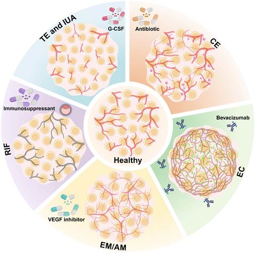
Vascularized endometrial organoids may support drug development and safety assessment (Figure 4). They can be used to evaluate the impact of treatment on epithelial/stromal cells and vascular networks within the same system. For endometriosis, the relevant endpoints include the inhibition of ectopic epithelial proliferation and changes in vascular tissue and function. Toxicity studies could assess vascular barrier damage caused by environmental exposure (including micro- and nanoplastics) or pharmaceuticals, as well as the impact on implantation-related functions. Biobanks containing different patient models could also support the detection of patient-specific drug responses [28]. Whether these responses can predict clinical treatment outcomes still requires prospective assessment.
Personalized drug screening in vascularized endometrial organoid models of vascular-related diseases. The outer portion shows organoid models derived from patients with Thin Endometrium (TE)/Intrauterine Adhesion (IUA), associated with insufficient angiogenesis; Chronic Endometritis (CE), associated with inflammatory vascular dysfunction; Recurrent Implantation Failure (RIF), associated with functional vascular insufficiency; and Endometriosis (EM)/Adenomyosis (AM), associated with pathological angiogenesis. The proposed workflow screens pro-angiogenic factors, such as G-CSF; anti-inflammatory agents and antibiotics; immunomodulators; and anti-angiogenic therapies, such as bevacizumab and VEGF inhibitors. Organoid response profiles could inform patient stratification and candidate individualized treatments aimed at restoring vascular function (center) and improving reproductive outcomes. Clinical prediction and treatment selection require validation.
In microfluidic vascularized endometrial models, fluid shear stress should be regarded as an active mechanical signal rather than merely a perfusion parameter [124]. Shear stress regulates endothelial barrier integrity, vascular gene expression, and paracrine signaling, and thus may indirectly affect epithelial receptivity through endothelial-stromal-epithelial crosstalk. However, direct evidence that specific shear stress levels regulate classical receptivity markers (LIF, ITGAV/ITGB3, HOXA10, MUC1) remains limited. Future research should systematically alter shear stress and simultaneously evaluate endothelial barrier function and the expression of epithelial receptivity markers to clarify the interaction between mechanical force and biochemical signals.
The future horizon: Vascularized organoids as building blocks for endometrial regeneration and transplantation
Tissue-engineered endometrial transplantation is being studied for IUA and infertility related to severe endometrial injury. Establishing a functional vascular system remains a core challenge. Pre-vascularization strategies combine endometrial organoids with biocompatible hydrogels (including gelatin methacryloyl (GelMA) and endometrial decellularized extracellular matrix (dECM) hydrogels) to form constructs containing vascular networks [125,126]. After transplantation, preformed vessels can connect to the host circulation and improve graft survival, integration, and recovery in experimental systems [54]. Endometrial organoids and their derivatives (including conditioned medium, EVs, and functional mitochondria) have also promoted repair and improved reproductive outcomes in animal models [57,69]. Combining these components with pro-angiogenic strategies and injectable sustained-release hydrogels may support local epithelial regeneration and angiogenesis [127]. In preclinical studies, transplanted organoids have colonized the recipient's uterus, differentiated into glandular structures, and restored fertility [56]. Further optimization should evaluate transplantation efficiency, durable function, and safety. Clinical translation also requires compliance with good manufacturing practice (GMP), regulatory assessment, and ethical oversight of human-derived materials.
Long-term graft survival and function may depend on stable neurovascular integration and control of fibrosis. Pre-vascularization does not guarantee the continuous existence of the engineered network. Implanted vessels may undergo remodeling and degeneration, connect to host vessels through vascular anastomosis, or be partially replaced by the host vascular system. These outcomes depend on perfusion stability, inflammation, pericyte coverage, and host-graft vascular competition. Therefore, pre-vascularization may mainly accelerate early perfusion and improve initial survival. Sustained efficacy requires stable anastomosis, maturation, and appropriate endothelial identity in the host environment [128]. Microneedles loaded with En-ADV spheres offer a minimally invasive approach for local delivery of angiogenic cells and may address some limitations of intrauterine infusion [89]. Regenerative effects may also involve EVs and mitochondrial transfer rather than relying solely on long-term cell implantation. Distinguishing paracrine activity from cell replacement will help guide experimental design and regulatory evaluation.
Current challenges, limitations, and future directions
Technical challenges: Scalability, reproducibility, and long-term stability
Vascularized endometrial organoids face technical obstacles in their wider application. Batch variability of cell sources, matrix lots, and culture conditions (including medium composition and fluid shear stress) can affect the results. The addition of vascular and immune cell populations further increases variability. Therefore, it is necessary to standardize and quantitatively characterize features such as vascular density and permeability [65]. Complex preparation methods and costs also limit large-scale expansion and high-throughput screening, prompting efforts to develop automated culture and measurement workflows. Maintaining vascular stability for longer than several weeks remains difficult. Preventing degeneration and excessive fibrosis is necessary for reliable long-term experiments.
Biological limitations: Achieving full maturity, incorporating immune cells, and replicating hormonal cycling
Current models have several biological limitations. Endothelial cells of non-uterine origin, such as HUVECs, may not be able to fully reproduce the microvascular features of the endometrium, including hormone responses and expression of adhesion molecules. Endometrial endothelial cells have tissue-specific characteristics, including ERβ and progesterone receptor membrane components, and respond to decidualization signals that regulate permeability and remodeling. It remains uncertain to what extent HUVECs can reproduce these reactions. Models based on HUVECs and those based on human endometrial microvascular endothelial cells (HEMECs) should be directly compared under the same estradiol and progesterone conditions to assess permeability and angiogenesis. Alternative options include patient-derived HEMEC cultures and co-assembly of vascular organoids with endometrial organoids or stromal-epithelial models. These methods may better preserve donor-specific responses and support the study of endothelial identity, remodeling, and vascular-immune interactions under hormone regulation [129-131]. It remains difficult to obtain patient-specific and functionally mature HEMECs. Directed differentiation of induced pluripotent stem cells (iPSCs) into uterus-specific populations is another strategy that needs to be evaluated [132]. Studies on immunodeficient mice provided proof-of-concept evidence, but failed to establish immune compatibility for human transplantation. In allogeneic constructs, endothelial cells may become immune targets because they express MHC/HLA class I and can upregulate MHC/HLA class II and adhesion molecules during inflammation. These changes can promote T cell activation, leukocyte adhesion, and antibody-mediated damage. Autologous, HLA-matched, or iPSC-derived endothelial sources may reduce immunogenicity but they introduce challenges related to manufacturing, scale-up, quality control, and long-term safety [133]. Current models are also unable to fully reproduce the interactions between immune cells (including uNKs and macrophages) and vascular, epithelial, and stromal populations. Immune-vascularized assembloids may fill this gap [134]. Static hormonal conditions cannot reproduce the dynamic 28-day cycle, and lack of neural innervation limits the research on the contribution of neurovascular systems to homeostasis and disease.
Extrapolation from murine systems to human endometrial biology is another limitation. Mice lack spontaneous menstruation and the related periodic vascular disintegration and remodeling [1]. Therefore, rodent models cannot fully reproduce the hypoxia-reoxygenation, shedding, and inflammatory-angiogenic interactions during the human menstrual period. Although the VEGF/VEGFR, Ang/Tie2, and Dll4/Notch signaling pathways are conserved, there may be differences in angiogenic rates, microvascular density, pericyte coverage, extracellular matrix composition, and hemodynamic regulation among different species [1]. Differences in inflammatory responses may also affect the formation of new blood vessels. When interpreting mouse data, these factors should be taken into account, and the use of human models as supplementary experimental systems should be supported.
Biomaterials also restrict clinical translation. Matrigel is widely used in organoid culture, but its murine sarcoma origin, batch variability, mechanical mismatch with human endometrium, and immunogenicity concerns limit its clinical applicability [135,136]. Semi-synthetic hydrogels such as GelMA have the advantages of controllable mechanical properties and repeatable preparation and have supported the growth of endometrial cells and organoid formation [126]. Endometrial dECM hydrogels retain tissue-specific composition and bioactive signals, have supported microvascularization and have been reported to have low immunogenicity [137]. However, GelMA cannot reproduce the full complexity of natural ECM, and the preparation of dECM varies from donor to donor, making it difficult to standardize. Clinical-grade formulations customized for vascularized endometrial constructs still need further development and safety assessment.
Future directions: Integrating the nervous system, co-culture with embryos, and personalized medicine approaches
Future research can develop higher-order “uterine assembloids” by integrating vascularized and immune-functional endometrial models with myometrial and neural components. Such constructs can help build multi-organ chips of the female reproductive tract for studying organ-level interactions [138].
We propose a phased method for gradually integrating immune cells into a vascularized endometrial model [1,139,140]. First, a baseline model including epithelial, stromal, and endothelial compartments should be established under E2/P4 stimulation to evaluate vascular density, pericyte coverage, barrier integrity, and receptivity markers. Subsequently, uterine natural killer (uNK) cells could be introduced during the secretory phase given their role in spiral artery remodeling [1,139], to detect remodeling, extravasation, and cytokine secretion. The next step is to add macrophages and study their polarization and interaction with uNK cells in vascular regulation [140]. Subsequently, T cells can be incorporated to simulate the participation of adaptive immunity in diseases such as CE. This phased design can evaluate the contribution of each immune cell population to immune-vascular-stromal signaling and vascular function.
Multi-organ chips and immune-vascularized models address the issue of complementarity. The former captures the interactions between the reproductive tract and related systems. The latter adds cellular and molecular details to the endometrial compartment. The integration of the two can support research on systemic and multi-system diseases including endometriosis-related inflammation and hormone-dependent diseases.
Neural components could expand models of neurovascular interactions associated with endometriosis-related pain and adenomyosis. The sensory and sympathetic nerve fibers of the endometrium affect vascular tension, permeability, and angiogenesis through mediators such as substance P and CGRP. Potential methods include co-differentiating neural crest, endometrial, and endothelial lineages; incorporating dorsal root ganglia for pain-signaling research; and using microfluidic systems to achieve intercompartmental communication among neural, vascular, and endometrial compartments. Neurovascular endometrial models could be helpful for studying nerve-dependent vascular remodeling, pathological nerve budding and candidate treatment methods [141].
Integrating multi-omics data (including scRNA-seq and spatial transcriptomics) with AI-assisted image analysis provides a way to associate molecular profiles with organoid function and identify candidate biomarkers and therapeutic targets [65]. Machine learning can be used to comprehensively analyze vascular density, barrier integrity, and metabolic indicators, aiming to characterize drug responses and explore differences among patients. Physics-based models offer a complementary approach that can guide the selection of cell ratios, hydrogel properties, and culture conditions for experimental testing [142]. Clinical translation will also rely on scalable, repeatable and GMP-compliant automated culture methods. As these systems become increasingly complex, especially through co-culture with embryonic models, ethical oversight will need to keep pace with their development [76].
Patient-derived vascularized endometrium-on-a-chip systems equipped with biosensors and automated imaging offer opportunities to track hormonal responses, implantation-related changes, and treatment outcomes over time. Whether these measurements can predict clinical outcomes remains to be established. Incorporating nerve cells and tissue-specific immune cell populations (such as uterine NK cells) will enable researchers to examine the interactions that are lacking in the current models. Co-culture with human embryonic stem cell-derived blastoids or donated embryos could extend these studies to the early implantation stage, but appropriate ethical approval is required. Endometrial assembloids, vascularized platforms containing immune components, and stem cell-based embryo co-cultures can address issues related to receptivity, immune tolerance, and vascular remodeling. Combining uterine assembloids with blastoid-like structures requires particularly careful review. Such studies should clearly define developmental endpoints and justified culture durations, prohibit reproductive use or transfer of the resulting constructs, and comply with local regulations and updated ISSCR guidelines [143].
Together, patient-specific cells, controllable perfusion, multi-omics measurements, and machine-learning analyses provide an experimental basis for studying functional variations of the endometrium [22,75,76]. This work can provide a basis for patient-specific drug screening and, with further verification, offer a reference for diagnosis and treatment strategies. Progress toward clinical application will depend on reproducible results, functional comparison with human tissue, and prospective evidence that model findings are relevant to patient outcomes.
Conclusion
Vascularized endometrial organoids bring the vascular components into multicellular models of the human endometrium. Combining developmental biology with tissue engineering, microfluidics, and biomaterials makes it possible to study the physiological and disease-specific aspects of the endometrium that cannot be captured by simple epithelial culture.
Patient-derived models retain the genetic background of their source cells and can reproduce certain donor-associated phenotypes. They provide a means of examining individual differences and studying processes such as implantation failure. Their suitability for diagnostic, therapeutic, or regenerative applications must be evaluated for each intended use.
Several obstacles remain, including endothelial cell specificity, incomplete cellular composition, variable culture outcomes, and difficulties with scale-up. Further work should establish reliable perfusion, improve reproducibility, and compare model function with primary human tissue. These systems have value for mechanistic studies, patient-specific drug testing, and regenerative research; their role in clinical care remains to be determined through prospective evaluation.
Abbreviations
α-SMA: alpha-smooth muscle actin; 2D: Two-dimensional; 3D: three-dimensional; AI: Artificial intelligence; Ang: Angiopoietin; AO-EMO: Apical-out endometrial organoid; BMP: Bone morphogenetic protein; CE: Chronic endometritis; dECM: Decellularized extracellular matrix; E2: Estrogen; ECM: Extracellular matrix; EEO: Endometrial epithelial organoid; EH: Endometrial hyperplasia; En-ADV: Endometrial adventitial cell; En-PSC: Endometrial progenitor/stem cell; EndMT: Endothelial-mesenchymal transition; EoC: Endometrium-on-a-chip; ER: Estrogen receptor; ERS²: Endometrial Receptivity Scoring System; ESCs: Endometrial stromal cells; EV: Extracellular vesicle; EVT: Extravillous trophoblast; FITC: Fluorescein isothiocyanate; G-CSF: Granulocyte colony-stimulating factor; GelMA: Gelatin methacryloyl; GMP: Good Manufacturing Practice; hCG: Human chorionic gonadotropin; HEMEC: Human endometrial microvascular endothelial cell; HIF-2α: Hypoxia-inducible factor-2α; HSYA: Hydroxysafflor yellow A; HUVEC: Human umbilical vein endothelial cell; IUA: Intrauterine adhesion; IVF: In vitro fertilization; iPSC: Induced pluripotent stem cell; MMP: Matrix metalloproteinase; MSC: Mesenchymal stem cell; MUC1: Mucin 1; NG2: neuron-glial antigen 2; OPN: Osteopontin; PAEP: Progestagen-associated endometrial protein; PAS: Periodic acid-Schiff; PDMS: Polydimethylsiloxane; PDGFRβ: platelet-derived growth factor receptor beta; PRL: Prolactin; PRP: Platelet-rich plasma; PV MYH11: myometrial myosin heavy chain 11-positive perivascular cells; PV-STEAP4: six-transmembrane epithelial antigen of prostate 4-positive perivascular cells; PμSL: Projection micro-stereolithography; RIF: Recurrent implantation failure; scRNA-seq: Single-cell RNA sequencing; SNORD14E: small nucleolar RNA, C/D Box 14E; SPP1: Secreted phosphoprotein 1; TE: Thin endometrium; TEER: Trans-endothelial electrical resistance; TEM: Transmission electron microscopy; TGF-β: Transforming growth factor-β; TNF-α: Tumor necrosis factor-α; uNK: Uterine natural killer; VEGF: Vascular endothelial growth factor; VEGFR: Vascular endothelial growth factor receptor; vWF: von Willebrand factor; PGR: progesterone receptor gene; PR: progesterone receptor.
Acknowledgements
The figures were created with BioRender.com. This work was supported by the National Natural Science Foundation of China (82288102 and 82571918), the Beijing Natural Science Foundation (7264352), the Innovation Team and Talents Cultivation Program of National Administration of Traditional Chinese Medicine (ZYYCXTD-C-202404), the Key Projects of Yunnan Province Science and Technology Department (202302AA310044), the Open Research Project in State Key Laboratory of Vascular Homeostasis and Remodeling, Peking University (2025-VHR-O-SY-11), and the Clinical Cohort Construction Program of Peking University Third Hospital (No.BYSYDL2024001). We thank all the staff of the Reproductive Medicine Center, Department of Obstetrics and Gynecology, Peking University Third Hospital for their contributions. OpenAI Codex (OpenAI, San Francisco, CA, USA) was used solely for English-language editing during preparation of the final manuscript. No scientific data, analyses, or figures were generated by the tool. The authors reviewed and approved all changes and take full responsibility for the manuscript.
Competing Interests
The authors have declared that no competing interest exists.
References
1. Critchley HOD, Maybin JA, Armstrong GM, Williams ARW. Physiology of the Endometrium and Regulation of Menstruation. Physiol Rev. 2020;100:1149-1179
2. Diedrich K, Fauser BC, Devroey P, Griesinger G. The role of the endometrium and embryo in human implantation. Hum Reprod Update. 2007;13:365-377
3. Zhang L, Xiong W, Xiong Y, Liu H, Liu Y. 17 β-Estradiol promotes vascular endothelial growth factor expression via the Wnt/β-catenin pathway during the pathogenesis of endometriosis. Mol Hum Reprod. 2016;22:526-535
4. Xu QX, Madhavan M, Wei SW, Zhang WQ, Lu L, Wang KZ. et al. Aberrant activation of Notch1 signaling in the mouse uterine epithelium promotes hyper-proliferation by increasing estrogen sensitivity. FASEB J. 2023;37:e22983
5. Hou S, Xu H, Lei S, Zhao D. Overexpressed nicotinamide N-methyltransferase in endometrial stromal cells induced by macrophages and estradiol contributes to cell proliferation in endometriosis. Cell Death Discov. 2024;10:463
6. Zhang G, Cheng Y, Zhang Q, Li X, Zhou J, Wang J. et al. ATX-LPA axis facilitates estrogen-induced endometrial cancer cell proliferation via MAPK/ERK signaling pathway. Mol Med Rep. 2018;17:4245-4252
7. Wei D, Su Y, Leung PCK, Li Y, Chen ZJ. Roles of bone morphogenetic proteins in endometrial remodeling during the human menstrual cycle and pregnancy. Hum Reprod Update. 2024;30:215-237
8. Zhai L, Yang X, Dong J, Qian L, Gao Y, Lv Y. et al. O-GlcNAcylation mediates endometrial cancer progression by regulating the Hippo-YAP pathway. Int J Oncol. 2023;63:90
9. Hong IS. Endometrial stem/progenitor cells: Properties, origins, and functions. Genes Dis. 2023;10:931-947
10. Cao C, Li J, Zhang Y, Wu S, Liu Q, Xi Y. et al. Bushen Tiaojing Decoction promotes endometrial angiogenesis to improve endometrial receptivity by regulating KLF4-mediated mitochondrial function and transcription activity. J Ethnopharmacol. 2026;355:120658
11. Maybin JA, Critchley HO. Menstrual physiology: implications for endometrial pathology and beyond. Hum Reprod Update. 2015;21:748-761
12. Plaisier M. Decidualisation and angiogenesis. Best Pract Res Clin Obstet Gynaecol. 2011;25:259-271
13. Mo B, Vendrov AE, Palomino WA, DuPont BR, Apparao KB, Lessey BA. ECC-1 cells: a well-differentiated steroid-responsive endometrial cell line with characteristics of luminal epithelium. Biol Reprod. 2006;75:387-394
14. Chan RW, Schwab KE, Gargett CE. Clonogenicity of human endometrial epithelial and stromal cells. Biol Reprod. 2004;70:1738-1750
15. Chan HY, Robertson SA. Seminal fluid effects on uterine receptivity to embryo implantation: transcriptomic strategies to define molecular mechanisms. Reprod Fertil Dev. 2025;37:RD24162
16. Turco MY, Gardner L, Hughes J, Cindrova-Davies T, Gomez MJ, Farrell L. et al. Long-term, hormone-responsive organoid cultures of human endometrium in a chemically defined medium. Nat Cell Biol. 2017;19:568-577
17. Cai Y, Li N, Li H. Combining Endometrial Assembloids and Blastoids to Delineate the Molecular Roadmap of Implantation. Stem Cell Rev Rep. 2023;19:1268-1282
18. Luongo FP, Ortega Baño I, Belmonte G, Gentile M, Paccagnini E, Salumets A. et al. Modeling the window of implantation: insights from endometrial biopsy and menstrual blood-derived organoids and endometrial stromal cells. Hum Reprod Open. 2025;2025:hoaf063
19. Ye Z, Cheng M, Lian W, Leng Y, Qin X, Wang Y. et al. GPX4 deficiency-induced ferroptosis drives endometrial epithelial fibrosis in polycystic ovary syndrome. Redox Biol. 2025;83:103615
20. Kanton S, Paşca SP. Human assembloids. Development. 2022 149
21. Dai Y, Zhao F, Chen Q, Zeng B, Gu W, Zhang Y. et al. Microfluidic chip-integrated vascularized endometrial complexes: Mitochondrial function and paracrine crosstalk enhance regenerative potential. Bioact Mater. 2025;54:551-569
22. Lee G, Lee YG, Koo HS, Hwang SY, Lee D, Lee J. et al. Microengineered patient-derived endometrium-on-a-chip for the evaluation of endometrial receptivity and personalised translational medicine. Nat Commun. 2025;16:10439
23. Gong X, Yang C, Peng J, Ding X, Yang H, Wang AG. et al. Vascularized organoid-on-a-chip for centimeter-scale organoid cultivation. Bio-Design and Manufacturing. 2025;8:410-22
24. Tian J, Yang J, Chen T, Yin Y, Li N, Li Y. et al. Generation of Human Endometrial Assembloids with a Luminal Epithelium using Air-Liquid Interface Culture Methods. Adv Sci (Weinh). 2023;10:e2301868
25. Shibata S, Endo S, Nagai LAE, H Kobayashi E, Oike A, Kobayashi N. et al. Modeling embryo-endometrial interface recapitulating human embryo implantation. Sci Adv. 2024;10:eadi4819
26. Quintard C, Tubbs E, Jonsson G, Jiao J, Wang J, Werschler N. et al. A microfluidic platform integrating functional vascularized organoids-on-chip. Nat Commun. 2024;15:1452
27. Gu ZY, Jia SZ, Liu S, Leng JH. Endometrial Organoids: A New Model for the Research of Endometrial-Related Diseases†. Biol Reprod. 2020;103:918-926
28. Jabri A, Alsharif M, Abbad T, Taftafa B, Mhannayeh A, Elsalti A. et al. Endometrial Organoids and Their Role in Modeling Human Infertility. Cells. 2025;14:829
29. Burton GJ, Jauniaux E. The human placenta: new perspectives on its formation and function during early pregnancy. Proc Biol Sci. 2023;290:20230191
30. Bo C, Wang Y. Angiogenesis signaling in endometriosis: Molecules, diagnosis and treatment (Review). Mol Med Rep. 2024;29:43
31. Ma H, Weng F, Tong X, Li H, Yao Y, Yuan J. LncRNA TRPM2-AS promotes endometrial carcinoma progression and angiogenesis via targeting miR-497-5p/SPP1 axis. Cell Mol Biol Lett. 2024;29:93
32. Choi YM, Lee H, Ann M, Song M, Rheey J, Jang J. 3D bioprinted vascularized lung cancer organoid models with underlying disease capable of more precise drug evaluation. Biofabrication. 2023;15:034104
33. Shi Y, Sun L, Wang M, Liu J, Zhong S, Li R. et al. Vascularized human cortical organoids (vOrganoids) model cortical development in vivo. PLoS Biol. 2020;18:e3000705
34. Chai YC, To SK, Simorgh S, Zaunz S, Zhu Y, Ahuja K. et al. Spatially Self-Organized 3D Neural Concentroid as a Novel Reductionist Humanized Model to Study Neurovascular Development. Adv Sci (Weinh). 2024;11:e2304421
35. Stevens KR, Kreutziger KL, Dupras SK, Korte FS, Regnier M, Muskheli V. et al. Physiological function and transplantation of scaffold-free and vascularized human cardiac muscle tissue. Proc Natl Acad Sci U S A. 2009;106:16568-16573
36. Zhu X, Yu F, Yan G, Hu Y, Sun H, Ding L. Human endometrial perivascular stem cells exhibit a limited potential to regenerate endometrium after xenotransplantation. Hum Reprod. 2021;36:145-159
37. Li S, Ding L. Endometrial Perivascular Progenitor Cells and Uterus Regeneration. J Pers Med. 2021;11:477
38. Liang B, Qi L, Chen M, Mai C, Diao L, Ding L. et al. Single-cell RNA sequencing of endometrium uncovers dynamic characteristics and dysregulation of perivascular CD9(+)SUSD2(+) cells in thin endometrium. Stem Cell Res Ther. 2025;16:515
39. Gu W, Dai Y, Zhao F, Sun F, Zeng B, Lin X. et al. A microfluidic-engineered vascularized endometrium micro-organoid platform for functional repair of intrauterine adhesion. Biofabrication. 2026;18:025011
40. van den Berg CW, Ritsma L, Avramut MC, Wiersma LE, van den Berg BM, Leuning DG. et al. Renal Subcapsular Transplantation of PSC-Derived Kidney Organoids Induces Neo-vasculogenesis and Significant Glomerular and Tubular Maturation In Vivo. Stem Cell Reports. 2018;10:751-765
41. Rodríguez-Eguren A, Bueno-Fernandez C, Gómez-Álvarez M, Francés-Herrero E, Pellicer A, Bellver J. et al. Evolution of biotechnological advances and regenerative therapies for endometrial disorders: a systematic review. Hum Reprod Update. 2024;30:584-613
42. Shirure VS, Hughes CCW, George SC. Engineering Vascularized Organoid-on-a-Chip Models. Annu Rev Biomed Eng. 2021;23:141-167
43. Wörsdörfer P, Dalda N, Kern A, Krüger S, Wagner N, Kwok CK. et al. Generation of complex human organoid models including vascular networks by incorporation of mesodermal progenitor cells. Sci Rep. 2019;9:15663
44. Wang H, Ning X, Zhao F, Zhao H, Li D. Human organoids-on-chips for biomedical research and applications. Theranostics. 2024;14:788-818
45. Maiullari F, Ceraolo MG, Presutti D, Fratini N, Galbiati M, Fasciani A. et al. Modeling breast cancer dynamics through modulable small Vessel Environment Bioreactor (sVEB). Biomaterials. 2025;323:123441
46. Kroll KT, Mata MM, Homan KA, Micallef V, Carpy A, Hiratsuka K. et al. Immune-infiltrated kidney organoid-on-chip model for assessing T cell bispecific antibodies. Proc Natl Acad Sci U S A. 2023;120:e2305322120
47. Lee D, Kang YJ, Song H. Regenerating the uterus: translational advances in endometrial bioengineering and immunotherapeutics. Semin Immunopathol. 2025;47:36
48. Juraski AC, Sharma S, Sparanese S, da Silva VA, Wong J, Laksman Z. et al. 3D bioprinting for organ and organoid models and disease modeling. Expert Opin Drug Discov. 2023;18:1043-1059
49. Revokatova D, Bikmulina P, Heydari Z, Solovieva A, Vosough M, Shpichka A. et al. Getting Blood out of a Stone: Vascularization via Spheroids and Organoids in 3D Bioprinting. Cells. 2025;14:665
50. Caliari SR, Burdick JA. A practical guide to hydrogels for cell culture. Nat Methods. 2016;13:405-414
51. Zhang YS, Arneri A, Bersini S, Shin SR, Zhu K, Goli-Malekabadi Z. et al. Bioprinting 3D microfibrous scaffolds for engineering endothelialized myocardium and heart-on-a-chip. Biomaterials. 2016;110:45-59
52. Kolesky DB, Truby RL, Gladman AS, Busbee TA, Homan KA, Lewis JA. 3D bioprinting of vascularized, heterogeneous cell-laden tissue constructs. Adv Mater. 2014;26:3124-3130
53. Hinton TJ, Jallerat Q, Palchesko RN, Park JH, Grodzicki MS, Shue HJ. et al. 3D printing of complex biological structures by freeform reversible embedding of suspended hydrogels. Sci Adv. 2015;1:e1500758
54. Zhang H, Xu D, Li Y, Lan J, Zhu Y, Cao J. et al. Organoid Transplantation Can Improve Reproductive Prognosis by Promoting Endometrial Repair in Mice. Int J Biol Sci. 2022;18:2627-2638
55. Wimmer RA, Leopoldi A, Aichinger M, Wick N, Hantusch B, Novatchkova M. et al. Human blood vessel organoids as a model of diabetic vasculopathy. Nature. 2019;565:505-510
56. Ma Y, Qian J, Xu X, Wei C, Wang M, Zhang P. et al. Engraftment of self-renewing endometrial epithelial organoids promotes endometrial regeneration by differentiating into functional glands in rats. Front Bioeng Biotechnol. 2024;12:1449955
57. Hwang SY, Lee D, Lee G, Ahn J, Lee YG, Koo HS. et al. Endometrial organoids: a reservoir of functional mitochondria for uterine repair. Theranostics. 2024;14:954-972
58. Zhou CF, Liu MJ, Wang W, Wu S, Huang YX, Chen GB. et al. miR-205-5p inhibits human endometriosis progression by targeting ANGPT2 in endometrial stromal cells. Stem Cell Res Ther. 2019;10:287
59. Hur SE, Lee JY, Moon HS, Chung HW. Angiopoietin-1, angiopoietin-2 and Tie-2 expression in eutopic endometrium in advanced endometriosis. Mol Hum Reprod. 2006;12:421-426
60. Cho S, Kim HK, Choi YS, Park JH. The Levonorgestrel Intrauterine System Attenuates the Expression of Angiopoietin-1, Angiopoietin-2, and Vascular Endothelial Growth Factor in Adenomyosis. J Clin Med. 2025;14:8629
61. Albrecht ED, Pepe GJ. Steroid hormone regulation of angiogenesis in the primate endometrium. Front Biosci. 2003;8:d416-29
62. Ferreira GD, Capp E, Jauckus J, Strowitzki T, Germeyer A. Expression of semaphorin class 3 is higher in the proliferative phase on the human endometrium. Arch Gynecol Obstet. 2018;297:1175-1179
63. Adu-Gyamfi EA, Czika A, Liu TH, Gorleku PN, Fondjo LA, Djankpa FT. et al. Ephrin and Eph receptor signaling in female reproductive physiology and pathology†. Biol Reprod. 2021;104:71-82
64. Sivridis E, Giatromanolaki A, Gatter KC, Harris AL, Koukourakis MI. Association of hypoxia-inducible factors 1alpha and 2alpha with activated angiogenic pathways and prognosis in patients with endometrial carcinoma. Cancer. 2002;95:1055-1063
65. Nahon DM, Moerkens R, Aydogmus H, Lendemeijer B, Martínez-Silgado A, Stein JM. et al. Standardizing designed and emergent quantitative features in microphysiological systems. Nat Biomed Eng. 2024;8:941-962
66. Garcia-Alonso L, Handfield LF, Roberts K, Nikolakopoulou K, Fernando RC, Gardner L. et al. Mapping the temporal and spatial dynamics of the human endometrium in vivo and in vitro. Nat Genet. 2021;53:1698-1711
67. Dong Y, Li J, Cao D, Zhong J, Liu X, Duan YG. et al. Integrated MicroRNA and Secretome Analysis of Human Endometrial Organoids Reveal the miR-3194-5p/Aquaporin/S100A9 Module in Regulating Trophoblast Functions. Mol Cell Proteomics. 2023;22:100526
68. Juárez-Barber E, Segura-Benítez M, Carbajo-García MC, Bas-Rivas A, Faus A, Vidal C. et al. Extracellular vesicles secreted by adenomyosis endometrial organoids contain miRNAs involved in embryo implantation and pregnancy. Reprod Biomed Online. 2023;46:470-481
69. Qin X, Hu KL, Li Q, Sun Y, Peng T, Liu X. et al. In Situ Sprayed Hydrogel Delivers Extracellular Vesicles Derived from Human Endometrial Organoids for Uterine Function Preservation and Fertility Restoration. Adv Healthc Mater. 2025;14:e2403604
70. Lessey BA, Young SL. What exactly is endometrial receptivity? Fertility and sterility. 2019;111:611-617
71. Teh WT, McBain J, Rogers P. What is the contribution of embryo-endometrial asynchrony to implantation failure? Journal of assisted reproduction and genetics. 2016;33:1419-1430
72. Craciunas L, Gallos I, Chu J, Bourne T, Quenby S, Brosens JJ. et al. Conventional and modern markers of endometrial receptivity: a systematic review and meta-analysis. Hum Reprod Update. 2019;25:202-223
73. Evans J, Salamonsen LA, Winship A, Menkhorst E, Nie G, Gargett CE. et al. Fertile ground: human endometrial programming and lessons in health and disease. Nat Rev Endocrinol. 2016;12:654-667
74. Evans J, Salamonsen LA. Inflammation, leukocytes and menstruation. Rev Endocr Metab Disord. 2012;13:277-288
75. Song J, Zhao R, Zhang Y, Lu M, Liu P, Li T. et al. 3D post-implantation co-culture of human embryo and endometrium. Cell Stem Cell. 2026;33:58-72.e7
76. Molè MA, Elderkin S, Zorzan I, Penfold C, Horsley N, Pokhilko A. et al. Modeling human embryo implantation in vitro. Cell. 2026;189:87-105.e28
77. Rawlings TM, Makwana K, Taylor DM, Molè MA, Fishwick KJ, Tryfonos M. et al. Modelling the impact of decidual senescence on embryo implantation in human endometrial assembloids. Elife. 2021;10:e69603
78. Bischoff I, Hornburger MC, Mayer BA, Beyerle A, Wegener J, Fürst R. Pitfalls in assessing microvascular endothelial barrier function: impedance-based devices versus the classic macromolecular tracer assay. Sci Rep. 2016;6:23671
79. Zhang Y, Shi L, Lin X, Zhou F, Xin L, Xu W. et al. Unresponsive thin endometrium caused by Asherman syndrome treated with umbilical cord mesenchymal stem cells on collagen scaffolds: a pilot study. Stem Cell Res Ther. 2021;12:420
80. Azizi R, Aghebati-Maleki L, Nouri M, Marofi F, Negargar S, Yousefi M. Stem cell therapy in Asherman syndrome and thin endometrium: Stem cell- based therapy. Biomed Pharmacother. 2018;102:333-343
81. Zhang X, Lv H, Weng Q, Jiang P, Dai C, Zhao G. et al. "Thin endometrium" at single-cell resolution. Am J Obstet Gynecol. 2025;232:S135-S148
82. Santamaria X, Roson B, Perez-Moraga R, Venkatesan N, Pardo-Figuerez M, Gonzalez-Fernandez J. et al. Decoding the endometrial niche of Asherman's Syndrome at single-cell resolution. Nat Commun. 2023;14:5890
83. Yan ZY, Zhou WJ, Ye JF, Xie F, Zhang CX, Li MQ. Cellular Senescence in Endometrium: A Pivotal Regulator in Physiological Remodeling and Pathological Disorders. Int J Biol Sci. 2025;21:6745-6758
84. Zhao G, Dai J, Hu Y. Development of regenerative therapies targeting fibrotic endometrium in intrauterine adhesion or thin endometrium to restore uterine function. Sci China Life Sci. 2025;68:2264-2276
85. Niu Y, Le A. Advances in the Pathophysiology of Thin Endometrium. Reprod Sci. 2025;32:3807-3815
86. Asadikalameh Z, Esgandari M, Panahinia P, Ghayur S, Fazeli R, Etezadi A. Endometrial regeneration and the role of the extracellular matrix: mechanisms, challenges, and future perspectives. Tissue Cell. 2025;97:103074
87. Nwokoye PN, Abilez OJ. Bioengineering methods for vascularizing organoids. Cell Rep Methods. 2024;4:100779
88. Peng T, Yang S, Lian W, Liu X, Zheng P, Qin X. et al. Cytoskeletal and inter-cellular junction remodelling in endometrial organoids under oxygen-glucose deprivation: a new potential pathological mechanism for thin endometria. Hum Reprod. 2024;39:1778-1793
89. Li S, Li Y, Yu F, Li N, Liu C, Mao J. et al. Human Endometrium-Derived Adventitial Cell Spheroid-Loaded Antimicrobial Microneedles for Uterine Regeneration. Small. 2022;18:e2201225
90. Al-Lamee H, Soul J, Green D, Drury J, Hill CJ, Vasieva O. et al. Transcriptional Profiling of SSEA-1(+) Endometrial Epithelial Progenitor Cells Highlights Their Role in Endometrial Regeneration, Remodeling, and Homeostasis. FASEB J. 2025;39:e70578
91. Li N, Mao J, Wang M, Qi J, Jiang Z, Li Y. et al. Transplantation of human endometrial perivascular stem cells with hydroxy saffron yellow A promotes uterine repair in rats. Stem Cell Res Ther. 2024;15:217
92. Xu L, Ding L, Wang L, Cao Y, Zhu H, Lu J. et al. Umbilical cord-derived mesenchymal stem cells on scaffolds facilitate collagen degradation via upregulation of MMP-9 in rat uterine scars. Stem Cell Res Ther. 2017;8:84
93. Yasuo T, Kitaya K. Challenges in Clinical Diagnosis and Management of Chronic Endometritis. Diagnostics (Basel). 2022;12:2711
94. Amrani M, Renné C, Blaschke V, Schlautmann E, Schaffrath M, Linek B. et al. Chronic endometritis identified by plasma cells can often be diagnosed in patients with recurrent implantation failure. Eur J Obstet Gynecol Reprod Biol. 2025;312:114092
95. Carvalho FM, Aguiar FN, Tomioka R, de Oliveira RM, Frantz N, Ueno J. Functional endometrial polyps in infertile asymptomatic patients: a possible evolution of vascular changes secondary to endometritis. Eur J Obstet Gynecol Reprod Biol. 2013;170:152-156
96. Lindsay CV, Potter JA, Grimshaw AA, Abrahams VM, Tong M. Endometrial responses to bacterial and viral infection: a scoping review. Hum Reprod Update. 2023;29:675-693
97. Rostami R, Jahanbakhsh J, Mahdinia E, Rezaei A, Yarahmadi S, Omodiani N. et al. Endometrial Steroid Receptor Dysregulation and Its Association with Vitamin D, AMH, and Inflammation in Recurrent Implantation Failure: A Case-Control Study. Int J Fertil Steril. 2025;19:411-420
98. Li M, Hu J, Yao L, Gao M. Decreased ANGPTL4 impairs endometrial angiogenesis during peri-implantation period in patients with recurrent implantation failure. J Cell Mol Med. 2020;24:10730-10743
99. Yang H, Zhang J, Yan F, Chen Y, Wu Y, Luo J. et al. Ciliary IFT-B Transportation Plays an Important Role in Human Endometrial Receptivity Establishment and is Disrupted in Recurrent Implantation Failure Patients. Cell Prolif. 2025;58:e13819
100. Hu C, Deng J, Liu M, Ni T, Chen ZJ, Yan J. et al. Endometrial BMP2 Deficiency Impairs ITGB3-Mediated Trophoblast Invasion in Women With Repeated Implantation Failure. Endocrinology. 2024;165:bqae002
101. Gutiérrez Sierra MJ, Bolaños Bermúdez IA, Rodríguez Pinto LP, Torres Castiblanco JL, Torres Cortés DF, Rivero Rapalino OM. Hepatic endometriosis causing hemoperitoneum in a woman with deep endometriosis. Int J Gynaecol Obstet. 2026 doi: 10.1002/ijgo.71343. Online ahead of print
102. Horne AW, Saunders PTK. SnapShot: Endometriosis. Cell. 2019;179:1677-1677.e1
103. Shi J, Qi Y, Sun Y, Huang Y. Kallikrein-Related Peptidase 4 Promotes Proliferation, Migration, Invasion, and Pro-Angiogenesis of Endometrial Stromal Cells via Regulation of Brain-Derived Neurotrophic Factor Production in Endometriosis. Am J Pathol. 2024;194:121-134
104. Peng Y, Ding S, Xu P, Zhang X, Wang J, Li T. et al. CCL18 promotes endometriosis by increasing endometrial cell migration and neuroangiogenesis. Eur J Histochem. 2024 68
105. Guo X, Ding S, Li T, Wang J, Yu Q, Zhu L. et al. Macrophage-derived netrin-1 is critical for neuroangiogenesis in endometriosis. Int J Biol Macromol. 2020;148:226-237
106. Esfandiari F, Mansouri N, Shahhoseini M, Heidari Khoei H, Mikaeeli G, Vankelecom H. et al. Endometriosis organoids: prospects and challenges. Reprod Biomed Online. 2022;45:5-9
107. Tan Y, Flynn WF, Sivajothi S, Luo D, Bozal SB, Davé M. et al. Single-cell analysis of endometriosis reveals a coordinated transcriptional programme driving immunotolerance and angiogenesis across eutopic and ectopic tissues. Nat Cell Biol. 2022;24:1306-1318
108. Laschke MW, Menger MD. In vitro and in vivo approaches to study angiogenesis in the pathophysiology and therapy of endometriosis. Hum Reprod Update. 2007;13:331-342
109. Zhang C, Shi J, Dai Y, Li X, Leng J. Progress of the study of pericytes and their potential research value in adenomyosis. Sci Prog. 2024;107:368504241257126
110. Li B, Qi J, Cao Y, Long Y, Wei Z, Wang WS. et al. From Invaginating Site to Deep Lesion: Spatial Transcriptomics Unravels Ectopic Endometrial Penetration Features in Adenomyosis. Adv Sci (Weinh). 2025;12:e2411752
111. Stratopoulou CA, Rossi M, Beaussart C, Zipponi M, Camboni A, Donnez J. et al. Generation of epithelial-stromal assembloids as an advanced in vitro model of impaired adenomyosis-related endometrial receptivity. Fertil Steril. 2025;123:350-360
112. Wang J, Ni Y, Sun J, Qiu Y, Wei X, Wang B. et al. Ectopic endometrial mesenchymal stem cell-derived exosomal miR-4466 promotes angiogenesis by targeting RUNX1 in adenomyosis. Biochem Biophys Rep. 2026;45:102457 doi:10.1016/j.bbrep.2026.102457
113. Xu Y, Cheng T, Wang J, Qiu Z, Guan X, Yu Y. et al. Decoding adenomyosis pathogenesis using an assembloid model. Sci China Life Sci. 2026;69:136-150
114. SenthilKumar G, Zirgibel Z, Jaramillo-Torres MJ, Limpert RH, Cohen KE, Shult C. et al. Estrogen Influences Human Microvascular Endothelial Function Via Sex-Specific Regulation of Sphingolipids. JACC Basic Transl Sci. 2026;11:101389
115. Mittermeier T, Farrant C, Wise MR. Levonorgestrel-releasing intrauterine system for endometrial hyperplasia. Cochrane Database Syst Rev. 2020;9:CD012658
116. Zhou Y, Liu M, Duan J, He C, Xu M, Huang J. et al. Injectable self-expanding short-fiber scaffold reduces endometrial hyperplasia. J Control Release. 2025;387:114172
117. Kamat AA, Merritt WM, Coffey D, Lin YG, Patel PR, Broaddus R. et al. Clinical and biological significance of vascular endothelial growth factor in endometrial cancer. Clin Cancer Res. 2007Dec15;13:7487-7495
118. Gonzalez-Fernandez J, Zaragozano S, Monteagudo-Sánchez A, Simon C, Vilella F. Single-cell technology: the key to an improved understanding of the human endometrium in health and disease. Am J Obstet Gynecol. 2025;232:S43-S53
119. Gaffney D, Matias-Guiu X, Mutch D, Scambia G, Creutzberg C, Fotopoulou C. et al. 2023 FIGO staging system for endometrial cancer: The evolution of the revolution. Gynecol Oncol. 2024;184:245-253
120. Aloisi A, Sonoda Y, Gardner GJ, Park KJ, Elliott SL, Zhou QC. et al. Prospective Comparative Study of Laparoscopic Narrow Band Imaging (NBI) Versus Standard Imaging in Gynecologic Oncology. Ann Surg Oncol. 2018;25:984-990
121. Cabanos HF, Hata AN. Emerging Insights into Targeted Therapy-Tolerant Persister Cells in Cancer. Cancers (Basel). 2021;13:2666
122. Chen X, Liu X, Li QH, Lu BF, Xie BM, Ji YM. et al. A patient-derived organoid-based study identified an ASO targeting SNORD14E for endometrial cancer through reducing aberrant FOXM1 Expression and β-catenin nuclear accumulation. J Exp Clin Cancer Res. 2023;42:230
123. Thiel KW, Newtson AM, Devor EJ, Zhang Y, Malmrose PK, Bi J. et al. Global expression analysis of endometrial cancer cells in response to progesterone identifies new therapeutic targets. J Steroid Biochem Mol Biol. 2023;234:106399
124. Morigi M, Zoja C, Figliuzzi M, Foppolo M, Micheletti G, Bontempelli M. et al. Fluid shear stress modulates surface expression of adhesion molecules by endothelial cells. Blood. 1995;85:1696-1703
125. Gómez-Álvarez M, Bueno-Fernandez C, Rodríguez-Eguren A, Francés-Herrero E, Agustina-Hernández M, Faus A. et al. Hybrid Endometrial-Derived Hydrogels: Human Organoid Culture Models and In Vivo Perspectives. Adv Healthc Mater. 2024;13:e2303838
126. Salisbury E, Rawlings TM, Efstathiou S, Tryfonos M, Makwana K, Fitzgerald HC. et al. Photo-Cross-linked Gelatin Methacryloyl Hydrogels Enable the Growth of Primary Human Endometrial Stromal Cells and Epithelial Gland Organoids. ACS Appl Mater Interfaces. 2024;16:39140-39152
127. Liu Y, Jia D, Li L, Wang M. Advances in Nanomedicine and Biomaterials for Endometrial Regeneration: A Comprehensive Review. Int J Nanomedicine. 2024;19:8285-8308
128. Laschke MW, Menger MD. Prevascularization in tissue engineering: Current concepts and future directions. Biotechnol Adv. 2016;34:112-121
129. Kayisli UA, Luk J, Guzeloglu-Kayisli O. et al. Regulation of angiogenic activity of human endometrial endothelial cells in culture by ovarian steroids. J Clin Endocrinol Metab. 2004;89:5794-802
130. Wang X, Hawkins SM. Using advanced spatial and single-cell transcriptomics to characterize the human endometrium. Nat Genet. 2021;53:1628-1630
131. Albrecht ED, Babischkin JS, Lidor Y. et al. Effect of estrogen on angiogenesis in co-cultures of human endometrial cells and microvascular endothelial cells. Hum Reprod. 2003;18:2039-47
132. Cheung VC, Peng CY, Marinić M, Sakabe NJ, Aneas I, Lynch VJ. et al. Pluripotent stem cell-derived endometrial stromal fibroblasts in a cyclic, hormone-responsive, coculture model of human decidua. Cell Rep. 2021;35:109138
133. Pashneh-Tala S, MacNeil S, Claeyssens F. The Tissue-Engineered Vascular Graft-Past, Present, and Future. Tissue Eng Part B Rev. 2016;22:68-100
134. Hibaoui Y, Feki A. Organoid Models of Human Endometrial Development and Disease. Front Cell Dev Biol. 2020;8:84
135. Aisenbrey EA, Murphy WL. Synthetic alternatives to Matrigel. Nat Rev Mater. 2020;5:539-551
136. Giles R, Meijer EM, Maas RGC, van Dijk CGM, Verhaar MC, Cheng C. Animal-free alternatives for Matrigel in human iPSC-derived blood vessel organoid culture. Sci Rep. 2025;15:36042
137. Brown M, Li J, Moraes C, Tabrizian M, Li-Jessen NYK. Decellularized extracellular matrix: New promising and challenging biomaterials for regenerative medicine. Biomaterials. 2022;289:121786
138. Kleinová M, Varga I, Čeháková M, Valent M, Klein M. Exploring the black box of human reproduction: endometrial organoids and assembloids - generation, implantation modeling, and future clinical perspectives. Front Cell Dev Biol. 2024;12:1482054
139. Moffett A, Colucci F. Uterine NK cells: active regulators at the maternal-fetal interface. J Clin Invest. 2014;124:1872-1879
140. Vento-Tormo R, Efremova M, Botting RA, Turco MY, Vento-Tormo M, Meyer KB. et al. Single-cell reconstruction of the early maternal-fetal interface in humans. Nature. 2018;563:347-353
141. Rizzuti M, Melzi V, Brambilla L, Quetti L, Sali L, Ottoboni L. et al. Shaping the Neurovascular Unit Exploiting Human Brain Organoids. Mol Neurobiol. 2024;61:6642-6657
142. Carrasco-Mantis A, Reina-Romo E, Sanz-Herrera JA. A multiphysics hybrid continuum - agent-based model of in vitro vascularized organoids. Comput Biol Med. 2025;185:109559
143. Wu H, Wang H. Progress in stem cell-based embryo models and their applications in developmental biology and biomedicine. Nat Rev Mol Cell Biol. 2026;27:178-193
144. Miwa I, Tamura H, Takasaki A, Yamagata Y, Shimamura K, Sugino N. Pathophysiologic features of "thin" endometrium. Fertil Steril. 2009;91:998-1004
145. Wu J, Wang J, Pei Z, Zhu Y, Zhang X, Zhou Z. et al. Endothelial senescence induced by PAI-1 promotes endometrial fibrosis. Cell Death Discov. 2025;11:89
146. Lv H, Zhao G, Jiang P, Wang H, Wang Z, Yao S. et al. Deciphering the endometrial niche of human thin endometrium at single-cell resolution. Proc Natl Acad Sci U S A. 2022;119:e2115912119
147. Hu J, Zeng B, Jiang X, Hu L, Meng Y, Zhu Y. et al. The expression of marker for endometrial stem cell and fibrosis was increased in intrauterine adhesious. Int J Clin Exp Pathol. 2015;8:1525-1534
148. Weizel I, Lasri D, Hersko Klement A, Bentov Y. Endometrial hypoperfusion: the missing link in refractory thin endometrium. Front Reprod Health. 2025;7:1732672
149. Nazari L, Salehpour S, Hoseini S, Zadehmodarres S, Azargashb E. Effects of autologous platelet-rich plasma on endometrial expansion in patients undergoing frozen-thawed embryo transfer: A double-blind RCT. Int J Reprod Biomed. 2019;17:443-448
150. Haghighi L, Ajdary M, Govahi A, Chaichian S, Kashi AM, Derakhshan R. et al. Angiogenic lipid-based drug delivery system (PhytoSolve) for treatment of a thin endometrium in animal model. Tissue Cell. 2024;90:102481
151. Gao L, Jiang G, Liang E, Zhang Y, Cheng B, Zhang X. et al. Collagen Scaffold Augments the Therapeutic Effect of Human Umbilical Cord Mesenchymal Stem Cells in a Rat Model of Intrauterine Adhesion. Int J Stem Cells. 2025;18:135-145
Author contact
Corresponding authors: Rong Li (Email: roseli001com), Xunsi Qin (Email: qinxunsicom), and Yang Yu (Email: yuyang5012com).