Impact Factor ISSN: 1449-2288




Int J Biol Sci 2027; 23(1):407-426. doi:10.7150/ijbs.130487 This issue Cite
Review
Metabolic Syndrome-Associated Osteoarthritis: Linking Systemic Metabolic Dysregulation to Disease Pathogenesis and Therapeutic Perspectives
Xinyu Zhang1,2,3, Guangyu Ding1, Bowei Ni1, Liyuan Jia3  , Liu Yang1,2,4
1. Institute of Orthopedic Surgery, Xijing Hospital, Fourth Military Medical University, Xi'an City, Shaanxi Province 710032, China.
2. Medical Research Institute, Northwestern Polytechnical University, Xi'an City, Shaanxi Province 710072, China.
3. School of Medicine, Faculty of Life Science & Medicine, Northwest University, Xi'an City, Shaanxi Province 710069, China.
4. Research Institute for Innovation, Xijing Hospital, Fourth Military Medical University, Xi'an City, Shaanxi Province 710032, China.
Received 2025-12-24; Accepted 2026-10-4; Published 2027-1-1
Abstract
Osteoarthritis (OA) is a complex, whole-joint disorder that initially manifests within joint tissues, progressing to structural damage and loss of function. This is typically accompanied by clinical symptoms, including pain, and restricted mobility. The development of OA is influenced by factors like aging, metabolic disturbances, joint injury, and excessive physical activity. Among these, metabolic disturbances as a systemic alteration cause insidious and continuously progressive joint damage, compared with post-traumatic OA with joint instability and acute inflammation. Meanwhile, with the rising prevalence of metabolic diseases, metabolic disturbances have emerged as a key driver of OA, contributing to a progressively younger age at onset in aging OA patients with metabolic disorders. Thus, metabolic syndrome-associated osteoarthritis (MetS-associated OA), which refers to OA occurring in the context of systemic metabolic disturbances associated with metabolic syndrome, is considered as a distinct phenotype. Investigating the role of metabolic dysfunction in the initiation and progression of MetS-associated OA is crucial for identifying early preventive measures and therapeutic targets. This review provides a comprehensive overview of the epidemiology, clinicopathological features, underlying mechanisms, and potential interventions for MetS-associated OA. We discuss the relationships between systemic metabolic dysregulation and joint degeneration, while identifying current challenges and future research directions.
Keywords: osteoarthritis, articular cartilage, metabolic dysfunction, obesity
Introduction
Osteoarthritis (OA) is a chronic and progressive disease characterized by brief morning stiffness, joint pain, and exacerbation of pain during physical activity [1]. In adults over 70 years old, OA ranks as the seventh leading cause of disability, posing a substantial societal burden [2]. Driven by an aging population and the increasing prevalence of metabolic disorders, the global incidence of OA continues to rise [3]. Conservative estimates suggest that approximately 600 million individuals worldwide—7.6% of the global population—are affected by the condition [2]. Established risk factors for OA include aging, metabolic disturbances, joint injury, and high-intensity sports with sustained abnormal joint loading, such as marathon running, speed skating, and weightlifting. Notably, metabolic syndrome (MetS), particularly as induced by obesity, has been recognized as a key driver of OA onset [4]. Epidemiological data indicate that a high body mass index (BMI) accounts for 20.4% of OA cases, with each 5-unit increase in BMI correlating with a 35% higher risk of developing OA [5]. Beyond obesity, the severity of hypertension as well as the presence of hyperglycemia or dyslipidemia is independently linked to more severe functional impairment and advanced radiographic changes in OA joints [6-8]. Moreover, studies indicate that diabetes, hypertension, overweight, and obesity remain significantly associated with greater joint pain both pre- and post-intervention even after mutual adjustment for these factors [9]. In these subgroups, first-line interventions based on education and exercise also appear less effective [10]. Metabolic syndrome-associated osteoarthritis (MetS-associated OA) is increasingly recognized as a distinct clinical phenotype of OA, highlighting the contribution of systemic metabolic disturbances associated with MetS to disease pathogenesis [11]. In this review, we summarize current evidence regarding the epidemiology, clinical characteristics, pathogenic mechanisms, and therapeutic strategies of MetS-associated OA. To facilitate a mechanistic understanding, we discuss how systemic metabolic disturbances contribute to OA progression across multiple joint tissues and highlight current knowledge gaps and future research directions. Furthermore, we outline emerging approaches involving biomarkers and imaging modalities to facilitate future personalized management strategies for MetS-associated OA.
1. Epidemiological evidence linking MetS to OA
According to the 2005 International Diabetes Federation (IDF) criteria, MetS is diagnosed when waist circumference exceeds 94 cm in men or 80 cm in women, accompanied by at least two of the following conditions: (1) fasting glucose ≥ 5.6 mmol/L (100 mg/dL) or a diagnosis of type 2 diabetes; (2) high-density lipoprotein-cholesterol (HDL)-C < 1.0 mmol/L (40 mg/dL) in men or < 1.3 mmol/L (50 mg/dL) in women, or current treatment for reduced HDL-C; (3) triglycerides (TG) > 1.7 mmol/L (150 mg/dL) or treatment for hypertriglyceridemia; and (4) blood pressure ≥ 130/85 mmHg or antihypertensive therapy [12]. Numerous clinical studies report a strong association between metabolic abnormalities and OA onset and progression.
Among the various metabolic derangements, dysfunction of lipid metabolism is strongly implicated in OA development and is also associated with more severe joint pain [13]. For instance, researchers observed a significant positive correlation between elevated total cholesterol levels and OA in a clinical cohort of patients with metabolic comorbidities [14]. Similarly, the study team employed a machine-learning algorithm to construct a predictive model and identified serum total cholesterol as a significant risk factor for Kellgren-Lawrence grade progression in knee OA through cross-sectional analysis [15]. Additional studies also reported that serum low-density lipoprotein (LDL) and TG levels are markedly higher in OA patients compared with non-OA controls [16], and elevated TG levels are specifically associated with hand OA [17, 18]. Additionally, a recent large-scale cohort study reported that higher levels of HDL are inversely associated with the risk of developing knee OA and are further linked to the incidence of knee replacement surgery [19].
Beyond dysregulated lipid metabolism, glucose disturbances in diabetes—another major clinical metabolic disorder—are also implicated in OA development and progression. A comprehensive meta-analysis of 49 studies involving 1.19 million participants reported that the prevalence of OA among individuals with diabetes mellitus was 29.5%, compared with 14.4% in non-diabetic controls, representing a 46% higher risk of OA associated with diabetes [20]. Moreover, the expression of matrix metalloproteinases (MMPs) in synovial fluid has been shown to be significantly higher in diabetic OA than in non-diabetic OA or healthy controls, suggesting that diabetes may be a pivotal contributor to articular cartilage degeneration [21].
Hypertension represents another common comorbidity in OA. Clinical data indicate that hypertension is the most prevalent metabolic comorbidity among OA patients [22], and after adjusting for body mass index, it remains an independent risk factor for both radiographic and symptomatic knee OA [23, 24]. Elevated systolic and pulse pressures are linked to a higher incidence of radiographic knee osteoarthritis, whereas antihypertensive medication use is associated with a reduced incidence in OA patients [25].
Collectively, these metabolic abnormalities initiate and exacerbate OA-related functional structural damage and pain levels, as well as lead to worse clinical outcomes [9]. Further, elevated blood pressure, dyslipidemia, elevated fasting blood glucose, and central obesity are recognized as the primary risk factors for MetS. In 2009, both IDF and the American Heart Association/National Heart, Lung, and Blood Institute (AHA/NHLBI) reached a consensus that the presence of any three of these factors is sufficient to establish a diagnosis of MetS [26]. Thus, OA occurring in the context of systemic metabolic disturbances associated with MetS is referred to as MetS-associated OA. And a recent prospective cohort study by Yang et al. further reported that OA patients with a metabolically unhealthy status are associated with higher all-cause mortality [27]. In conclusion, each component of MetS, both independently and synergistically, contributes to the initiation and progression of MetS-associated OA, ultimately affecting patient quality of life.
2. Clinical features and etiological factors of MetS-associated OA
Imaging of MetS-associated OA commonly displays the common MRI signs of OA, such as joint-space narrowing accompanied by reduced tibial cartilage thickness. However, due to the presence of systemic metabolic disturbances, MetS-associated OA exhibits more severe degeneration across various joint tissues, accompanied by distinctive manifestations [28]. Those with MetS-associated OA tend to have higher cartilage damage scores and more pronounced osteophytes compared with OA patients with normal metabolism [29]. And accumulating evidence indicates that synovitis is one of the typical pathological manifestations of MetS-associated OA patients [30], characterized by synovial hypertrophy and joint effusion that impair mobility, induce swelling and pain, and ultimately reduce functional scores [31, 32]. Recent studies have found that increased MRI signal intensity of the infrapatellar fat pad (IPFP) in OA patients correlates with tibiofemoral cartilage structural defects and bone marrow lesions (BMLs) [33]. Furthermore, three-dimensional textural analysis of the IPFP provides more predictive and reliable assessments of knee OA progression than traditional clinical MRI scores [34]. These findings highlight the potential of MRI-detected IPFP characteristics as enhanced biomarkers for monitoring disease advancement. In obese patients, elevated circulating levels of leptin and resistin have been shown to accelerate OA progression and exacerbate joint pain, correlating with more severe radiographic changes in advanced disease [35]. And the increased incidence of joint replacement associated with diabetes and high BMI is also linked to a poorer prognosis in OA patients, which may be partly attributable to non-metabolic confounders such as increased surgical complexity, higher perioperative infection rates, and suboptimal adherence to rehabilitation [36, 37].
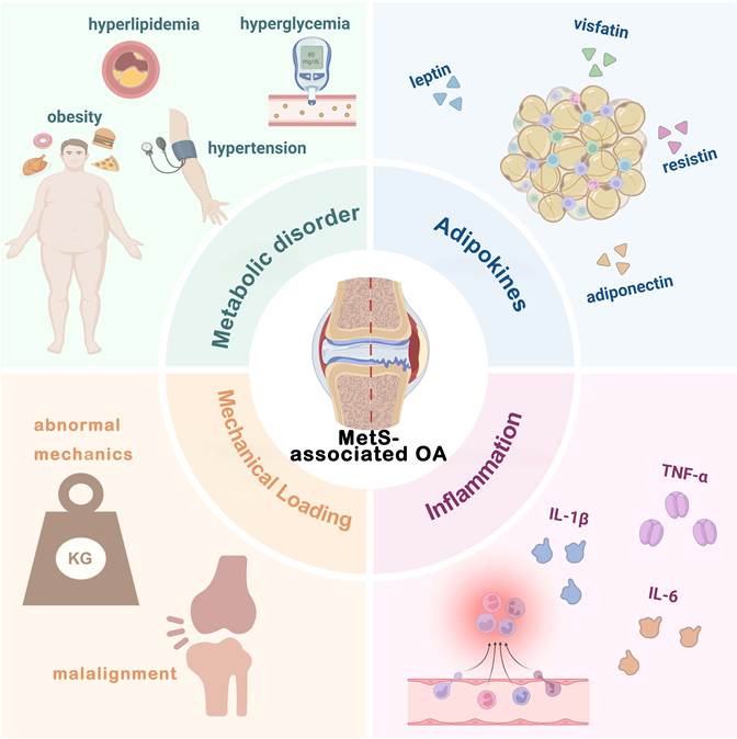
Therefore, MetS not only increases susceptibility to OA and accelerates its progression but also gives rise to distinct clinical manifestations, primarily driven by systemic metabolic disturbances affecting the joints (Figure 1). Comprehensive investigation of these etiological factors may deepen our understanding of MetS-associated OA and inform early prevention and targeted intervention strategies aimed at slowing or even halting disease progression.
Etiological factors of MetS-associated OA. Metabolic Syndrome-Associated Osteoarthritis (MetS-associated OA) arises from the complex interplay of systemic and local factors. Metabolic disturbances, including obesity, hyperlipidemia, hyperglycemia, and hypertension, establish a pro-degenerative milieu. Adipokines such as leptin, resistin, visfatin, and adiponectin, secreted by adipose tissue, regulate cartilage metabolism and joint inflammation. Pro-inflammatory cytokines (IL-1β, IL-6, and TNF-α) further amplify synovial inflammation and extracellular matrix degradation. Moreover, abnormal mechanical loading and malalignment exacerbate cartilage stress and structural damage. Collectively, these factors synergistically contribute to the onset and progression of MetS-associated OA.
2.1 Role of systemic metabolic factors
Metabolic disturbances induce systemic alterations that can reach the joint through blood circulation, thereby exerting pathogenic effects that contribute to both the onset and progression of OA. The following sections detail the clinical and biochemical evidence supporting the role of these metabolic factors in MetS-associated OA [38].
Dysregulated lipid metabolism in MetS, marked by triglycerides and cholesterol accumulation, leads to elevated circulating TG and LDL-C, alongside reduced HDL-C, thereby promoting OA initiation and progression [39, 40]. Clinical studies have reported that reduced HDL-C levels in both serum and synovial fluid of patients with OA are inversely correlated with cartilage damage and symptom severity, suggesting a potential protective role for HDL-C in OA pathogenesis [41-43]. At the mechanistic level, cholesterol reverse transport (CRT) is the process by which cholesterol effluxes from peripheral cells and is transported to the liver for metabolic clearance [44]. The preclinical study demonstrated that impaired CRT in chondrocytes results in intracellular cholesterol accumulation, inducing hypertrophic differentiation and subsequent ossification [45]. Furthermore, LDL primarily transports cholesterol from the liver to peripheral tissues [46], and elevated LDL levels may also result in excessive cholesterol accumulation within chondrocytes. In obese individuals, excessive TG storage in adipose tissue also disrupts lipid homeostasis and impairs the anti-lipolytic function of adipocytes, resulting in the hydrolysis of TG into glycerol and free fatty acids (FFAs) [47-49]. These FFAs subsequently reach the joint via systemic circulation, where elevated synovial FFAs levels have been strongly associated with OA development in obese mice [50].
Insulin resistance (IR) underlies the development of type 2 diabetes and exerts a more pronounced effect on knee OA severity in diabetic patients compared to non-diabetic individuals with KOA [51, 52]. Insulin receptors are widely expressed in the synovium of both healthy humans and mice [53], mediating insulin's inhibitory effects on synovial inflammation and catabolic activity. However, insulin resistance in diabetic patients [54] impairs this protective signaling pathway, resulting in pronounced synovitis or synovial hypertrophy that directly accelerates OA progression [55, 56]. Concurrently, clinical studies report significantly elevated levels of advanced glycation end-products (AGEs) in the cartilage, synovium, and subchondral bone of MetS-associated OA patients relative to healthy controls [55, 57]. Persistent hyperglycemia associated with MetS promotes excessive intra-articular glucose accumulation, thereby intensifying AGEs formation and deposition [58]. The accumulated AGEs amplify inflammatory signaling and facilitate collagen degradation within articular cartilage, indirectly increasing the OA risk [59, 60].
Articular cartilage is avascular and aneural, limiting chondrocyte access to oxygen and glucose [49]. More than 50% of chondrocytes' requirements for glucose, oxygen, and water are supplied by the subchondral vasculature [61, 62]. Studies have demonstrated that hypertensive rats exhibit more severe subchondral bone damage compared to normotensive controls. Hypertension-induced impairment of subchondral blood flow disrupts nutrient and gas exchange within articular cartilage, which may be a primary contributor to subchondral bone injury and subsequent cartilage lesions [63-65]. Moreover, ischemia in the subchondral bone can induce metabolic dysfunction in osteoblasts, increase osteoclastic resorption, and trigger osteocyte apoptosis, ultimately compromising subchondral bone structure and function. This leads to diminished mechanical support for the overlying cartilage and accelerates OA progression [66-68].
2.2 Role of adipose tissue-derived adipokines
Beyond the impact of systemic metabolic disturbances, local adipose tissue in obese individuals also releases systemic factors that act on joints, triggering and exacerbating OA [69, 70]. Adipose tissue produces diverse bioactive molecules collectively termed adipokines. In obesity or MetS, adipokines secretion is upregulated, and these factors can promote OA development both through systemic circulation and local joint effects. Key adipokines implicated include leptin, adiponectin, resistin, among others.
Comprehensive overview of metabolic exposures and mechanistic evidence in MetS-associated osteoarthritis
| Metabolic exposure | Evidence | Pathological features | Mechanisms | Limitations | References |
|---|---|---|---|---|---|
| Obesity/MetS | HFD animal models, chondrocytes in vitro | Cartilage degeneration, subchondral bone calcification | Lipotoxicity, inflammatory activation | Primarily focused on single molecular targets and established inflammatory pathways | [30, 109, 113, 116] |
| HFD animal models, chondrocytes in vitro | Cartilage degeneration | Lipotoxicity, lipid metabolism dysregulation, epigenetic regulation, ECM degradation | Most mechanistic evidence is derived from HFD-DMM models potentially limiting their applicability to MetS-associated OA | [119] | |
| HFD animal models, chondrocytes in vitro | Cartilage degeneration, osteophyte formation, synovial lesion, and pain sensitivity | Lipotoxicity, inflammatory activation, mitochondrial dysfunction, ECM degradation | The upstream metabolic drivers of mitochondrial dysfunction remain incompletely understood | [114, 127, 128] | |
| HFD mice, chondrocytes in vitro | Cartilage degeneration | Lipotoxicity, mitochondrial structural impairment, autophagy inhibition | The mechanisms linking metabolic stress-induced mitochondrial structural impairment to ECM degradation remain unclear | [123, 126] | |
| HFD mice | Cartilage degeneration, synovitis | Synovial macrophage activation, synovial immune cell remodeling | Experimental synovial immune landscapes may not fully recapitulate spontaneous MetS-associated OA | [131] | |
| HFD rats, chondrocytes in vitro | Cartilage degeneration | Adipokine imbalance, inflammatory activation | Single adipokine effects may not fully represent the metabolic complexity of MetS-associated OA | [110] | |
| Clinical trial, IPFP tissues in vitro | Cartilage degeneration, IPFP hypertrophy, intercellular fibrosis and angiogenesis | Adipose tissue heterogeneity, IPFP dysfunction, inflammation | The functional contribution of depot-specific adipose alterations to OA progression remains to be determined | [143, 145, 146, 150] | |
| HFD mice, osteoclasts and osteoblast in vitro | Cartilage degeneration, subchondral bone thickening and sclerosis | Osteoblast differentiation, osteoclast differentiation, subchondral bone remodeling | The interactions among bone-resident cells remain incompletely understood | [106, 153, 155] | |
| HFD mice, MSCs and endothelial progenitor cells in vitro | Cartilage degeneration, subchondral bone sclerosis and type H vessel formation | Adipokine-mediated signaling, aberrant osteogenesis and angiogenesis | The link between aberrant angiogenesis and bone remodeling under metabolic dysregulation remains poorly understood | [156, 157] | |
| Clinical trial, HFHC-fed mice, BMAds in vitro | Subchondral BMAd size was positively correlated with BMI in hand OA. MAT expansion, bone formation | Bone MAT remodeling, complement-mediated metabolic signaling, osteogenesis | The mechanisms linking bone marrow adipose tissue remodeling to coordinated osteochondral remodeling have not been fully elucidated | [154, 166] | |
| Dyslipidemia | Clinical trial, high-cholesterol diet (HCD) mice, chondrocytes in vitro | Elevated serum total cholesterol and LDL levels in OA patients with a higher BMI. Cartilage degeneration | Cholesterol metabolism, LRP family-mediated signaling, ECM degradation | Cholesterol-loading models typically evaluate isolated lipid abnormalities and may not fully represent the complex metabolic environment of MetS-associated OA | [16, 120] |
| Type 2 diabetes mellitus / Hyperglycemia | Clinical trial, hyperglycemic OA mice model, synovial macrophage and FLSs in vitro | More severe synovial hyperplasia and synovitis in patients with diabetic OA. Cartilage degeneration, synovitis | Synovial metabolic reprogramming, synovial immunometabolic inflammation | The crosstalk between synovial immunometabolic reprogramming and other joint tissues remains unclear | [134, 138] |
| Hypertension | Clinical trial | Cartilage degeneration, subchondral bone remodeling | Subchondral vascular dysfunction, hemodynamic stress, microcirculatory impairment | Limited mechanistic studies | [160-162] |
| Metabolic stressors (SFAs, adipokine, AGEs, insulin) | Chondrocytes in vitro | Chondrocyte apoptosis, cartilage degeneration | Lipotoxicity, ER stress, apoptosis, adipokine imbalance, inflammatory activation | Validation in in vivo OA models is limited | [107, 108, 139, 219] |
Leptin is the most extensively studied adipokine in OA. In patients with MetS-associated OA, leptin concentrations in both serum and synovial fluid are higher than those in non-MetS-associated OA patients [71], which has been well documented as being positively associated with disease severity and pain intensity. [72, 73]. The researchers reported that leptin-deficient mice developed morbid obesity but did not exhibit systemic inflammation or knee OA [74]. Conversely, implantation of leptin-containing fat pads into lipodystrophic (LD) mice induces both structural OA and pain [75], further underscoring the pivotal role of leptin in OA pathogenesis and its potential utility as a biomarker.
Adiponectin is the most abundant adipokine in human plasma and is primarily secreted by white adipose tissue. Early studies reported reduced adiponectin levels in the synovial fluid of OA patients, suggesting a negative correlation with synovial inflammation [76, 77]. However, a cross-sectional study by Orellana et al. demonstrated that, compared to synovial leptin, synovial adiponectin exhibits a stronger positive correlation with the severity of KOA in women [78]. Evidence shows that adiponectin levels correlate with patient BMI and correspond to OA severity. Interestingly, adiponectin levels do not increase further when OA patients damage becomes more severe [79]. More recent investigations have revealed increased adiponectin expression in OA joint synovial cells, IPFP, osteophytes, cartilage, and bone tissue in OA rats [80]. The current body of research on adiponectin remains controversial, potentially due to variations in detection methods, stages of OA progression, and sampling sites in patients across studies.
Resistin also has been demonstrated to exert effects on OA development comparable to those of leptin. Studies have reported a positive correlation between resistin levels and elevated BMI in OA patients [35]. Clinical investigations further indicate that resistin concentrations are positively correlated with OA severity, synovitis, and joint structural abnormalities [81-83]. Mechanistically, resistin may promote OA progression by upregulating pro-inflammatory cytokines and inducing MMPs synthesis in human chondrocytes and synovial cells [84].
Visfatin is an adipokine predominantly secreted by visceral adipose tissue and is known to promote B-cell maturation [85]. Clinical studies have reported that visfatin signaling is highly expressed in the hip synovial tissue of obese OA patients, co-localizing with regions of cartilage damage [86], and is also present in OA-associated osteophytes [87], indicating its potential involvement in multiple pathogenic processes in OA [88]. However, the association between elevated visfatin levels and the presence of MetS or increased BMI in OA still warrants further in-depth investigation.
2.3 Role of low-grade inflammation
Studies have demonstrated that obese OA patients exhibit systemic low-grade inflammation, characterized by elevated levels of pro-inflammatory cytokines such as interleukin (IL)-1, tumor necrosis factor (TNF)-α, and IL-6 in the synovial fluid, synovium, subchondral bone, and cartilage of affected joints [89]. Furthermore, animal studies have shown that OA mice fed a high-fat diet (HFD) present significantly increased serum levels of IL-1β and TNF-α compared to controls, accompanied by more severe cartilage degeneration [90, 91]. Collectively, both clinical and preclinical evidence underscores the critical role of inflammatory mediators in MetS-associated OA pathogenesis.
The mechanisms underlying obesity-induced systemic low-grade inflammation, beyond alterations in adipokine profiles, also involve macrophage activation. Activated macrophages enhance the expression and secretion of inflammatory cytokines in both human tissues and animal models of OA, thereby driving systemic chronic inflammation. These inflammatory mediators subsequently act locally within the joint, increasing the catabolic activity of chondrocytes, accelerating extracellular matrix (ECM) degradation, and ultimately contributing indirectly to OA pathogenesis [92-94]. Hyperglycemia-induced reactive oxygen species (ROS) production promotes macrophages polarization toward the M1 pro-inflammatory phenotype during diabetes onset and progression, further promoting chronic inflammation [95]. Moreover, Sun et al. demonstrated that a HFD increases macrophage infiltration into the synovium, with a pronounced shift toward the M1 phenotype, thereby exacerbating synovitis and promoting OA progression [96].
2.4 Role of mechanical loading factors
In obese individuals, muscle weakness, joint malalignment, and postural alterations contribute to slower walking speeds, shorter and wider strides, and prolonged stance phases compared with individuals of normal weight, leading to abnormal mechanical joints loading and an increased OA risk [97, 98]. Clinically, even a modest weight loss of 5 kg or a 5% reduction in body weight in obese patients has been associated with a 90% lower probability of knee damage over a six-year period [99]. Furthermore, excess weight leads to joint malalignment and abnormal joint mechanics, particularly knee malalignment and hyperextension, which further increase mechanical stress and accelerate cartilage degeneration, ultimately leading to OA [100, 101].
Although obesity-induced excessive mechanical loading contributes to OA development, altered biomechanics alone cannot fully account for the increased OA risk observed in non-weight-bearing joints such as the hands and wrists [102, 103]. This suggests that mechanical stress represents only one component of OA pathogenesis in obesity, and the development of MetS-associated OA must also involve systemic factors such as metabolic dysfunction.
3. Pathological features and underlying mechanisms of MetS-associated OA
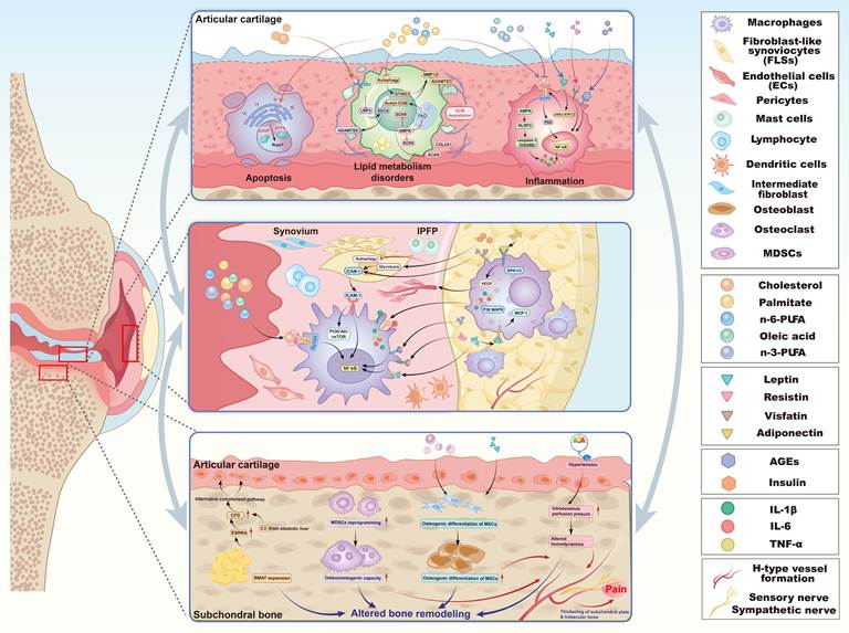
Regarding OA pathological features, articular cartilage displays early degenerative changes characterized by surface fibrillation, reduced and eventually lost safranin O staining intensity, and focal fissures [104]. The team of Zheng observed characteristic histomorphological changes of horizontal fissures at the osteochondral interface between the tibial plateau articular cartilage and subchondral bone in patients with grade III obesity [105]. In addition, tissue inflammation, fibrosis, and vascularization are hallmark pathological changes observed in the synovium and IPFP of MetS-associated OA animal models. Micro-computed tomography (micro-CT) analyses reveal a rapid increase in subchondral bone plate and trabecular thickness, accompanied by elevated bone mineral density [106]. Overall, elucidating the pathogenesis of MetS-associated OA is crucial for uncovering the origins of its distinct pathological alterations and may provide a scientific foundation for identifying potential therapeutic targets and developing precision treatments (Figure 2).
Underlying mechanisms of MetS-associated OA. Excessive lipid metabolic by-products, adipokines, diabetes-related advanced glycation end products, and hyperinsulinemia induce pathological changes in articular chondrocytes, synovium, the infrapatellar fat pad (IPFP) and subchondral bone. These metabolic disturbances trigger chondrocyte apoptosis, extracellular matrix degradation, chronic inflammation, synovial immune cell activation, and IPFP remodeling, thereby fostering local inflammation, fibrosis, altered bone remodeling and angiogenesis, and ultimately driving osteoarthritis progression.
3.1 Articular cartilage degeneration in MetS-associated OA
Chondrocyte apoptosis is a hallmark feature of articular cartilage degeneration. Palmitic acid (PA), one of the saturated fatty acids (SFAs), has been reported to induce endoplasmic reticulum (ER) stress by activating the protein kinase RNA-like ER kinase (PERK) pathway, which subsequently upregulates ER stress markers such as activating transcription factor 4 (ATF4) and C/EBP homologous protein (CHOP). This cascade further modulates the expression of nuclear protein 1 (Nupr1), ultimately triggering chondrocyte apoptosis via ER stress mechanisms, thereby leading to cartilage degeneration [107, 108].
In addition to this, chronic inflammation is a critical pathological hallmark of MetS-associated OA. Recent studies have demonstrated that PA and n-6 polyunsaturated fatty acids (n-6 PUFA) facilitate the interaction between Toll-like receptor 4 (TLR4) and myeloid differentiation-2 (MD-2), activating TLR4 indirectly. This subsequently triggers the nuclear factor κB (NF-κB) signaling within chondrocyte nuclei, thereby contributing to inflammatory responses [50, 109]. Additionally, the adipokine leptin can activate the Janus kinase 2-signal transducer and activator of transcription 3 (JAK2-STAT3) signaling pathway in chondrocytes by binding to its receptor (leptin receptor, LepR) in HFD-fed animal models, thereby modulating TLR4 expression [110]. Similarly, via binding to the cell-surface receptor RAGE, AGEs produced under chronic hyperglycemia can also activate the NF-κB signaling pathway, triggering inflammatory responses in human chondrocytes [111, 112]. Additional studies have demonstrated that PA and n-6 PUFA also induce inactivation of AMP-activated protein kinase alpha (AMPKα), which subsequently activates the NOD-like receptor protein 3 (NLRP3) inflammasome in chondrocytes. This activation leads to the production of IL-1β and IL-18 via the NLRP3/caspase-1/GSDMD signaling pathway, resulting in pyroptosis of human chondrocytes and increased severity of OA in HFD mice [113, 114].
Conversely, the study team compared the effects of PA and oleic acid (OLA), a monounsaturated fatty acid (MUFA), demonstrating that PA induces human chondrocyte matrix degradation and pro-inflammatory responses, whereas OLA does not [115]. MUFAs and n-3 polyunsaturated fatty acids (n-3 PUFA) have been reported to inhibit activation of both the TLR4/NF-κB signaling pathway and the NLRP3 inflammasome, thereby mitigating the pro-inflammatory responses in both chondrocytes in vitro and in obesity-related PTOA mice [113, 116]. As noted above, the chronic inflammatory milieu resulting from metabolic dysregulation promotes chondrocyte senescence, thereby accelerating articular cartilage degeneration as well as the onset and progression of osteoarthritis [117].
Studies have shown that metabolic dysregulation, particularly disturbances in lipid metabolism, can also influence the process of cellular senescence [118]. Therefore, latest studies have investigated lipid metabolic dysfunction within chondrocytes from OA cartilage of obese individuals, revealing that excess FFAs increase fatty acid oxidation (FAO) in chondrocytes. Excessive FAO leads to the accumulation of acetyl-CoA, which in turn further promotes FAO activity to inhibit AMPK activity, induce SRY-box transcription factor 9 (SOX9) degradation, thereby impairing cartilage ECM anabolism. On the other hand, acetyl-CoA coordinates epigenetic regulation, affecting the MMP13 and ADAMTS7 transcriptional activation, ultimately contributing to chondrocyte senescence and OA development [119]. As a metabolic intermediate, acetyl-CoA serves both as a key substrate in lipid metabolism and as an acetyl donor for epigenetic modifications such as histone acetylation, highlighting its potential as an emerging therapeutic target in MetS-associated OA. Cao et al. reported that elevated cholesterol levels in both high-cholesterol diet (HCD)-fed mice and human cartilage explants downregulate the low-density lipoprotein receptor-related protein 3 (LRP3) expression in chondrocytes, disrupting cartilage ECM metabolism and promoting OA cartilage degeneration via the cholesterol-LRP3-SDC4 signaling axis [16]. Additionally, the study demonstrated that cholesterol treatment significantly decreases LRP3 and LRP4 expression while increasing LRP5 and LRP6 expression in chondrocytes. These findings align with prior research on the LDL receptor-related protein (LRP) family's involvement in OA progression [120], underscoring the crucial role of LRPs in cholesterol metabolism within OA chondrocytes and offering new insights for future investigations [121].
Chronic inflammation, oxidative stress, and defective autophagy in OA are now recognized to exert pleiotropic effects on mitochondrial structure and function, thereby contributing significantly to chondrocyte degeneration [122]. As a critical signaling hub, Mitochondria convert upstream metabolic stress signals into structural damage in chondrocytes, underscoring their centrality in MetS-associated OA pathogenesis.
Mitochondrial structural impairment is tightly linked to autophagy activity. Lipid overload in chondrocytes can also suppress autophagic activity, thereby reducing STING1 degradation. The consequent STING1 accumulation activates the STING1-TBK1-IRF3 and STING1-TBK1-MAP2K3-MAPK14 signaling cascades, ultimately driving extracellular matrix degradation [123]. In addition, in vitro studies have demonstrated that cholesterol and palmitic acid increase cytosolic mtDNA levels while impairing mitochondrial coupling efficiency, maximal respiratory capacity, and spare respiratory capacity, all of which are essential for ATP production [124-126]. These observations suggest that lipotoxicity directly induces mitochondrial structural impairment and bioenergetic dysfunction, ultimately leading to chondrocyte injury.
Moreover, Sirtuin 3 (Sirt3), a mitochondrial deacetylase, has been found to promote chondrocyte fatty acid oxidation in HFD-induced obese mice, thereby aggravating mitochondrial oxidative stress and driving MetS-associated OA progression via metabolic dysregulation of glycolysis and oxidative phosphorylation (OXPHOS). Of note, this pathogenic effect contrasts with the previously documented chondroprotective role of Sirt3 in PT-OA and aging-associated OA [127]. By contrast, semaglutide has recently been shown to attenuate OA progression in obese mice through local modulation of the "GLP-1R-AMPK-PFKFB3" axis in joints. This axis redirects chondrocyte metabolism toward OXPHOS, ensuring adequate mitochondrial ATP production and thereby ameliorating metabolic dysfunction in cartilage [128].
A synthesis of current studies on the mechanistic framework of chondrocyte degeneration reveals that systemic metabolic dysregulation drives abnormal lipid signals that act on local chondrocytes, such as FFAs, LDL, adipokines, and AGEs. These signals release lipotoxic mediators, which in turn trigger inflammatory stress and cell injury cascades, culminating in ECM degradation and cartilage degeneration.
3.2 Synovial macrophage infiltration and fibroproliferation in MetS-associated OA
Synovitis is characterized by synovial cells proliferation and aggregation, intimal lining hyperplasia, and fibrosis within the subintimal layer [129, 130]. Harasymowicz NS and colleagues demonstrated that obesity in mice with joint injury reduces the number of synovial patrolling monocytes, thereby disrupting immune homeostasis and promoting the infiltration of pro-inflammatory macrophages. Furthermore, obesity impedes the restoration of barrier-forming lining macrophages during the late stages of OA, exacerbating disease progression [131]. The polarization of synovial macrophages toward a pro-inflammatory M1-like phenotype underlies the chronic synovial inflammation in OA [132]. This shift is characterized by a metabolic reprogramming from OXPHOS to aerobic glycolysis, a phenomenon known as the Warburg effect [133]. Studies have shown that hyperglycemia enhances macrophage glycolysis and impairs synovial macrophage generation through CD11b lactylation, thereby exacerbating inflammation and accelerating OA progression [134]. Metabolic products associated with MetS activate pattern recognition receptors (PRRs) on the synovial surface, including Toll-like receptors (TLRs) and the receptor for advanced glycation end-products (RAGE), which serve as key mediators driving synovial macrophage polarization and activation [135-137].
Additionally, Yang et al. employed single-cell RNA sequencing (scRNA-seq) to identify eight distinct synovial cell clusters in humans, revealing a significantly elevated proportion of macrophages in the synovium of patients with diabetic osteoarthritis (DOA). They attributed this increase primarily to diabetes-induced glycolysis regulation in fibroblast-like synoviocytes (FLSs), which leads to intercellular adhesion molecule-1 (ICAM-1) release. In both DOA patients and db/db diabetic OA mouse models, ICAM-1 further facilitates M1-polarized synovial macrophages infiltration and drives late-stage FLSs fibroproliferation [138]. Similarly, the adipokine visfatin enhances synovial ICAM-1 production, thereby promoting macrophage infiltration and FLS fibroproliferation [139]. Moreover, findings reported that elevated insulin levels in diabetes activate the PI3K/Akt/mTOR and NF-κB signaling pathways in the synovium, inhibit FLS autophagy, and promote macrophage infiltration [140]. However, further studies are warranted to validate these proposed mechanisms in human DOA and further elucidate their relevance to disease progression.
3.3 Hypertrophy, fibrosis, and angiogenesis of IPFP in MetS-associated OA
Alterations in the IPFP are a hallmark pathological feature in both patients and animal models of MetS-associated OA [141, 142]. Biochemical markers in IPFP of obese OA patients exhibit markedly elevated levels of IL-6, IL-1β, and TNF-α [143], which are closely associated with the pronounced activation of NF-κB, p38 MAPK, and ERK1/3 signaling pathways in human primary chondrocytes induced by IPFP [144, 145]. Moreover, adipocytes within the IPFP exhibit characteristic hypertrophy, along with prominent intercellular fibrosis and angiogenesis in obese models [146, 147]. These findings suggest that the IPFP may serve as a predictive indicator of MetS-associated OA related inflammatory activation.
IPFP and the posterior synovium are widely recognized as an integrated anatomical and functional unit [148]. Tang et al. reported that these two tissues share a highly similar immune cell composition [149]. Inflammatory cytokines secreted by the IPFP are thought to drive inflammation and fibrosis in the adjacent synovium in knee OA via paracrine mechanisms [150, 151], while elevated vascular endothelial growth factor (VEGF) levels in the IPFP are positively associated with increased synovial vascularity [152]. Collectively, MRI characteristics and biochemical profiles position IPFP as a potentially valuable biomarker for OA diagnosis and prevention, and its crosstalk with the synovium in MetS-associated OA warrants further investigation.
3.4 Abnormal remodeling of subchondral bone in MetS-associated OA
Subchondral bone pathology is not merely a secondary feature of OA, but a critical factor in OA pathogenesis and severity. Studies have shown that MetS-associated OA mice exhibit abnormal bone remodeling characterized by subchondral bone thickening and sclerosis prior to articular cartilage degeneration, accompanied by aberrant angiogenesis and bone marrow adiposity [153, 154].
MetS-associated OA subchondral bone exhibits high-turnover bone metabolism, in which the dynamic balance between bone resorption and formation is disrupted. During obesity in both humans and mice, myeloid-derived suppressor cells (MDSCs) increase and undergo reprogramming to enhance osteoclastogenic capacity, leading to increased osteoclast numbers in the subchondral bone and exacerbating OA in obese mice [155]. And a study has demonstrated that subchondral osteoclasts in MetS-associated OA mice undergo senescence at the pre-OA or early OA stage. The preosteoclast secretome promotes osteoblast differentiation while suppressing osteoclast differentiation via COX2-PGE2 signaling, thereby accelerating subchondral plate and trabecular thickening [106]. These findings indicate that metabolic disturbances drive rapid osteoclast reprogramming and uncouple resorption from formation, resulting in abnormal bone remodeling. In addition, fatty acid binding protein 4 (FABP4) and leptin have been reported to drive the osteogenic differentiation of bone marrow mesenchymal stem cells (MSCs) through PI3K/Akt signaling, while knockout of these genes significantly attenuates subchondral bone sclerosis and H-type vessel formation in HFD-fed mice [156, 157]. Currently, it is recognized that increased H-type vessel formation in the subchondral bone can further allow the penetration of sensory nerve endings and sympathetic nerves [158], thereby generating pain. However, whether distinct mechanisms exist in metabolic disturbances to mediate vascular-neural coupling remains to be further explored.
Furthermore, local microvascular function plays a pivotal role in subchondral bone remodeling, with hypertension constituting a significant factor as a systemic vascular disorder [159]. Mechanistically, hypertension disrupts joint homeostasis through both biomechanical and biochemical pathways [160]. Elevated intraosseous perfusion pressure and altered hemodynamics can modify subchondral bone mineral density, inducing a rod-to-plate transition in subchondral trabecular bone prior to cartilage degeneration [161]. This remodeling disrupts the homogeneous stiffness distribution at the osteochondral interface and impairs normal vascular nutrient supply, thereby promoting degeneration of the overlying articular cartilage matrix [162, 163].
Beyond this, bone marrow adipose tissue (BMAT) is considered the only tissue that closely links adipocytes and bone cells, and is therefore particularly relevant in the context of metabolic disturbances [164, 165]. A study analyzing BMAT explants from clinical OA patients found that increased lipolysis in BMAT promotes fatty acid oxidation and potentially regulates bone remodeling via osteoblast activity, a process modulated by BMI [166]. And recent evidence has demonstrated that a high-fat diet drives robust ESRRA estrogen-related receptor α (ESRRA) expression in murine bone marrow adipose tissue (BMAT), which in turn enhances the transcription of BMAT-derived complement factor D (CFD). In concert with component 3 (C3) derived from steatotic liver, CFD activates the alternative complement pathway, facilitating ERK1/2 phosphorylation and mitochondrial dysfunction, which culminate in chondrocyte senescence and catabolism [154]. The uniqueness of BMAT highlights its promising potential as an emerging regulator of subchondral bone remodeling and OA pathogenesis.
4. Therapeutic strategies and interventions for MetS-associated OA
4.1 Mechanism-based interventions and targeted therapies for MetS-associated OA
Diet and lifestyle modifications represent the most specific and effective early strategies for MetS-associated OA intervention and management [167]. A network meta-analysis by Shahid et al. demonstrated that the combination of psychology (P), diet (D), and exercise (E) yields the greatest efficacy in weight reduction. When overweight/obese patients lose more than 7% of their body weight, pain and physical function impairment caused by OA are improved [168]. Consistently, animal studies in HFD models have shown that dietary normalization or increased physical activity can gradually restore body composition and metabolic homeostasis, thereby slowing OA progression [169, 170].
Glucagon-like peptide-1 receptor agonists (GLP-1RAs), which mimic the action of the natural incretin hormone glucagon-like peptide-1(GLP-1), include semaglutide, whereas tirzepatide is a dual glucose-dependent insulinotropic polypeptide (GIP)/GLP-1 receptor agonist. These agents are currently among the most effective, safe, and well-tolerated anti-obesity medications (AOMs) that can improve nearly all adverse health consequences associated with obesity [171, 172]. In a multi-country, multicenter clinical trial conducted by Copenhagen University Hospital, weekly administration of semaglutide over 68 weeks led to substantial reductions in both mean body weight and pain severity in patients with obesity-associated knee OA [173]. For individuals at risk of joint replacement, treatment with semaglutide or tirzepatide can delay or even eliminate the need for surgery by reducing body weight [174]. Beyond weight loss, GLP-1RAs have demonstrated anti-inflammatory activity [175] and the capacity to mitigate ECM degradation by inhibiting AGEs [176]. These findings suggest that GLP-1RAs-mediated knee OA symptoms may be partly due to anti-inflammatory effects and ECM degradation inhibition, rather than merely due to reduced joint loading.
Besides pharmacological interventions targeting MetS-associated OA, correcting the underlying metabolic disturbances of MetS can disrupt OA progression at its source. Metformin, the first-line therapy for type 2 diabetes, has been shown to reduce OA incidence in diabetic patients [177] and to attenuate cartilage loss as well as lower joint replacement rate in individuals with concurrent type 2 diabetes and OA [178, 179]. Emerging evidence further indicates that metformin can promote weight reduction in obese individuals [180] and alleviate knee pain and stiffness while improving joint function in overweight or obese patients with symptomatic knee OA [181, 182]. Although gastrointestinal side effects may occur, these typically diminish with continued use [183]. Overall, metformin appears to be an effective and well-tolerated option for both MetS-associated OA prevention and management.
Lipid-lowering agents are a class of medications that reduce circulating triglyceride and cholesterol levels. Statins, the first-line therapy in this class, are widely prescribed for the management of hypercholesterolemia and coronary artery disease (CAD) [184]. Experimental studies have demonstrated that statins not only decrease serum triglyceride and cholesterol concentrations in mice fed a HCD but also markedly attenuate synovial inflammation and cartilage degeneration [185]. In vitro, statins have been shown to substantially mitigate chondrocyte damage under high-glucose culture conditions [186, 187]. Thus, in patients with OA complicated by hypercholesterolemia and diabetes, statins may offer considerable therapeutic benefit. Nevertheless, clinical evidence also suggests that statin use may increase the risk of developing type 2 diabetes [188], highlighting the need for careful patient stratification and treatment planning based on individual patient circumstances.
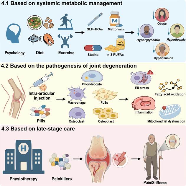
Finally, as previously noted, n-3 PUFAs exert inhibitory effects on the development of MetS-associated OA, and their anti-inflammatory and antioxidant properties may help prevent or attenuate cartilage loss [189]. In a randomized clinical trial, Hill et al. reported that OA patients receiving low-dose n-3 PUFA supplementation (fish oil) experienced greater improvements in pain and physical function after two years of treatment compared with controls [190]. Nevertheless, the use of n-3 PUFAs remains controversial due to potential adverse effects, including gastrointestinal discomfort and possible increased risks of cancer and cardiovascular disease. Determining the optimal balance between n-3 PUFA selection and the ideal anti-inflammatory dosage still requires further investigation [191]. These interventions and targeted therapies are summarized in Table 2 and Figure 3, which outlines their mechanisms of action, potential roles in OA, and adverse reactions reported in clinical trials or basic research.
Therapeutic strategies and interventions for MetS-associated OA
| Preventions and treatments | Targets of action | Type of study | Potential roles in OA | Representative drugs | Adverse reactions | References |
|---|---|---|---|---|---|---|
| Non-pharmacological interventions | Lose weight | Clinical trial | Effectively enhances joint function and reduces pain in individuals with OA | Exercise (E) | NA | [220] |
| Clinical trial | Significantly enhance joint mechanical efficiency during gait, 6-minute walk test (6MWT) distance, and balance for obese OA patients; Alleviate joint pain, improve joint function, and enhance physical and mental health-related quality of life (HRQoL) in OA patients | Diet + Exercise (D+E) | [221, 222] | |||
| Clinical trial | A weight loss of at least 7% can improve pain and physical function in overweight/obese individuals | Psychology, Diet + Exercise (PDE) | [168] | |||
| Basic research | Both aerobic and endurance training can alleviate joint damage in high-fat/high-sucrose (HFS) rats | Exercise (E) | [169] | |||
| GLP-1RAs | Lose weight | Clinical trial | Markedly reduce cartilage loss and pain in knee joints of obese patients; Delay or cancel joint replacement surgery | Tirzepatide, Semaglutide | Increase in gastrointestinal events, eating disorders, and the risk of depression/suicidal ideation | [173, 223-226] |
| Metabolic reprogramming from glycolysis to OXPHOS | Basic research | Attenuate cartilage degeneration, osteophyte formation, synovial pathology, and pain sensitivity in obese mice | Semaglutide | [128] | ||
| Reduce levels of inflammatory factors and inhibit the effects of AGEs | Basic research | Improves the deterioration of cartilage ECM components | Dulaglutide | [176] | ||
| Biguanide antidiabetic drugs | Lose weight | Clinical trial | Alleviates joint pain and improves joint function in overweight or obese patients | Metformin | Gastrointestinal symptoms are common and tend to decrease over time | [178, 181, 182] |
| Reduce glycemic levels | Clinical trial | The rate of joint replacement in patients with DOA decreased over a period of 10 years. | [179] | |||
| Clinical trial | Reduces the risk of OA development in diabetic patients and prevent OA | [177] | ||||
| Inhibit oxidative stress and inflammatory responses | Basic research | Protects diabetic rats from secondary articular cartilage damage | [227, 228] | |||
| Regulate osteoprogenitor cells and type H vessel formation | Basic research | Mitigates aberrant osteogenesis and pain responses | [198] | |||
| Statins | Reduce serum cholesterol and triglyceride concentrations | Clinical trial | Significantly reduces the risk of knee pain in patients with OA. Mitigates the longitudinal progression of femorotibial joint bone marrow lesion (BML) volume | Atorvastatin (ATO), Simvastatin | Increase the risk of developing type 2 diabetes in the general population | [188, 229, 230] |
| Basic research | In mice with dyslipidemia, the thickening of the synovium and the degeneration of cartilage were significantly alleviated | [185, 231, 232] | ||||
| Basic research | Significantly reduces chondrocyte inflammation induced by high glucose (HG) and prevent cartilage degradation | [186, 187] | ||||
| n-3-PUFA | Inhibit oxidative stress and inflammatory responses | Clinical trial | Improve joint pain, stiffness, and physical function in patients with OA | Fish oil, Krill oil | Gastrointestinal events, and increased incidence of infections, cancers, and cardiocerebrovascular diseases | [190, 191, 233] |
| Basic research | Reduces the number and infiltration of pro-inflammatory synovial macrophages, thereby improving synovitis and cartilage degeneration in obese OA mice | Resolvin D1 (RvD1) | [195] | |||
| Lipotoxicity-targeted therapies | Inhibit ER stress-mediated cell death | Basic research | Attenuate palmitic acid-induced chondrocyte apoptosis | Target Nupr1 or aberrant ER stress signaling | Many potential therapeutic agents remain at the preclinical stage, and their long-term efficacy for MetS-associated OA requires further investigation | [107, 108] |
| Suppress aberrant FAO activation | Basic research | Alleviates cartilage degeneration in metabolically challenged PTOA mice | FAO inhibitor trimetazidine | [119] | ||
| Modulating lipid metabolism and suppressing chondrocyte ferroptosis | Basic research | Preserves articular cartilage homeostasis in HFD-fed rats | miR-34a-5p inhibitors | [197] | ||
| Anti-inflammatory immune-metabolic therapy | Inhibition of MAPK and NF-κB activation | Basic research | Attenuates AGEs-induced human chondrocyte apoptosis | PPARγ agonist pioglitazone | [194] | |
| Inhibit CDK5-mediated PPARγ/NF-κB activation | Basic research | Reverse M1 macrophage polarization and chondrocyte apoptosis in vitro and alleviate cartilage damage in obese mice in vivo | PPARγ agonist rosiglitazone | [234] | ||
| Inhibit NLRP3 inflammasome and activate AMPK | Basic research | Attenuates inflammation and cartilage matrix degradation in palmitic acid-treated human chondrocytes | Xanthohumol | [114] | ||
| Reduce synovial macrophage infiltration | Basic research | Alleviate persistent synovitis in obese mice | Clodronate-loaded liposomes (CL), GAS6 | [195] | ||
| PGE₂ pathway-targeted therapies | Suppress PGE₂ synthesis or block EP4-mediated signaling | Basic research | Alleviating pain responses in MetS-associated OA mice | Imrecoxib, HL-43 | Increase the risk of cardiovascular events during OA treatment | [106, 202] |
Therapeutic strategies and interventions for MetS-associated OA. Therapeutic approaches include (4.1) systemic metabolic management targeting obesity, insulin resistance, dyslipidemia, and related metabolic disturbances; (4.2) mechanism-based targeted therapies addressing downstream processes linking metabolic dysregulation to OA progression, including immunometabolic inflammation, lipotoxic stress, oxidative stress, and other metabolic stress responses within joint tissues; and (4.3) late-stage care for symptom relief and functional improvement. This framework highlights the mechanistic alignment between metabolic abnormalities, joint pathogenesis, and therapeutic strategies.
4.2 Targeted therapies for MetS-associated OA based on the pathogenesis of joint degeneration
Given that intracellular damage and metabolic reprogramming within the joint have already occurred by the time most OA patients with MetS develop articular symptoms, it is also necessary to simultaneously target local degenerative mechanisms for therapeutic intervention, in order to delay irreversible structural and functional damage. Nupr1 knockdown suppresses metabolic dysregulation-induced activation of ER stress-mediated pro-apoptotic pathways [107, 108], suggesting that targeting Nupr1 or aberrant ER stress signaling may represent a potential strategy for alleviating chondrocyte apoptosis in MetS-associated OA.
In addition, lipotoxic products derived from systemic metabolic dysregulation drive chronic inflammatory responses in joints by broadly activating the TLR4/NF-κB signaling pathway and the AMPK/NLRP3 axis. Peroxisome proliferator-activated receptor γ (PPARγ) has been demonstrated to reduce circulating TG, TC, and LDL-C levels while also acting as a critical anti-inflammatory transcription factor in OA [192, 193]. Studies have demonstrated that the PPARγ agonist pioglitazone attenuates AGEs-induced human chondrocyte apoptosis through inhibition of MAPK and NF-κB activation [194]. And Xanthohumol attenuates mitochondrial dysfunction through AMPK activation and inhibits NLRP3 inflammasome-mediated inflammation and cartilage matrix degradation in palmitic acid-treated human chondrocytes [114]. Chronic inflammation is also a key driver of synovitis. One study used clodronate-loaded liposomes (CL) to deplete activated macrophages in the synovium of HFD-fed PTOA mice, thereby alleviating persistent synovial inflammation [195]. In addition, intra-articular administration of GAS6 restores macrophage efferocytosis in obese mice and prevents MetS-associated OA development [196]. Although many potential therapeutic agents remain at the preclinical stage, they hold considerable promise as anti-inflammatory strategies for MetS-associated OA treatment.
Systemic metabolic dysregulation affects joint tissues not only by indirectly activating inflammatory and cellular stress responses but also by directly driving intracellular metabolic reprogramming to facilitate local adaptation to metabolic perturbations. Previous studies have demonstrated that metabolic dysregulation-driven FAO activation contributes to OA pathogenesis, whereas cartilage-targeted delivery of the FAO inhibitor trimetazidine alleviates cartilage degeneration in metabolically challenged PTOA mice [119]. Furthermore, a novel nanocomplex (P-T-M) enables efficient chondrocyte-targeted delivery of miR-34a-5p inhibitors, which preserves articular cartilage homeostasis in HFD-fed rats by modulating lipid metabolism and suppressing chondrocyte ferroptosis [197].
4.3 Therapies for MetS-associated OA based on late-stage care
Finally, once irreversible joint damage has developed in advanced MetS-associated OA, metabolic and anti-inflammatory therapies may have limited efficacy. Therefore, management should shift toward alleviating pain, preserving function, and improving quality of life. Metformin alleviates cartilage damage in high-fat diet-induced MetS-associated OA by reducing subchondral bone macrophage accumulation and prostaglandin production. In addition, metformin mitigates aberrant osteogenesis and pain responses in MetS-associated OA through the regulation of osteoprogenitor cells and type H vessel formation [198]. Selective COX-2 inhibitors have been shown to mitigate pathological subchondral bone remodeling and OA progression in MetS-associated OA mice by targeting osteoclast senescence [106], while also alleviating pain responses through suppression of PGE2 production [199]. However, despite their therapeutic potential, currently available COX-2 inhibitors increase the risk of cardiovascular events during OA treatment and exhibit a less favorable risk-benefit profile in patients with pre-existing MetS [200, 201]. Conversely, studies have also shown that the EP4 antagonist HL-43 attenuates OA progression and pain in mice by suppressing COX-2-derived PGE2/EP4 signaling-mediated subchondral bone angiogenesis and sensory nerve innervation [202].
Taking together, a combination of systemic metabolic correction, joint-specific intervention, and symptomatic management may represent a rational therapeutic framework for MetS-associated OA. Early intervention targeting upstream metabolic abnormalities may help limit metabolic stress and delay joint degeneration, whereas patients with established local tissue damage may require joint-specific therapies in combination with metabolic management and symptomatic treatment.
5. Summary and future prospects
Taken together, MetS induces systemic metabolic disturbances, which subsequently act on local joint tissues to cause OA, thereby forming the MetS-associated OA phenotype characterized by distinct clinical manifestations and pathological features. Recent studies suggest that meniscal hypertrophy, extrusion, and tears are also recognized contributors to structural progression and disease severity in knee OA [203]. Longitudinal clinical data demonstrated that medial meniscus volume and extrusion in overweight and obese patients were associated with the incidence of radiographic knee OA at 30 months [204, 205]. Concurrently, preliminary MRI and micro-CT observations in HFD-fed mice revealed more marked reductions in meniscal density and an increased fragmentation rate [206]. Thus, the meniscus may serve as a key imaging-based target for early detection and intervention in asymptomatic MetS-associated OA [207].
Previous studies have demonstrated that metabolic dysregulation can drive macrophage infiltration and M1 polarization within the synovium through mechanisms such as inducing glycolytic reprogramming of FLS and upregulating the OA-associated protease HtrA1 [138, 208]. In addition, recent investigations have identified RCAN1+IL1α+ senescent synovial fibroblasts within the OA synovial lining layer, which secrete excessive SFAs that act on cartilage to accelerate chondrocyte senescence and ECM degradation [209]. Meanwhile, it has become increasingly recognized that subchondral bone lesions in MetS-associated OA may precede cartilage degeneration [142, 153]. Such crosstalk may involve the reciprocal exchange of inflammatory mediators, adipokines, lipid metabolites, extracellular vesicles, and vascular and neural signals, thereby creating a feed-forward network that links systemic metabolic stress to multi-tissue joint degeneration. Thus, beyond focusing on the dysfunctional mechanisms of individual joint tissues in MetS-associated OA, it is essential to recognize that, given its nature as a whole-joint disease, the cellular crosstalk within and across joint tissues may play a pivotal role in driving its initiation and progression. This broader perspective may ultimately allow us to intervene at the very root of the disease process. Moreover, when other pathogenic factors of OA are superimposed on MetS, the specific alterations in systemic and local joint metabolism, as well as their synergistic contributions to OA development, remain incompletely understood. The ambiguity regarding whether metabolic dysregulation serves as an initiating factor or a contributing factor has hindered both a comprehensive understanding of MetS-associated OA and the development of effective targeted interventions.
Patients with MetS-associated OA frequently experience exacerbated joint pain, representing a significant clinical concern. A large-scale cross-sectional cohort study in South Korea demonstrated a positive correlation between elevated cholesterol levels in MetS and knee OA pain [14]. Dyslipidemia-induced increases in sphingomyelin 34:1 also have been shown to elicit arthritic pain in murine models [210]. And sphingomyelin is hydrolyzed to ceramide by acid sphingomyelinase. Studies have found that palmitic acid treatment induces ceramide accumulation in human chondrocytes, potentially contributing to OA development through dysregulation of ECM catabolism [211]. Subchondral bone ischemia resulting from hypertension is also implicated as a risk factor for joint pain [23, 212, 213]. Furthermore, the coexistence of diabetes, hypertension, and obesity with OA is associated with heightened joint pain severity [9]. Collectively, components of MetS may independently or synergistically lower pain thresholds and exacerbate joint pain severity [214, 215]. The pathogenesis of joint pain in MetS-associated OA is likely mediated by various metabolic factors and adipokines circulating in the blood that act on macrophage-rich regions such as the synovium and IPFP, promoting the accumulation of inflammatory mediators and triggering inflammatory responses [216-218], but the precise mechanisms remain to be fully elucidated. A comprehensive understanding of pain etiology and pathogenesis is essential for facilitating early intervention and effective pain management.
Research has demonstrated the critical role of biomarkers in osteoarthritis research, clinical trials, and therapeutic development. However, validated indicators for early and targeted screening or longitudinal monitoring of MetS-associated OA remain scarce. Nevertheless, although bioinformatic and machine learning analyses have identified a limited number of peripheral blood-based diagnostic gene signatures for MetS-associated OA, their functional relevance to local joint pathology has not yet been experimentally confirmed. Consequently, there is an urgent need to establish a comprehensive monitoring framework that integrates biomarkers reflecting systemic metabolism, cellular senescence, and local joint alterations. Such an integrated approach would provide a robust foundation for the development of effective strategies for early detection, risk stratification, and targeted intervention in MetS-associated OA. Therefore, future research should more actively leverage advanced technologies such as genomics, proteomics, and metabolomics to comprehensively investigate characteristic pathological alterations in MetS-associated OA and their interplay with OA driven by other endotypes. Such efforts can substantially enhance our understanding of OA pathogenesis and allow MetS-associated OA research to transition from phenomenological description to a new phase characterized by mechanism driven insights and precision targeted interventions to facilitate the development of more targeted therapeutic strategies.
Abbreviations
OA: Osteoarthritis; MetS: Metabolic syndrome; MetS-associated OA: Metabolic syndrome-associated Osteoarthritis; DOA: Diabetic osteoarthritis; BMI: Body mass index; TG: Triglycerides; HDL: High-density lipoprotein; LDL: Low-density lipoprotein; FFAs: Free fatty acids; SFAs: Saturated fatty acids; MUFA: Monounsaturated fatty acid; PUFA: Polyunsaturated fatty acids; PA: Palmitic acid; OLA: Oleic acid; LD: Lipodystrophic; HFD: High-fat diet; HCD: High-cholesterol diet; ECM: Extracellular matrix; MMPs: Matrix metalloproteinases; ADAMTS: A disintegrin and metalloproteinase with thrombospondin motifs; BMLs: Bone marrow lesions; IPFP: Infrapatellar fat pad; CRT: Cholesterol reverse transport; IR: Insulin resistance; AGEs: Advanced glycation end-products; RAGE: Receptor for advanced glycation end-products; IL: Interleukin; TNF-α: Tumor necrosis factor-α; COX-2: Cyclooxygenase-2; LepR: Leptin receptor; ROS: Reactive oxygen species; micro-CT: Micro-computed tomography; ER: Endoplasmic reticulum; CHOP: C/EBP homologous protein; ATF4: Activating transcription factor 4; PERK: Protein kinase RNA-like ER kinase; Nupr1: Nuclear protein 1; TLR4: Toll-like receptor 4; MD-2: Myeloid differentiation-2; NF-κB: Nuclear factor κB; AMPKα: AMP-activated protein kinase alpha; NLRP3: NOD-like receptor protein 3; JAK2-STAT3: Janus kinase 2-signal transducer and activator of transcription 3; FAO: Fatty acid oxidation; SOX9: SRY-box transcription factor 9; LRP3: Lipoprotein receptor-related protein 3; PRRs: Pattern recognition receptors; scRNA-seq: single-cell RNA sequencing; FLSs: Fibroblast-like synoviocytes; ICAM-1: Intercellular adhesion molecule-1; VEGF: Vascular endothelial growth factor; P: Psychology; D: Diet; E: Exercise; GLP-1: Glucagon-like peptide-1; GLP-1RAs: Glucagon-like peptide-1 receptor agonists; AOMs: Anti-obesity medications; CAD: Coronary artery disease.
Acknowledgements
Funding
This work was supported by these programs: National Natural Science Foundation of China, 82130070. National Natural Science Foundation of China, 82394442. National Natural Science Foundation of China, 82002361. Shaanxi Province Health Science and Technology Innovation Capability Enhancement Program, 2025TD-1.
Authorship contributions
Writing - review & editing the manuscript: Xinyu Zhang, Validation & Visualization: Guangyu Ding & Bowei Ni, Conceptualization, review & editing: Liu Yang & Liyuan Jia. All authors read and approved of the final manuscript.
Al usage statement
During the preparation of this manuscript, ChatGPT (OpenAI) was used for language polishing, and Recraft AI was used to assist in creating selected graphical elements in Figure 3. The features and scientific content of these elements were determined by the authors based on the cited literature. The authors reviewed and edited all AI-assisted outputs and take full responsibility for the accuracy, completeness, and originality of the manuscript.
Competing Interests
The authors have declared that no competing interest exists.
References
1. Gelber AC. Knee Osteoarthritis. Annals of Internal Medicine. 2024;177:ITC129-ITC44
2. Steinmetz JD, Culbreth GT, Haile LM, Rafferty Q, Lo J, Fukutaki KG. et al. Global, regional, and national burden of osteoarthritis, 1990-2020 and projections to 2050: a systematic analysis for the Global Burden of Disease Study 2021. The Lancet Rheumatology. 2023;5:e508-e22
3. Wallace IJ, Felson DT, Worthington S, Duryea J, Clancy M, Aliabadi P. et al. Knee osteoarthritis risk in non-industrial societies undergoing an energy balance transition: evidence from the indigenous Tarahumara of Mexico. Annals of the Rheumatic Diseases. 2019;78:1693-8
4. Sun H, Sun L, Liu M, Zhuang Y, Ning X, Yang H. et al. Global burden of high BMI-Related osteoarthritis and causal effects of obesity: a GBD 1990-2021 and bidirectional two-sample mendelian randomization study. International Journal of Surgery. 2025
5. Voinier D, Neogi T, Stefanik JJ, Guermazi A, Roemer FW, Thoma LM. et al. Using Cumulative Load to Explain How Body Mass Index and Daily Walking Relate to Worsening Knee Cartilage Damage Over Two Years: The MOST Study. Arthritis Rheumatology. 2020;72:957-65
6. Li H, George DM, Jaarsma RL, Mao X. Metabolic syndrome and components exacerbate osteoarthritis symptoms of pain, depression and reduced knee function. Annals of Translational Medicine. 2016;4:133 -
7. Afifi AE-MA, Shaat RM, Gharbia OM, Boghdadi YE, Eshmawy MME, El-Emam OA. Osteoarthritis of knee joint in metabolic syndrome. Clinical Rheumatology. 2018;37:2855-61
8. Yasuda E, Nakamura R, Matsugi R, Goto S, Ikenaga Y, Kuroda K. et al. Association between the severity of symptomatic knee osteoarthritis and cumulative metabolic factors. Aging Clinical and Experimental Research. 2018;30:481-8
9. Dell'Isola A, Vinblad J, Turkiewicz A, Kiadaliri A, Abbott A, Rolfson O. et al. The coexistence of diabetes, hypertension and obesity is associated with worse pain outcomes following exercise for osteoarthritis: A cohort study on 80,893 patients. Osteoarthritis and Cartilage. 2024;32:1308-18
10. Recenti F, Battista S, Lohmander S, Vinblad J, Kiadaliri A, Abbott A. et al. Association between metabolic conditions, physical activity and self-efficacy before and after a first-line exercise and education intervention for osteoarthritis: a longitudinal register study using the SOAD cohort. RMD Open. 2025;11:e005804
11. Zhuo Q, Yang W, Chen J, Wang Y. Metabolic syndrome meets osteoarthritis. Nature Reviews Rheumatology. 2012;8:729-37
12. Alberti KGM, Zimmet P, Shaw J. The metabolic syndrome-a new worldwide definition. The Lancet. 2005;366:1059-62
13. Sanchez-Santos M, Judge A, Gulati M, Spector T, Hart D, Newton J. et al. Association of metabolic syndrome with knee and hand osteoarthritis: A community-based study of women. Seminars in Arthritis and Rheumatism. 2019;48:791-8
14. Cho BW, Kim DS, Kwon HM, Yang IH, Lee W-S, Park KK. Cross-Sectional Association between Hypercholesterolemia and Knee Pain in the Elderly with Radiographic Knee Osteoarthritis: Data from the Korean National Health and Nutritional Examination Survey. Journal of Clinical Medicine. 2021;10:933
15. Li H-B, Du Y-J, Kenmegne GR, Kang C-W. Machine learning analysis of serum cholesterol's impact on knee osteoarthritis progression. Scientific Reports. 2024 14
16. Cao C, Shi Y, Zhang X, Li Q, Zhang J, Zhao F. et al. Cholesterol-induced LRP3 downregulation promotes cartilage degeneration in osteoarthritis by targeting Syndecan-4. Nature Communications. 2022 13
17. Garcia-Gil M, Reyes C, Ramos R, Sanchez-Santos MT, Prieto-Alhambra D, Spector TD. et al. Serum Lipid Levels and Risk of Hand Osteoarthritis: The Chingford Prospective Cohort Study. Scientific Reports. 2017 7
18. Hsueh T-Y, Chen Y-P. The association between dyslipidemia and hand osteoarthritis: a systematic review and meta-analysis. Clinical Rheumatology. 2025;44:1439-48
19. Li C, Wu J, Zhang Y, He H, Hu Y, Wei J. et al. Plasma lipoprotein subclasses and risk of incident knee osteoarthritis: A population-based cohort study. Osteoarthritis and Cartilage. 2025
20. Louati K, Vidal C, Berenbaum F, Sellam J. Association between diabetes mellitus and osteoarthritis: systematic literature review and meta-analysis. RMD Open. 2015;1:e000077
21. Luo S, Shi Q, Chen J, Wang H, Wu W, Zha Z. Expression and Significance of MMPs in Synovial Fluid, Serum and PBMC Culture Supernatant Stimulated by LPS in Osteoarthritis Patients with or Without Diabetes. Experimental and Clinical Endocrinology Diabetes. 2019;127:195-202
22. Law L, Heerey JJ, Devlin BL, Brukner P, De Livera AM, Attanayake A. et al. The Effect of an Anti-Inflammatory Diet for Knee oSTeoarthritis (FEAST) Trial: Baseline Characteristics and Relationships with Dietary Inflammatory Index. Journal of the American Nutrition Association. 2025:1-10
23. Lo K, Au M, Ni J, Wen C. Association between hypertension and osteoarthritis: A systematic review and meta-analysis of observational studies. Journal of Orthopaedic Translation. 2022;32:12-20
24. Zhang Y-M, Wang J, Liu X-G. Association between hypertension and risk of knee osteoarthritis. Medicine. 2017;96:e7584
25. Lo GH, McAlindon TE, Katz JN, Driban JB, Price LL, Eaton CB. et al. Systolic and pulse pressure associate with incident knee osteoarthritis: data from the Osteoarthritis Initiative. Clinical Rheumatology. 2017;36:2121-8
26. Alberti KGMM, Eckel RH, Grundy SM, Zimmet PZ, Cleeman JI, Donato KA. et al. Harmonizing the Metabolic Syndrome. Circulation. 2009;120:1640-5
27. Yang H, Muhui Z, Fan T, Chen H, Fang X, Li ZA. et al. Associations of metabolic status with all-cause mortality among individuals with osteoarthritis: A prospective cohort study. Journal of Orthopaedic Translation. 2025;51:207-17
28. Berenbaum F, Wallace IJ, Lieberman DE, Felson DT. Modern-day environmental factors in the pathogenesis of osteoarthritis. Nature Reviews Rheumatology. 2018;14:674-81
29. Roze RH, Bierma-Zeinstra SMA, Agricola R, Oei EHG, Waarsing JH. Differences in MRI features between two different osteoarthritis subpopulations: data from the Osteoarthritis Initiative. Osteoarthritis and Cartilage. 2016;24:822-6
30. Cao X, Cui Z, Ding Z, Chen Y, Wu S, Wang X. et al. An osteoarthritis subtype characterized by synovial lipid metabolism disorder and fibroblast-like synoviocyte dysfunction. Journal of Orthopaedic Translation. 2022;33:142-52
31. Mathiessen A, Conaghan PG. Synovitis in osteoarthritis: current understanding with therapeutic implications. Arthritis Research & Therapy. 2017 19
32. Yao Q, Wu X, Tao C, Gong W, Chen M, Qu M. et al. Osteoarthritis: pathogenic signaling pathways and therapeutic targets. Signal Transduction and Targeted Therapy. 2023 8
33. Han W, Aitken D, Zheng S, Wluka AE, Zhu Z, Blizzard L. et al. Association Between Quantitatively Measured Infrapatellar Fat Pad High Signal-Intensity Alteration and Magnetic Resonance Imaging-Assessed Progression of Knee Osteoarthritis. Arthritis Care Research. 2019;71:638-46
34. Li J, Fu S, Gong Z, Zhu Z, Zeng D, Cao P. et al. MRI-based Texture Analysis of Infrapatellar Fat Pad to Predict Knee Osteoarthritis Incidence. Radiology. 2022;304:611-21
35. Lambova SN, Batsalova T, Moten D, Stoyanova S, Georgieva E, Belenska-Todorova L. et al. Serum Leptin and Resistin Levels in Knee Osteoarthritis-Clinical and Radiologic Links: Towards Precise Definition of Metabolic Type Knee Osteoarthritis. Biomedicines. 2021;9:1019
36. Munugoda IP, Wills K, Cicuttini F, Graves SE, Lorimer M, Jones G. et al. The association between ambulatory activity, body composition and hip or knee joint replacement due to osteoarthritis: a prospective cohort study. Osteoarthritis and Cartilage. 2018;26:671-9
37. Blankstein M, Browne JA, Sonn KA, Ashkenazi I, Schwarzkopf R. Go Big or Go Home: Obesity and Total Joint Arthroplasty. The Journal of Arthroplasty. 2023;38:1928-37
38. Lu B, Driban JB, Xu C, Lapane KL, McAlindon TE, Eaton CB. Dietary Fat Intake and Radiographic Progression of Knee Osteoarthritis: Data From the Osteoarthritis Initiative. Arthritis Care Research. 2017;69:368-75
39. Courties A, Sellam J, Berenbaum F. Metabolic syndrome-associated osteoarthritis. Current Opinion in Rheumatology. 2017;29:214-22
40. Thijssen E, Van Caam A, Van Der Kraan PM. Obesity and osteoarthritis, more than just wear and tear: pivotal roles for inflamed adipose tissue and dyslipidaemia in obesity-induced osteoarthritis. Rheumatology. 2015;54:588-600
41. Zhang K, Ji Y, Dai H, Khan AA, Zhou Y, Chen R. et al. High-Density Lipoprotein Cholesterol and Apolipoprotein A1 in Synovial Fluid: Potential Predictors of Disease Severity of Primary Knee Osteoarthritis. CARTILAGE. 2021;13:1465S-73S
42. Triantaphyllidou IE, Kalyvioti E, Karavia E, Lilis I, Kypreos KE, Papachristou DJ. Perturbations in the HDL metabolic pathway predispose to the development of osteoarthritis in mice following long-term exposure to western-type diet. Osteoarthritis and Cartilage. 2013;21:322-30
43. Herrero-Manley L, Alabajos-Cea A, Suso-Martí L, Cuenca-Martínez F, Calatayud J, Casaña J. et al. Serum lipid biomarkers and inflammatory cytokines associated with onset and clinical status of patients with early knee osteoarthritis. Frontiers in Nutrition. 2023 10
44. Chawla A, Boisvert WA, Lee C-H, Laffitte BA, Barak Y, Joseph SB. et al. A PPARγ-LXR-ABCA1 Pathway in Macrophages Is Involved in Cholesterol Efflux and Atherogenesis. Molecular Cell. 2001;7:161-71
45. Kostopoulou F, Malizos KN, Papathanasiou I, Tsezou A. MicroRNA-33a regulates cholesterol synthesis and cholesterol efflux-related genes in osteoarthritic chondrocytes. Arthritis Research Therapy. 2015 17
46. Ward NC, Watts GF, Eckel RH. Statin Toxicity. Circulation Research. 2019;124:328-50
47. Wei G, Lu K, Umar M, Zhu Z, Lu WW, Speakman JR. et al. Risk of metabolic abnormalities in osteoarthritis: a new perspective to understand its pathological mechanisms. Bone Research. 2023 11
48. Sekar S, Shafie SR, Prasadam I, Crawford R, Panchal SK, Brown L. et al. Saturated fatty acids induce development of both metabolic syndrome and osteoarthritis in rats. Scientific Reports. 2017 7
49. Wu X, Fan X, Crawford R, Xiao Y, Prasadam I. The Metabolic Landscape in Osteoarthritis. Aging and disease. 2022;13:1166
50. Yu X, Zheng G, Hu Z, Tang S, Xu J, Shang P. et al. Asiatic acid ameliorates obesity-related osteoarthritis by inhibiting myeloid differentiation protein-2. Food Function. 2020;11:5513-24
51. Schett G, Kleyer A, Perricone C, Sahinbegovic E, Iagnocco A, Zwerina J. et al. Diabetes Is an Independent Predictor for Severe Osteoarthritis. Diabetes Care. 2013;36:403-9
52. Eymard F, Parsons C, Edwards MH, Petit-Dop F, Reginster JY, Bruyère O. et al. Diabetes is a risk factor for knee osteoarthritis progression. Osteoarthritis and Cartilage. 2015;23:851-9
53. Ribeiro M, López De Figueroa P, Nogueira-Recalde U, Centeno A, Mendes AF, Blanco FJ. et al. Diabetes-accelerated experimental osteoarthritis is prevented by autophagy activation. Osteoarthritis and Cartilage. 2016;24:2116-25
54. Hamada D, Maynard R, Schott E, Drinkwater CJ, Ketz JP, Kates SL. et al. Suppressive Effects of Insulin on Tumor Necrosis Factor-Dependent Early Osteoarthritic Changes Associated with Obesity and Type 2 Diabetes Mellitus. Arthritis Rheumatology. 2016;68:1392-402
55. Veronese N, Cooper C, Reginster J-Y, Hochberg M, Branco J, Bruyère O. et al. Type 2 diabetes mellitus and osteoarthritis. Seminars in Arthritis and Rheumatism. 2019;49:9-19
56. Griffin TM, Huffman KM. Editorial: Insulin Resistance: Releasing the Brakes on Synovial Inflammation and Osteoarthritis? Arthritis Rheumatology. 2016;68:1330-3
57. Li Q, Wen Y, Wang L, Chen B, Chen J, Wang H. et al. Hyperglycemia-induced accumulation of advanced glycosylation end products in fibroblast-like synoviocytes promotes knee osteoarthritis. Experimental Molecular Medicine. 2021;53:1735-47
58. Jørgensen AEM, Kjær M, Heinemeier KM. The Effect of Aging and Mechanical Loading on the Metabolism of Articular Cartilage. The Journal of Rheumatology. 2017;44:410-7
59. Mobasheri A, Rayman MP, Gualillo O, Sellam J, Van Der Kraan P, Fearon U. The role of metabolism in the pathogenesis of osteoarthritis. Nature Reviews Rheumatology. 2017;13:302-11
60. Steenvoorden MMC, Huizinga TWJ, Verzijl N, Bank RA, Ronday HK, Luning HAF. et al. Activation of receptor for advanced glycation end products in osteoarthritis leads to increased stimulation of chondrocytes and synoviocytes. Arthritis Rheumatism. 2006;54:253-63
61. Imhof H, Sulzbacher I, Grampp S, Czerny C, Youssefzadeh S, Kainberger F. Subchondral Bone and Cartilage Disease. Investigative Radiology. 2000;35:581-8
62. Hu W, Chen Y, Dou C, Dong S. Microenvironment in subchondral bone: predominant regulator for the treatment of osteoarthritis. Annals of the Rheumatic Diseases. 2021;80:413-22
63. Chan PB, Wen C. Spontaneous hypertensive rat exhibits bone and meniscus phenotypes of osteoarthritis: is it an appropriate control for MetS-associated OA? Annals of the Rheumatic Diseases. 2018;77:e25
64. Sampath SJP, Venkatesan V, Ghosh S, Kotikalapudi N. Obesity, Metabolic Syndrome, and Osteoarthritis-An Updated Review. Current Obesity Reports. 2023;12:308-31
65. Tuckermann J, Adams RH. The endothelium-bone axis in development, homeostasis and bone and joint disease. Nature Reviews Rheumatology. 2021;17:608-20
66. Chadha R. Revealed aspect of metabolic osteoarthritis. Journal of Orthopaedics. 2016;13:347-51
67. Findlay DM. Vascular pathology and osteoarthritis. Rheumatology. 2007;46:1763-8
68. Azzini GOM, Santos GS, Visoni SBC, Azzini VOM, Santos RGD, Huber SC. et al. Metabolic syndrome and subchondral bone alterations: The rise of osteoarthritis - A review. Journal of Clinical Orthopaedics and Trauma. 2020;11:S849-S55
69. Xu H, Kang J-H, Choi S-E, Park D-J, Kweon S-S, Lee Y-H. et al. Increased adiponectin levels are associated with higher radiographic scores in the knee joint, but not in the hand joint. Scientific Reports. 2021 11
70. Economou A, Mallia I, Fioravanti A, Gentileschi S, Nacci F, Bellando Randone S. et al. The Role of Adipokines between Genders in the Pathogenesis of Osteoarthritis. International Journal of Molecular Sciences. 2024;25:10865
71. Liu B, Gao Y-H, Dong N, Zhao C-W, Huang Y-F, Liu J-G. et al. Differential expression of adipokines in the synovium and infrapatellar fat pad of osteoarthritis patients with and without metabolic syndrome. Connective Tissue Research. 2019;60:611-8
72. Kroon FPB, Veenbrink AI, De Mutsert R, Visser AW, Van Dijk KW, Le Cessie S. et al. The role of leptin and adiponectin as mediators in the relationship between adiposity and hand and knee osteoarthritis. Osteoarthritis and Cartilage. 2019;27:1761-7
73. Zhu J, Ruan G, Cen H, Meng T, Zheng S, Wang Y. et al. Association of serum levels of inflammatory markers and adipokines with joint symptoms and structures in participants with knee osteoarthritis. Rheumatology. 2022;61:1044-52
74. Griffin TM, Huebner JL, Kraus VB, Guilak F. Extreme obesity due to impaired leptin signaling in mice does not cause knee osteoarthritis. Arthritis Rheumatism. 2009;60:2935-44
75. Collins KH, Lenz KL, Welhaven HD, Ely E, Springer LE, Paradi S. et al. Adipose-derived leptin and complement factor D mediate osteoarthritis severity and pain. Science Advances. 2025 11
76. Hao D, Li M, Wu Z, Duan Y, Li D, Qiu G. Synovial fluid level of adiponectin correlated with levels of aggrecan degradation markers in osteoarthritis. Rheumatology International. 2011;31:1433-7
77. Louer CR, Furman BD, Huebner JL, Kraus VB, Olson SA, Guilak F. Diet-induced obesity significantly increases the severity of posttraumatic arthritis in mice. Arthritis Rheumatism. 2012;64:3220-30
78. Orellana C, Calvet J, Berenguer-Llergo A, Albiñana N, García Manrique M, Galisteo Lencastre C. et al. Synovial Adiponectin Was More Associated with Clinical Severity than Synovial Leptin in Women with Knee Osteoarthritis. CARTILAGE. 2021;13:1675S-83S
79. Francin PJ, Abot A, Guillaume C, Moulin D, Bianchi A, Gegout-Pottie P. et al. Association between adiponectin and cartilage degradation in human osteoarthritis. Osteoarthritis and Cartilage. 2014;22:519-26
80. Huang N-C, Yang T-S, Busa P, Lin C-L, Fang Y-C, Chen I-J. et al. Detection and Evaluation of Serological Biomarkers to Predict Osteoarthritis in Anterior Cruciate Ligament Transection Combined Medial Meniscectomy Rat Model. International Journal of Molecular Sciences. 2021;22:10179
81. Zhang Q, Zhao Y, Li L, Fan Q, Huang B, Du H. et al. Metabolism-Related Adipokines and Metabolic Diseases: Their Role in Osteoarthritis. Journal of Inflammation Research. 2025 Volume 18: 1207-33
82. Alissa EM, Alzughaibi LS, Marzouki ZM. Relationship between serum resistin, body fat and inflammatory markers in females with clinical knee osteoarthritis. The Knee. 2020;27:45-50
83. Han W, Aitken D, Zheng S, Wang B, Wluka AE, Zhu Z. et al. Higher Serum Levels of Resistin Are Associated with Knee Synovitis and Structural Abnormalities in Patients with Symptomatic Knee Osteoarthritis. Journal of the American Medical Directors Association. 2019;20:1242-6
84. Zhao C-W, Gao Y-H, Song W-X, Liu B, Ding L, Dong N. et al. An Update on the Emerging Role of Resistin on the Pathogenesis of Osteoarthritis. Mediators of Inflammation. 2019;2019:1-8
85. Franco-Trepat E, Guillán-Fresco M, Alonso-Pérez A, Jorge-Mora A, Francisco V, Gualillo O. et al. Visfatin Connection: Present and Future in Osteoarthritis and Osteoporosis. Journal of Clinical Medicine. 2019;8:1178
86. Philp AM, Butterworth S, Davis ET, Jones SW. eNAMPT Is Localised to Areas of Cartilage Damage in Patients with Hip Osteoarthritis and Promotes Cartilage Catabolism and Inflammation. International Journal of Molecular Sciences. 2021;22:6719
87. Junker S, Frommer KW, Krumbholz G, Tsiklauri L, Gerstberger R, Rehart S. et al. Expression of adipokines in osteoarthritis osteophytes and their effect on osteoblasts. Matrix Biology. 2017;62:75-91
88. Han D-F, Li Y, Xu H-Y, Li R-H, Zhao D. An Update on the Emerging Role of Visfatin in the Pathogenesis of Osteoarthritis and Pharmacological Intervention. Evidence-Based Complementary and Alternative Medicine. 2020;2020:1-7
89. Wang T, He C. Pro-inflammatory cytokines: The link between obesity and osteoarthritis. Cytokine Growth Factor Reviews. 2018;44:38-50
90. Jiang M, Li X, Yu X, Liu X, Xu X, He J. et al. Oral Administration of Resveratrol Alleviates Osteoarthritis Pathology in C57BL/6J Mice Model Induced by a High-Fat Diet. Mediators of Inflammation. 2017;2017:1-11
91. Guss JD, Ziemian SN, Luna M, Sandoval TN, Holyoak DT, Guisado GG. et al. The effects of metabolic syndrome, obesity, and the gut microbiome on load-induced osteoarthritis. Osteoarthritis and Cartilage. 2019;27:129-39
92. Warmink K, Vinod P, Korthagen NM, Weinans H, Rios JL. Macrophage-Driven Inflammation in Metabolic Osteoarthritis: Implications for Biomarker and Therapy Development. International Journal of Molecular Sciences. 2023;24:6112
93. Chen D, Shen J, Zhao W, Wang T, Han L, Hamilton JL. et al. Osteoarthritis: toward a comprehensive understanding of pathological mechanism. Bone Research. 2017;5:16044
94. Jrad AIS, Trad M, Bzeih W, El Hasbani G, Uthman I. Role of pro-inflammatory interleukins in osteoarthritis: a narrative review. Connective Tissue Research. 2023;64:238-47
95. Rendra E, Riabov V, Mossel DM, Sevastyanova T, Harmsen MC, Kzhyshkowska J. Reactive oxygen species (ROS) in macrophage activation and function in diabetes. Immunobiology. 2019;224:242-53
96. Sun AR, Panchal SK, Friis T, Sekar S, Crawford R, Brown L. et al. Obesity-associated metabolic syndrome spontaneously induces infiltration of pro-inflammatory macrophage in synovium and promotes osteoarthritis. PLOS ONE. 2017;12:e0183693
97. Felson DT, Goggins J, Niu J, Zhang Y, Hunter DJ. The effect of body weight on progression of knee osteoarthritis is dependent on alignment. Arthritis Rheumatism. 2004;50:3904-9
98. Runhaar J, Koes BW, Clockaerts S, Bierma-Zeinstra SMA. A systematic review on changed biomechanics of lower extremities in obese individuals: a possible role in development of osteoarthritis. Obesity Reviews. 2011;12:1071-82
99. Whittaker JL, Runhaar J, Bierma-Zeinstra S, Roos EM. A lifespan approach to osteoarthritis prevention. Osteoarthritis and Cartilage. 2021;29:1638-53
100. Chen L, Zheng JJY, Li G, Yuan J, Ebert JR, Li H. et al. Pathogenesis and clinical management of obesity-related knee osteoarthritis: Impact of mechanical loading. Journal of Orthopaedic Translation. 2020;24:66-75
101. Karimi MT, Hemati F. Knee joint osteoarthritis in obese subjects, effects of diet and exercise on knee-joint loading: a review of literature. Current Orthopaedic Practice. 2022;33:376-83
102. Gløersen M, Steen Pettersen P, Neogi T, Jafarzadeh SR, Vistnes M, Thudium CS. et al. Associations of Body Mass Index with Pain and the Mediating Role of Inflammatory Biomarkers in People with Hand Osteoarthritis. Arthritis Rheumatology. 2022;74:810-7
103. Loef M, Ioan-Facsinay A, Mook-Kanamori DO, Willems Van Dijk K, De Mutsert R, Kloppenburg M. et al. The association of plasma fatty acids with hand and knee osteoarthritis: the NEO study. Osteoarthritis and Cartilage. 2020;28:223-30
104. Pragasam SSJ, Venkatesan V. Metabolic Syndrome Predisposes to Osteoarthritis: Lessons from Model System. CARTILAGE. 2021;13:1598S-609S
105. Chen L, Yao F, Wang T, Li G, Chen P, Bulsara M. et al. Horizontal fissuring at the osteochondral interface: a novel and unique pathological feature in patients with obesity-related osteoarthritis. Annals of the Rheumatic Diseases. 2020;79:811-8
106. Su W, Liu G, Mohajer B, Wang J, Shen A, Zhang W. et al. Senescent preosteoclast secretome promotes metabolic syndrome associated osteoarthritis through cyclooxygenase 2. eLife. 2022 11
107. Tan L, Raghunatha. Nupr1 regulates palmitate-induced apoptosis in human articular chondrocytes. Bioscience Reports. 2019;39:BSR20181473
108. Tan L, Armstrong AR, Rosas S, Patel CM, Sabrina S, Willey JS. et al. Nuclear protein-1 is the common link for pathways activated by aging and obesity in chondrocytes: A potential therapeutic target for osteoarthritis. The FASEB Journal. 2023 37
109. Ma H, Xie C, He G, Chen Z, Lu H, Wu H. et al. Sparstolonin B suppresses free fatty acid palmitate-induced chondrocyte inflammation and mitigates post-traumatic arthritis in obese mice. Journal of Cellular and Molecular Medicine. 2022;26:725-35
110. Jiang M, He J, Sun Y, Dong X, Yao J, Gu H. et al. Leptin Induced TLR4 Expression via the JAK2-STAT3 Pathway in Obesity-Related Osteoarthritis. Oxidative Medicine and Cellular Longevity. 2021;2021:1-16
111. Gu J, Lin H, Zhang Y, Xu T, Wang T, Xue X. et al. Activation of GPR40 Suppresses AGE-Induced Reduction of Type II Collagen and Aggrecan in Human SW1353 Chondrocytes. Drug Design, Development and Therapy. 2020 Volume 14: 2371-9
112. Zhang Z, Zha Z, Zhao Z, Liu W, Li W. Lentinan Inhibits AGE-Induced Inflammation and the Expression of Matrix-Degrading Enzymes in Human Chondrocytes. Drug Design, Development and Therapy. 2020 Volume 14: 2819-29
113. Jin X, Dong X, Sun Y, Liu Z, Liu L, Gu H. Dietary Fatty Acid Regulation of the NLRP3 Inflammasome via the TLR4/NF-κB Signaling Pathway Affects Chondrocyte Pyroptosis. Oxidative Medicine and Cellular Longevity. 2022;2022:1-25
114. Sun W, Yue J, Xu T, Cui Y, Huang D, Shi H. et al. Xanthohumol alleviates palmitate-induced inflammation and prevents osteoarthritis progression by attenuating mitochondria dysfunction/NLRP3 inflammasome axis. Heliyon. 2023;9:e21282
115. Alvarez-Garcia O, Rogers NH, Smith RG, Lotz MK. Palmitate Has Proapoptotic and Proinflammatory Effects on Articular Cartilage and Synergizes with Interleukin-1. Arthritis Rheumatology. 2014;66:1779-88
116. Xiong T, Huang S, Wang X, Shi Y, He J, Yuan Y. et al. n-3 polyunsaturated fatty acids alleviate the progression of obesity-related osteoarthritis and protect cartilage through inhibiting the HMGB1-RAGE/TLR4 signaling pathway. International Immunopharmacology. 2024;128:111498
117. Wiley CD, Campisi J. The metabolic roots of senescence: mechanisms and opportunities for intervention. Nature Metabolism. 2021;3:1290-301
118. Zeng Q, Gong Y, Zhu N, Shi Y, Zhang C, Qin L. Lipids and lipid metabolism in cellular senescence: Emerging targets for age-related diseases. Ageing Research Reviews. 2024;97:102294
119. Mei Z, Yilamu K, Ni W, Shen P, Pan N, Chen H. et al. Chondrocyte fatty acid oxidation drives osteoarthritis via SOX9 degradation and epigenetic regulation. Nature Communications. 2025 16
120. Shin Y, Huh YH, Kim K, Kim S, Park KH, Koh J-T. et al. Low-density lipoprotein receptor-related protein 5 governs Wnt-mediated osteoarthritic cartilage destruction. Arthritis Research Therapy. 2014;16:R37
121. Naniwa S, Nishida K, Yoshida A, Nasu Y, Nakahara R, Ohtsuki T. et al. LRP4 and Agrin Are Modulated by Cartilage Degeneration and Involved in β-Catenin Signaling in Human Articular Chondrocytes. International Journal of Molecular Sciences. 2025;26:1007
122. Danet L, Guicheux J, Boutet M-A, Vinatier C. Exploring mitochondrial functions and dysfunctions in chondrocytes: toward the identification of novel therapies for osteoarthritis. Bone Research. 2026 14
123. Kang X, Liu W, Ma X, Feng D, Sun H, Wu S. Dysfunctional autophagy triggers STING1 activation to exacerbate cartilage degeneration in obesity-associated osteoarthritis. Autophagy. 2025;21:3344-60
124. Farnaghi S, Prasadam I, Cai G, Friis T, Du Z, Crawford R. et al. Protective effects of mitochondria-targeted antioxidants and statins on cholesterolinduced osteoarthritis. The FASEB Journal. 2017;31:356-67
125. Vázquez-Mosquera ME, Fernández-Moreno M, Cortés-Pereira E, Relaño S, Dalmao-Fernández A, Ramos-Louro P. et al. Oleate Prevents Palmitate-Induced Mitochondrial Dysfunction in Chondrocytes. Frontiers in Physiology. 2021 12
126. Ma Y, Pang Y, Liu C, Tian Y, Zheng K, Yao M. et al. Mitochondria relay cholesterol signal exacerbates osteoarthritis in mice. Nature Communications. 2025 16
127. Zhu S, Donovan EL, Makosa D, Mehta-D'Souza P, Jopkiewicz A, Batushansky A. et al. Sirt3 Promotes Chondrogenesis, Chondrocyte Mitochondrial Respiration and the Development of High-Fat Diet-Induced Osteoarthritis in Mice. Journal of Bone and Mineral Research. 2020;37:2531-47
128. Qin H, Yu J, Yu H, Zhou C, Yuan D, Wang Z. et al. Semaglutide ameliorates osteoarthritis progression through a weight loss-independent metabolic restoration mechanism. Cell Metabolism. 2026;38:582-97.e6
129. Sanchez-Lopez E, Coras R, Torres A, Lane NE, Guma M. Synovial inflammation in osteoarthritis progression. Nature Reviews Rheumatology. 2022;18:258-75
130. Sun AR, Wu X, Crawford R, Li H, Mei L, Luo Y. et al. Effects of Diet Induced Weight Reduction on Cartilage Pathology and Inflammatory Mediators in the Joint Tissues. Frontiers in Medicine. 2021 8
131. Harasymowicz NS, Harissa Z, Rashidi N, Lenz K, Tang R, Guilak F. Injury and obesity differentially and synergistically induce dysregulation of synovial immune cells in osteoarthritis. Annals of the Rheumatic Diseases. 2025;84:1033-44
132. Dickson BM, Roelofs AJ, Rochford JJ, Wilson HM, De Bari C. The burden of metabolic syndrome on osteoarthritic joints. Arthritis Research Therapy. 2019 21
133. O'Neill LAJ, Kishton RJ, Rathmell J. A guide to immunometabolism for immunologists. Nature Reviews Immunology. 2016;16:553-65
134. Zhou H, Xiao Y, Xue X, Liu J, Xu H, Wang J. et al. Hyperglycemia exacerbates osteoarthritis by impairing macrophage efferocytosis through modulation of CD11b lactylation. Nature Communications. 2025 17
135. Eitner A, Wildemann B. Diabetes - osteoarthritis and joint pain. Bone Joint Research. 2021;10:307-9
136. Scanzello CR. Role of low-grade inflammation in osteoarthritis. Current Opinion in Rheumatology. 2017;29:79-85
137. Barreto G, Senturk B, Colombo L, Brück O, Neidenbach P, Salzmann G. et al. Lumican is upregulated in osteoarthritis and contributes to TLR4-induced pro-inflammatory activation of cartilage degradation and macrophage polarization. Osteoarthritis and Cartilage. 2020;28:92-101
138. Yang J, Li S, Li Z, Yao L, Liu M, Tong KL. et al. Targeting YAP1-regulated Glycolysis in Fibroblast-Like Synoviocytes Impairs Macrophage Infiltration to Ameliorate Diabetic Osteoarthritis Progression. Advanced Science. 2024 11
139. Law Y-Y, Lin Y-M, Liu S-C, Wu M-H, Chung W-H, Tsai C-H. et al. Visfatin increases ICAM-1 expression and monocyte adhesion in human osteoarthritis synovial fibroblasts by reducing miR-320a expression. Aging. 2020;12:18635-48
140. Qiao L, Li Y, Sun S. Insulin Exacerbates Inflammation in Fibroblast-Like Synoviocytes. Inflammation. 2020;43:916-36
141. Terada H, Kojima T, Takasu C, Kawabata S, Shimada N, Nihei K. et al. Fibrosis of the infrapatellar fat pad induces gradual cartilage degeneration in a rat model. Tissue and Cell. 2025;95:102851
142. Iwata M, Ochi H, Hara Y, Tagawa M, Koga D, Okawa A. et al. Initial Responses of Articular Tissues in a Murine High-Fat Diet-Induced Osteoarthritis Model: Pivotal Role of the IPFP as a Cytokine Fountain. PLoS ONE. 2013;8:e60706
143. Duan L, Ma Y, Wang Y, Liu J, Tan Z, Wu Q. et al. Infrapatellar fat pads participate in the development of knee osteoarthritis in obese patients via the activation of the NF-κB signaling pathway. International Journal of Molecular Medicine. 2020;46:2260-70
144. Oliveira MC, Vullings J, Van De Loo FAJ. Osteoporosis and osteoarthritis are two sides of the same coin paid for obesity. Nutrition. 2020;70:110486
145. Zhou Z, Tang SA, Nie X, Zhang Y, Li D, Zhao Y. et al. Osteoarthritic infrapatellar fat pad aggravates cartilage degradation via activation of p38MAPK and ERK1/2 pathways. Inflammation Research. 2021;70:1129-39
146. Harasymowicz NS, Clement ND, Azfer A, Burnett R, Salter DM, Simpson AHWR. Regional Differences Between Perisynovial and Infrapatellar Adipose Tissue Depots and Their Response to Class II and Class III Obesity in Patients with Osteoarthritis. Arthritis Rheumatology. 2017;69:1396-406
147. Jiménez-Muro M, Soriano-Romaní L, Mora G, Ricciardelli D, Nieto JA. The microbiota-metabolic syndrome axis as a promoter of metabolic osteoarthritis. Life Sciences. 2023;329:121944
148. Macchi V, Stocco E, Stecco C, Belluzzi E, Favero M, Porzionato A. et al. The infrapatellar fat pad and the synovial membrane: an anatomo-functional unit. Journal of Anatomy. 2018;233:146-54
149. Tang SA, Yao L, Ruan J, Kang J, Cao Y, Nie X. et al. Single-cell atlas of human infrapatellar fat pad and synovium implicates APOE signaling in osteoarthritis pathology. Science Translational Medicine. 2024 16
150. Eymard F, Pigenet A, Citadelle D, Tordjman J, Foucher L, Rose C. et al. Knee and hip intra-articular adipose tissues (IAATs) compared with autologous subcutaneous adipose tissue: a specific phenotype for a central player in osteoarthritis. Annals of the Rheumatic Diseases. 2017;76:1142-8
151. Zeng N, Yan Z-P, Chen X-Y, Ni G-X. Infrapatellar Fat Pad and Knee Osteoarthritis. Aging and disease. 2020;11:1317
152. Favero M, El-Hadi H, Belluzzi E, Granzotto M, Porzionato A, Sarasin G. et al. Infrapatellar fat pad features in osteoarthritis: a histopathological and molecular study. Rheumatology. 2017;56:1784-93
153. Zhao C, Kong K, Liu P, Chen X, Rong K, Zhang P. et al. Regulating obesity-induced osteoarthritis by targeting p53-FOXO3, osteoclast ferroptosis, and mesenchymal stem cell adipogenesis. Nature Communications. 2025 16
154. Huang T, Wang Z, Gao L, Gao J, Lu Z, Li P. et al. Targeting adipocyte ESRRA alleviates osteoarthritis via interrupting inter-organelle crosstalk of complement C3-CFD-MAC cascade. Bone Research. 2026 14
155. Zhang L, Kirkwood CL, Sohn J, Lau A, Bayers-Thering M, Bali SK. et al. Expansion of myeloid-derived suppressor cells contributes to metabolic osteoarthritis through subchondral bone remodeling. Arthritis Research Therapy. 2021 23
156. Zhang C, Mao Y, Xin Y, Li H, Cai M, Lin Y. et al. Fatty acid binding protein 4 induces osteogenesis and angiogenesis as pathogenesis of metabolic osteoarthritis. Molecular Medicine. 2025 32
157. Li R, Xi Z, Luo S, Qin J, Liu T, Zhang J. Serum Leptin Exacerbates Osteoarthritis by Promoting Subchondral Bone H-Type Vessel Angiogenesis via Activation of the PI3K/ AKT Pathway. Journal of Cellular and Molecular Medicine. 2026 30
158. Burr DB, Gallant MA. Bone remodelling in osteoarthritis. Nature Reviews Rheumatology. 2012;8:665-73
159. Hussain SM, Dawson C, Wang Y, Tonkin AM, Chou L, Wluka AE. et al. Vascular Pathology and Osteoarthritis: A Systematic Review. The Journal of Rheumatology. 2020;47:748-60
160. Ching K, Houard X, Berenbaum F, Wen C. Hypertension meets osteoarthritis - revisiting the vascular aetiology hypothesis. Nature Reviews Rheumatology. 2021;17:533-49
161. Ni R, Guo XE, Yan C, Wen C. Hemodynamic stress shapes subchondral bone in osteoarthritis: An emerging hypothesis. Journal of Orthopaedic Translation. 2022;32:85-90
162. Ashmeik W, Joseph GB, Nevitt MC, Lane NE, McCulloch CE, Link TM. Association of blood pressure with knee cartilage composition and structural knee abnormalities: data from the osteoarthritis initiative. Skeletal Radiology. 2020;49:1359-68
163. Chen Y, Hu Y, Yu YE, Zhang X, Watts T, Zhou B. et al. Subchondral Trabecular Rod Loss and Plate Thickening in the Development of Osteoarthritis. Journal of Bone and Mineral Research. 2018;33:316-27
164. Zapata-Linares N, Toillon I, Wanherdrick K, Pigenet A, Duhalde F, Binvignat M. et al. Implication of bone marrow adipose tissue in bone homeostasis during osteoarthritis. Osteoarthritis and Cartilage. 2025;33:951-64
165. Bouagga S, Antoniadis A, Loisay L, Hugle T, Geurts J. Anti-diabetic drugs target subchondral marrow adipose tissue metabolism in a preclinical model of knee osteoarthritis. Osteoarthritis and Cartilage. 2024;32:S458
166. Maniglio M, Loisay L, De Haro D, Antoniadis A, Hügle T, Geurts J. Subchondral bone marrow adipose tissue lipolysis regulates bone formation in hand osteoarthritis. Osteoarthritis and Cartilage. 2025;33:322-9
167. Daugaard CL, Hangaard S, Bartels EM, Gudbergsen H, Christensen R, Bliddal H. et al. The effects of weight loss on imaging outcomes in osteoarthritis of the hip or knee in people who are overweight or obese: a systematic review. Osteoarthritis and Cartilage. 2020;28:10-21
168. Shahid A, Thirumaran AJ, Christensen R, Venkatesha V, Henriksen M, Bowden JL. et al. Comparison of weight loss interventions in overweight and obese adults with knee osteoarthritis: A systematic review and network meta-analysis of randomized trials. Osteoarthritis and Cartilage. 2025;33:518-29
169. Abughazaleh N, Boldt K, Rios JL, Mattiello SM, Collins KH, Seerattan R-A. et al. Aerobic and Resistance Training Attenuate Differently Knee Joint Damage Caused by a High-Fat-High-Sucrose Diet in a Rat Model. CARTILAGE. 2024;15:453-60
170. Datta P, Zhang Y, Parousis A, Sharma A, Rossomacha E, Endisha H. et al. High-fat diet-induced acceleration of osteoarthritis is associated with a distinct and sustained plasma metabolite signature. Scientific Reports. 2017 7
171. O'Keefe JH, Franco WG, O'Keefe EL. Anti-consumption agents: Tirzepatide and semaglutide for treating obesity-related diseases and addictions, and improving life expectancy. Progress in Cardiovascular Diseases. 2025;89:102-12
172. Shadbolt C, Dowsey MM. Optimising lifestyle modification for people with osteoarthritis and obesity in the era of GLP-1 receptor agonists. Osteoarthritis and Cartilage. 2025;33:397-8
173. Bliddal H, Bays H, Czernichow S, Uddén Hemmingsson J, Hjelmesæth J, Hoffmann Morville T. et al. Once-Weekly Semaglutide in Persons with Obesity and Knee Osteoarthritis. New England Journal of Medicine. 2024;391:1573-83
174. Baser O, Rodchenko K, Vivier E, Baser I, Lu Y, Mohamed M. The impact of approved anti-obesity medications on osteoarthritis. Expert Opinion on Pharmacotherapy. 2024;25:1565-73
175. Yaribeygi H, Maleki M, Jamialahmadi T, Sahebkar A. Anti-inflammatory benefits of semaglutide: State of the art. Journal of Clinical Translational Endocrinology. 2024;36:100340
176. Li H, Chen J, Li B, Fang X. The protective effects of dulaglutide against advanced glycation end products (AGEs)-induced degradation of type Ⅱ collagen and aggrecan in human SW1353 chondrocytes. Chemico-Biological Interactions. 2020;322:108968
177. Baker MC, Sheth K, Liu Y, Lu D, Lu R, Robinson WH. Development of Osteoarthritis in Adults with Type 2 Diabetes Treated with Metformin vs a Sulfonylurea. JAMA Network Open. 2023;6:e233646
178. Wang Y, Hussain SM, Wluka AE, Lim YZ, Abram F, Pelletier J-P. et al. Association between metformin use and disease progression in obese people with knee osteoarthritis: data from the Osteoarthritis Initiative-a prospective cohort study. Arthritis Research Therapy. 2019 21
179. Lu C-H, Chung C-H, Lee C-H, Hsieh C-H, Hung Y-J, Lin F-H. et al. Combination COX-2 inhibitor and metformin attenuate rate of joint replacement in osteoarthritis with diabetes: A nationwide, retrospective, matched-cohort study in Taiwan. PLOS ONE. 2018;13:e0191242
180. Apolzan JW, Venditti EM, Edelstein SL, Knowler WC, Dabelea D, Boyko EJ. et al. Long-Term Weight Loss with Metformin or Lifestyle Intervention in the Diabetes Prevention Program Outcomes Study. Annals of Internal Medicine. 2019;170:682-90
181. Pan F, Wang Y, Lim YZ, Urquhart DM, Estee MM, Wluka AE. et al. Metformin for Knee Osteoarthritis in Patients with Overweight or Obesity. JAMA. 2025;333:1804
182. Alimoradi N, Tahami M, Firouzabadi N, Haem E, Ramezani A. Metformin attenuates symptoms of osteoarthritis: role of genetic diversity of Bcl2 and CXCL16 in OA. Arthritis Research & Therapy. 2023 25
183. Group TDPPR. Long-Term Safety, Tolerability, and Weight Loss Associated with Metformin in the Diabetes Prevention Program Outcomes Study. Diabetes Care. 2012;35:731-7
184. Gai Z, Wang T, Visentin M, Kullak-Ublick G, Fu X, Wang Z. Lipid Accumulation and Chronic Kidney Disease. Nutrients. 2019;11:722
185. Van Gemert Y, Blom AB, Di Ceglie I, Walgreen B, Helsen M, Sloetjes A. et al. Intensive cholesterol-lowering treatment reduces synovial inflammation during early collagenase-induced osteoarthritis, but not pathology at end-stage disease in female dyslipidemic E3L.CETP mice. Osteoarthritis and Cartilage. 2023;31:934-43
186. Juybari KB, Hosseinzadeh A, Sharifi AM. Protective effects of atorvastatin against high glucose-induced nuclear factor-κB activation in cultured C28I2 chondrocytes. Journal of Receptors and Signal Transduction. 2019;39:1-8
187. Hosseinzadeh A, Bahrampour Juybari K, Kamarul T, Sharifi AM. Protective effects of atorvastatin on high glucose-induced oxidative stress and mitochondrial apoptotic signaling pathways in cultured chondrocytes. Journal of Physiology and Biochemistry. 2019;75:153-62
188. Wang H, Heizhati M, Li N, Gan L, Yao L, Li M. et al. The use of statins are associated with an increased risk of new-onset diabetes in patients with hypertension and obstructive sleep apnoea, a longitudinal study. Diabetology Metabolic Syndrome. 2025 17
189. Oppedisano F, Bulotta RM, Maiuolo J, Gliozzi M, Musolino V, Carresi C. et al. The Role of Nutraceuticals in Osteoarthritis Prevention and Treatment: Focus on n-3 PUFAs. Oxidative Medicine and Cellular Longevity. 2021;2021:1-12
190. Hill CL, March LM, Aitken D, Lester SE, Battersby R, Hynes K. et al. Fish oil in knee osteoarthritis: a randomised clinical trial of low dose versus high dose. Annals of the Rheumatic Diseases. 2016;75:23-9
191. Gao S-G, Zeng C, Wei J, Wang Y-L, Lei G-H. Paying attention to the safety and efficacy of fish oil in treatment of knee osteoarthritis. Annals of the Rheumatic Diseases. 2016;75:e13
192. Pian X, Tang Y, Sun Y, Sheng Y, Yan S, Yuan H. et al. Danggui Shaoyao powder improves hepatic lipid metabolism in atherosclerosis mice via PPARγ-LXRα-ABCA1 pathway regulation. Journal of Traditional Chinese Medical Sciences. 2024;11:199-206
193. Giaginis C, Giagini A, Theocharis S. Peroxisome proliferator-activated receptor-γ (PPAR-γ) ligands as potential therapeutic agents to treat arthritis. Pharmacological Research. 2009;60:160-9
194. Zhang H-B, Zhang Y, Chen C, Li Y-Q, Ma C, Wang Z-J. Pioglitazone inhibits advanced glycation end product-induced matrix metalloproteinases and apoptosis by suppressing the activation of MAPK and NF-κB. Apoptosis. 2016;21:1082-93
195. Sun AR, Wu X, Liu B, Chen Y, Armitage CW, Kollipara A. et al. Pro-resolving lipid mediator ameliorates obesity induced osteoarthritis by regulating synovial macrophage polarisation. Scientific Reports. 2019 9
196. Yao Z, Qi W, Zhang H, Zhang Z, Liu L, Shao Y. et al. Down-regulated GAS6 impairs synovial macrophage efferocytosis and promotes obesity-associated osteoarthritis. eLife. 2023 12
197. Zhang Z, Fu L, Zheng Y, Wu J, Zhang Z, Long C. et al. Antilipotoxic framework nucleic acid-loaded injectable hydrogel microspheres for the targeted delivery of miR-34a-5p inhibitors to attenuate dietary fat-associated osteoarthritis. Chemical Engineering Journal. 2025;522:168281
198. Liu X, Guo Q, Wang L, Gu Y, Meng S, Gu Y. et al. Metformin attenuates high-fat diet induced metabolic syndrome related osteoarthritis through inhibition of prostaglandins. Frontiers in Cell and Developmental Biology. 2023 11
199. Wang Y-H, Zhu L-L, Li T-L, Zhou Q. Imrecoxib: Advances in Pharmacology and Therapeutics. Drug Design, Development and Therapy. 2024 Volume 18: 1711-25
200. Solomon SD, Wittes J, Finn PV, Fowler R, Viner J, Bertagnolli MM. et al. Cardiovascular risk of celecoxib in 6 randomized placebo-controlled trials: the cross trial safety analysis. Circulation. 2008;117:2104-13
201. Cunnington M, Webb D, Qizilbash N, Blum D, Mander A, Funk MJ. et al. Risk of ischaemic cardiovascular events from selective cyclooxygenase-2 inhibitors in osteoarthritis. Pharmacoepidemiology and Drug Safety. 2008;17:601-8
202. Jiang W, Jin Y, Zhang S, Ding Y, Huo K, Yang J. et al. PGE2 activates EP4 in subchondral bone osteoclasts to regulate osteoarthritis. Bone Research. 2022 10
203. Ghouri A, Muzumdar S, Barr AJ, Robinson E, Murdoch C, Kingsbury SR. et al. The relationship between meniscal pathologies, cartilage loss, joint replacement and pain in knee osteoarthritis: a systematic review. Osteoarthritis and Cartilage. 2022;30:1287-327
204. Xu D, Van Der Voet J, Waarsing JH, Oei EH, Klein S, Englund M. et al. Are changes in meniscus volume and extrusion associated to knee osteoarthritis development? A structural equation model. Osteoarthritis and Cartilage. 2021;29:1426-31
205. Xu D, Van Der Voet J, Hansson NM, Klein S, Oei EHG, Wagner F. et al. Association between meniscal volume and development of knee osteoarthritis. Rheumatology. 2021;60:1392-9
206. Mohana-Borges A, Avula LV, South SM, Statum S, Ma Y, Cheng X. et al. Rethinking meniscal ossicles as imaging biomarkers for meniscal turnover, overload, and pathology in pre-clinical knee osteoarthritis research. Osteoarthritis and Cartilage. 2026;34:1184-94
207. Harvey JT, McAlindon TE, Baek J, Mackay J, Zhang M, Lo GH. et al. FROM MENISCAL DEGENERATION TO OSTEOARTHRITIS: TRACKING EARLY DISEASE PROGRESSION WITH MRI-BASED COMPOSITE SCORES: DATA FROM THE OSTEOARTHRITIS INITIATIVE. Osteoarthritis Imaging. 2025;5:100290
208. Blackler G, Klapak J, Guo Q, Philpott HT, Jiang H, Ocica D. et al. Metabolic Stress Accelerates Dysregulated Synovial Macrophage-Fibroblast Communication and Htra1 Overproduction in Osteoarthritis. openRxiv. 2024
209. Hu J, Yu D, Wang J, Guo P, Chen M, Wu D. et al. Targeting of hyperlipid-producing senescent synovial fibroblasts to ameliorate cartilage degeneration. Proceedings of the National Academy of Sciences. 2025
210. Rickman RH, Pattison LA, Qiu L, Dannawi M, Welsh F, Smith ESJ. The osteoarthritis associated sphingolipid sphingomyelin 34:1 causes inflammatory pain in mice. Osteoarthritis and Cartilage. 2025
211. Ma H, Liu CM, Shao SQ, Song YY, Zhou J, Cao LN, Yin HQ, Yin SL. Myriocin alleviates Oleic/Palmitate induced chondrocyte degeneration via the suppression of ceramide. Eur Rev Med Pharmacol Sci. 2020Dec;24(24):12938-12947
212. Shi X, Schlenk EA. Association of Hypertension with Knee Pain Severity Among People with Knee Osteoarthritis. Pain Management Nursing. 2022;23:135-41
213. Liu Y, Du G. Blood pressure is associated with knee pain severity in middle-aged and elderly individuals with or at risks for osteoarthritis: data from the Osteoarthritis Initiative. BMC Musculoskeletal Disorders. 2024 25
214. Eitner A, Culvenor AG, Wirth W, Schaible HG, Eckstein F. Impact of Diabetes Mellitus on Knee Osteoarthritis Pain and Physical and Mental Status: Data From the Osteoarthritis Initiative. Arthritis Care Research. 2021;73:540-8
215. Castano Betancourt MC, Morais CL, Vannucci Nunes Lipay M, Aragão J, De Azevedo E Souza Munhoz M, Gomes Machado E. et al. Gender differences in the effect of diabetes mellitus and its treatment on osteoarthritic pain. BMJ Open Diabetes Research Care. 2019;7:e000736
216. Zhang H, Lin C, Zeng C, Wang Z, Wang H, Lu J. et al. Synovial macrophage M1 polarisation exacerbates experimental osteoarthritis partially through R-spondin-2. Annals of the Rheumatic Diseases. 2018;77:1524-34
217. Satake Y, Izumi M, Aso K, Ikeuchi M. Association between infrapatellar fat pad ultrasound elasticity and anterior knee pain in patients with knee osteoarthritis. Scientific Reports. 2023 13
218. Dainese P, Wyngaert KV, De Mits S, Wittoek R, Van Ginckel A, Calders P. Association between knee inflammation and knee pain in patients with knee osteoarthritis: a systematic review. Osteoarthritis and Cartilage. 2022;30:516-34
219. Zhao C-W, Song W-X, Liu B, Gao Y-H, Ding L, Huang Y-F. et al. Resistin induces chemokine and matrix metalloproteinase production via CAP1 receptor and activation of p38-MAPK and NF-κB signalling pathways in human chondrocytes. Pathogenesis of rheumatoid arthritis: one year in review 2022. 2022;40:501-13
220. Kolasinski SL, Neogi T, Hochberg MC, Oatis C, Guyatt G, Block J. et al. 2019 American College of Rheumatology/Arthritis Foundation Guideline for the Management of Osteoarthritis of the Hand, Hip, and Knee. Arthritis Rheumatology. 2020;72:220-33
221. Mihalko SL, Cox P, Beavers DP, Miller GD, Nicklas BJ, Lyles M. et al. Effect of intensive diet and exercise on self-efficacy in overweight and obese adults with knee osteoarthritis: The IDEA randomized clinical trial. Translational Behavioral Medicine. 2019;9:227-35
222. Chu IJH, Lim AYT, Ng CLW. Effects of meaningful weight loss beyond symptomatic relief in adults with knee osteoarthritis and obesity: a systematic review and meta-analysis. Obes Rev. 2018;19:1597-607
223. Zhu H, Zhou L, Wang Q, Cai Q, Yang F, Jin H. et al. Glucagon-like peptide-1 receptor agonists as a disease-modifying therapy for knee osteoarthritis mediated by weight loss: findings from the Shanghai Osteoarthritis Cohort. Annals of the Rheumatic Diseases. 2023;82:1218-26
224. Yang Y, Hao C, Jiao T, Yang Z, Li H, Zhang Y. et al. Osteoarthritis treatment via the GLP-1-mediated gut-joint axis targets intestinal FXR signaling. Science. 2025 388
225. Ruggiero R, Longo M, Mascolo A, Di Nuzzo M, Laino LV, Caruso P. et al. Real-world safety comparison of liraglutide and semaglutide in weight management: Insights from European pharmacovigilance data. European Journal of Pharmacology. 2025;1004:178004
226. Kim TH, Lee K, Park S, Oh J, Park J, Jo H. et al. Adverse drug reaction patterns of GLP-1 receptor agonists approved for obesity treatment: Disproportionality analysis from global pharmacovigilance database. Diabetes, Obesity and Metabolism. 2025;27:3490-502
227. Halabitska I, Babinets L, Oksenych V, Kamyshnyi O. Diabetes and Osteoarthritis: Exploring the Interactions and Therapeutic Implications of Insulin, Metformin, and GLP-1-Based Interventions. Biomedicines. 2024;12:1630
228. Lim YZ, Wang Y, Estee M, Abidi J, Udaya Kumar M, Hussain SM. et al. Metformin as a potential disease-modifying drug in osteoarthritis: a systematic review of pre-clinical and human studies. Osteoarthritis and Cartilage. 2022;30:1434-42
229. Veronese N, Koyanagi A, Stubbs B, Cooper C, Guglielmi G, Rizzoli R. et al. Statin Use and Knee Osteoarthritis Outcomes: A Longitudinal Cohort Study. Arthritis Care Research. 2019;71:1052-8
230. Elshorbagy A, Lyons ARM, Vallejo-Vaz AJ, Stevens CAT, Dharmayat KI, Brandts J. et al. Association of BMI, lipid-lowering medication, and age with prevalence of type 2 diabetes in adults with heterozygous familial hypercholesterolaemia: a worldwide cross-sectional study. The Lancet Diabetes Endocrinology. 2024;12:811-23
231. Van Gemert Y, Kruisbergen NNL, Blom AB, Van Den Bosch MHJ, Van Der Kraan PM, Pieterman EJ. et al. IL-1β inhibition combined with cholesterol-lowering therapies decreases synovial lining thickness and spontaneous cartilage degeneration in a humanized dyslipidemia mouse model. Osteoarthritis and Cartilage. 2023;31:340-50
232. Wang X, Wang D, Xia P, Cheng K, Wang Q, Wang X. et al. Ultrasound-targeted simvastatin-loaded microbubble destruction promotes OA cartilage repair by modulating the cholesterol efflux pathway mediated by PPARγ in rabbits. Bone Joint Research. 2021;10:693-703
233. Stonehouse W, Benassi-Evans B, Bednarz J, Vincent AD, Hall S, Hill CL. Krill oil improved osteoarthritic knee pain in adults with mild to moderate knee osteoarthritis: a 6-month multicenter, randomized, double-blind, placebo-controlled trial. The American Journal of Clinical Nutrition. 2022;116:672-85
234. Li Y, Li H, Chen X, Li X, Su J, Yang S. et al. CDK5 regulates PPARγ/NF-κB signaling to exacerbate obesity-related osteoarthritis via modulating macrophage polarization and chondrocyte apoptosis. Cellular Molecular Biology Letters. 2025 3
Author contact
Corresponding author: Institute of Orthopedic Surgery, Xijing Hospital, Fourth Military Medical University, Xi'an City, Shaanxi Province 710032, China; School of Medicine, Faculty of Life Science & Medicine, Northwest University, Xi'an City, Shaanxi Province 710069, China. E-mail address: yangliuedu.cn (Liu Yang), jialyedu.cn (Liyuan Jia).