Impact Factor ISSN: 1449-2288




Int J Biol Sci 2027; 23(2):774-808. doi:10.7150/ijbs.130805 This issue Cite
Review
Regulated Cell Death and Cancer Drug Resistance: From Molecular Mechanisms to Clinical Translations
Omid Vakili1, Daniel J. Klionsky2, Jun Ren3,4, Michael Aschner5, Wei Jet Oo6,7, Russel J. Reiter8, William C. Cho9, Kwang Seok Ahn10, Kiavash Hushmandi11  , Alan Prem Kumar6,7
1. Department of Clinical Biochemistry, School of Medicine, Shiraz University of Medical Sciences, Shiraz, Iran.
2. Life Sciences Institute, Department of Molecular, Cellular and Developmental Biology, University of Michigan, Ann Arbor, MI, USA.
3. Shanghai Institute of Cardiovascular Diseases, Department of Cardiology, Zhongshan Hospital Fudan University, Shanghai, China.
4. Department of Cardiology, Faculty of Medicine, Atatürk University, Erzurum 25240, Turkey.
5. Department of Molecular Pharmacology, Albert Einstein College of Medicine, Bronx, NY, 10461, USA.
6. Department of Pharmacology, Yong Loo Lin School of Medicine, National University of Singapore, Singapore, Singapore.
7. NUS Center for Cancer Research, Yong Loo Lin School of Medicine, National University of Singapore, Singapore, Singapore.
8. Department of Cell Systems and Anatomy, University of Texas Health Science Center at San Antonio, San Antonio, TX, 78229, USA.
9. Department of Clinical Oncology, Queen Elizabeth Hospital, Kowloon, Hong Kong, China.
10. Department of Science in Korean Medicine, Kyung Hee University, 24 Kyungheedae-ro, Dongdaemun-gu, Seoul 02447, Republic of Korea.
11. Department of Epidemiology, University of Tehran, Tehran, Iran.
Received 2025-12-31; Accepted 2026-9-15; Published 2027-1-1
Abstract
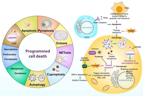
Cancer drug resistance remains a profound and multifaceted obstacle to the success of contemporary therapeutic regimens, substantially contributing to poor clinical outcomes and reduced patient survival. A central mechanism underlying this challenge is the remarkable ability of malignant cells to evade programmed and regulated cell death (RCD) pathways, thereby promoting cellular persistence, increased adaptability, increased aggressiveness, and metastatic dissemination. This comprehensive review highlights current knowledge on the canonical intrinsic and extrinsic apoptotic pathways, while other RCD modalities, including necroptosis, ferroptosis, pyroptosis, autophagy-dependent cell death, and cuproptosis, as well as alternative pathways, such as parthanatos, entosis, NETosis, lysosome-dependent cell death (LCD), alkaliptosis, and oxeiptosis, are thoroughly integrated. Emphasis is placed on the intricate molecular regulatory networks governing these death pathways and their dynamic crosstalk within the complex tumor microenvironment (TME). We provide an in-depth examination of pivotal signaling cascades, including those mediated by the BCL2 family, caspases, mitogen-activated protein kinase (MAPK), and JAK-STAT pathways, which collectively orchestrate the delicate equilibrium between cell survival and death. Furthermore, our review critically evaluates how cancer-associated factors, including epithelial-mesenchymal transition (EMT), multidrug resistance (MDR) via efflux transporters, and the immunomodulatory influence of the TME, converge to exacerbate therapeutic refractoriness. By integrating cutting-edge molecular insights with translational evidence, this review delineates promising therapeutic strategies that exploit RCD vulnerabilities to restore cell death sensitivity and dismantle resistance mechanisms, thereby catalyzing the advancement of precision oncology approaches grounded in mechanistic rigor and clinical relevance.
Keywords: regulated cell death (RCD), drug resistance, neoplasms, apoptosis, nonapoptotic cell death, tumor microenvironment, precision oncology
INTRODUCTION
Therapeutic resistance remains one of the most formidable challenges in modern oncology, substantially limiting the long-term efficacy of anticancer treatments [1, 2]. Although many malignancies initially respond to therapeutic interventions, tumor cells frequently acquire adaptive mechanisms that enable survival under therapeutic pressure, ultimately leading to disease recurrence, progression, and treatment failure [3, 4]. Despite remarkable advances in precision medicine and targeted therapies, resistance continues to represent a major clinical obstacle and is often associated with increased tumor aggressiveness and poor patient outcomes. Conventional treatment modalities, including surgery, chemotherapy, and radiotherapy, are similarly hindered by the remarkable plasticity of cancer cells, particularly cancer stem cells (CSCs), which contribute to tumor dormancy, relapse, and therapeutic resistance [5, 6]. Consequently, considerable efforts have been directed toward developing more effective therapeutic strategies, including novel chemotherapeutic agents, optimized treatment schedules, and drug repurposing methods designed to overcome resistance and improve clinical outcomes [7, 8]. Nevertheless, a comprehensive understanding of the molecular mechanisms underlying both intrinsic and acquired resistance remains an urgent unmet need in cancer therapy [9-11].
A fundamental determinant of tumor development and the therapeutic response is the balance between cellular proliferation and regulated cell death (RCD). Under physiological conditions, tightly controlled cell death programs maintain tissue homeostasis by eliminating damaged, aged, or potentially malignant cells [12]. Cancer disrupts this balance by promoting uncontrolled proliferation while simultaneously suppressing cell death pathways, thereby enabling malignant transformation and tumor progression [13]. Apoptosis, the most extensively characterized form of RCD, serves as a critical tumor-suppressive mechanism; however, cancer cells frequently acquire genetic and epigenetic alterations that disable apoptotic signaling and confer a survival advantage [14, 15]. Increasing evidence indicates that the ability to evade multiple forms of cell death represents a hallmark of cancer and contributes substantially to therapeutic resistance [16, 17].
In addition to its direct effects on tumor cell survival, RCD has emerged as a key regulator of the tumor microenvironment (TME) [18]. Recent advances have identified several nonapoptotic forms of RCD, including necroptosis, ferroptosis, pyroptosis, and autophagy-dependent cell death, each of which exerts distinct effects on both cancer cells and the surrounding stromal and immune compartments. These pathways not only influence tumor growth and therapeutic responsiveness but also actively shape the inflammatory and immunological landscape of tumors [19]. For example, nonapoptotic RCDs, such as necroptosis, are frequently accompanied by the release of danger/damage-associated molecular patterns (DAMPs) and proinflammatory mediators that can stimulate antitumor immunity, whereas chronic activation of inflammatory signaling may paradoxically promote tumor progression, immune evasion, and treatment resistance [20-22]. Similarly, ferroptosis has attracted considerable attention because of its ability to eliminate apoptosis-resistant cancer cells while simultaneously influencing immune cell function and metabolic interactions within the TME [23].
The dual role of RCD in regulating both tumor cell fate and the TME has profound implications for cancer therapy. By directly eliminating malignant cells and indirectly modulating immune surveillance, inflammation, stromal interactions, and therapeutic sensitivity, RCD pathways represent attractive targets for next-generation anticancer strategies [24, 25]. A growing body of experimental and clinical evidence has demonstrated that manipulation of these pathways can increase treatment efficacy, overcome resistance in refractory tumors, and potentially improve patient outcomes. Furthermore, the molecular components of RCD pathways are increasingly being explored as predictive biomarkers and therapeutic targets for personalized cancer treatment.
In the current review, we comprehensively discuss the diverse forms of RCD and their multifaceted roles in cancer biology. Particular emphasis is placed on the dual impact of RCD on tumor progression through the regulation of cancer cell survival and through remodeling of the tumor microenvironment, including immune and inflammatory responses. We also discuss the translational potential of targeting RCD pathways as innovative therapeutic approaches for overcoming resistance and improving the effectiveness of anticancer therapies.
ORIGINS AND MOLECULAR UNDERPINNINGS OF REGULATED CELL DEATH
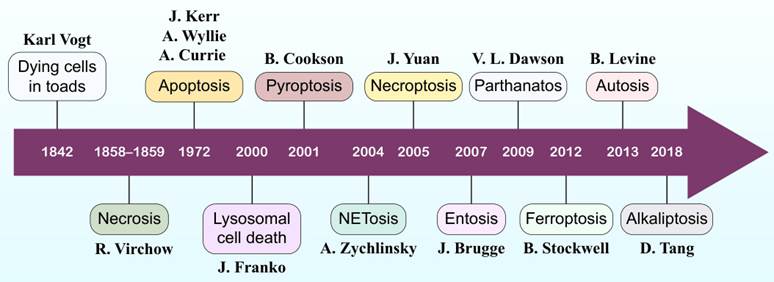
RCD encompasses a group of genetically controlled cellular processes that are essential for maintaining tissue homeostasis, development, and organismal health. Since the introduction of the concept of apoptosis by John Kerr, Andrew Wyllie, and Alastair Currie in 1972 [26] (Figure 1), extensive research has revealed that RCD comprises multiple distinct modalities beyond apoptosis, including necroptosis, pyroptosis, ferroptosis, entotic cell death, netotic cell death, parthanatos, lysosome-dependent cell death, autophagy-dependent cell death, alkaliptosis, and oxeiptosis [27]. Dysregulation of these pathways contributes to the development and progression of numerous diseases, particularly cancer [12].
Timeline of the discovery and conceptualization of regulated cell death modalities. The timeline depicts the historical progression of major forms of cell death from 1842-2018, which are aligned on a horizontal timeline and annotated with the researchers who first described or named each process.
In tumors, RCD plays a dual role; on the one hand, the activation of cell death pathways suppresses tumor initiation and growth by eliminating damaged or transformed cells, while on the other hand, alterations in RCD signaling can promote tumor survival, progression, immune evasion, and therapeutic failure [19]. Given the increasing recognition of the complex interactions between RCD and cancer, understanding the molecular characteristics of individual RCD pathways has become increasingly important. The following sections provide an overview of the major nonapoptotic RCD mechanisms and their relevance to cancer biology. Table 1 summarizes the key RCD modalities and their reported roles in cancer.
Classification of RCD Mechanisms and Their Functional Roles in Cancer
| RCD SUBTYPE | CORE MECHANISTIC PATHWAY | PROMINENT ROLE(S) IN CANCER | REFERENCE(S) |
|---|---|---|---|
| Apoptosis | Intrinsic (mitochondrial) and extrinsic (death receptor) pathways leading to caspase activation and cellular dismantling. | Loss of apoptosis contributes to tumor initiation, progression, and therapy resistance; restoring apoptotic signaling is a major therapeutic goal. | [51, 57, 69] |
| Autophagy | Lysosomal degradation of damaged organelles and proteins via ULK1-PtdIns3K-LC3 pathway. | Tumor suppression by removing damaged components in early stages Tumor promotion via metabolic adaptation and drug resistance in advanced cancer. | [114, 277, 278] |
| Necroptosis | Caspase-independent, RIPK1-, RIPK3- and MLKL-driven cell death causing membrane rupture and inflammation. | Exhibits context-dependent effects: anti-tumor immunity activation or pro-tumor inflammation and metastasis, depending on tumor microenvironment. | [141, 279, 280] |
| Ferroptosis | Iron-dependent oxidative death caused by lipid peroxidation and glutathione depletion. | Induces selective death in metabolically stressed tumors; resistance mechanisms (NFE2L2, MTOR) link ferroptosis suppression to tumor survival and drug resistance. | [281-283] |
| Pyroptosis | GSDM-mediated pore formation triggered by inflammasome-activated caspases (e.g., CASP1, CASP4, CASP5, CASP11). | Double-edged: promotes anti-tumor immunity via immunogenic death, but chronic pyroptosis-driven inflammation can foster tumor growth and metastasis. | [284-286] |
| Cuproptosis | Copper-dependent protein aggregation and mitochondrial dysfunction causing proteotoxic stress. | Copper overload promotes oxidative stress and oncogenic signaling; controlled induction of cuproptosis holds therapeutic promise in copper-addicted cancers. | [287-290] |
| Parthanatos | PARP1 hyperactivation leading to NAD⁺ and ATP depletion and AIFM1-mediated chromatinolysis. | Excessive PARP1 activity promotes cancer cell death under DNA damage; chronic activation linked to therapy-induced toxicity and resistance. | [229, 230, 232, 233, 291] |
| Entosis | Cell-in-cell engulfment and degradation through LC3-associated phagocytosis and lysosomal digestion. | Can suppress tumor growth by killing neighboring cells or promote progression through aneuploidy and nutrient scavenging. | [243, 249] |
| NETosis | Release of neutrophil extracellular traps (NETs) through ROS and histone citrullination. | NET-derived inflammation promotes tumor progression, metastasis, and immune evasion; NET inhibition may reduce metastatic spread. | [256, 261] |
| Alkaliptosis | NFKB-IKBKB-CA9 axis-driven intracellular alkalinization-induced death. | Emerging anti-tumor target; selective pH-dependent death in cancer cells resistant to apoptosis or ferroptosis. | [275] |
| Oxeiptosis | ROS-induced KEAP1-PGAM5-AIFM1-mediated caspase-independent cell death. | Anti-inflammatory cell death mechanism; may limit oxidative stress-induced inflammation in tumors. | [276] |
Apoptosis
Apoptosis is a precisely orchestrated biological process essential for maintaining tissue equilibrium in multicellular systems, facilitating the orderly elimination of superfluous or compromised cells [28]. This mechanism operates through multiple signaling cascades, which are primarily divided into two principal categories: the mitochondrial (intrinsic) route and the death receptor (extrinsic) route. A comprehensive understanding of these apoptotic systems is foundational for elucidating disease pathogenesis arising from their dysfunction and, subsequently, for creating targeted interventions addressing specific apoptotic mechanisms or genetic elements [29].
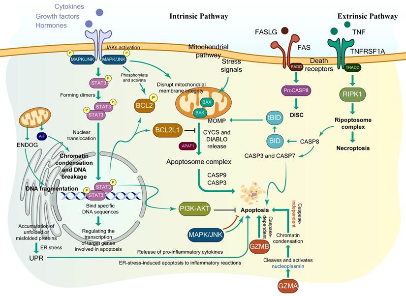
The mitochondrially mediated intrinsic apoptotic route functions under stringent regulation, responding to various cellular stressors through the liberation of death-promoting factors (see Figure 2 for more details). Maintaining mitochondrial structural integrity and determining cellular destiny depends critically on the precise quantitative equilibrium between death-promoting and death-inhibiting constituents of the BCL2 protein family. This mechanism culminates in cellular dismantling, producing apoptotic fragments with surface alterations that enable their rapid recognition and clearance by phagocytic cells [30, 31].
Schematic overview of major apoptotic signaling pathways and cross-talk mechanisms. The current diagram illustrates the core apoptotic cascades, including the intrinsic (mitochondrial) pathway, the extrinsic (death receptor) pathway, endoplasmic reticulum (ER) stress-induced apoptosis, and granzyme-mediated cell death: i) Intrinsic Pathway; intracellular stress signals induce mitochondrial outer membrane permeabilization (MOMP) via pro-apoptotic effectors BAX and BAK1. This triggers the release of cytochrome c (CYCS) and DIABLO/Smac, driving apoptosome formation alongside APAF1, which activates downstream executioner caspases (CASP9 and CASP3). Caspase-independent mitochondrial pathways are mediated by ENDOG and AIF, which translocate to the nucleus to induce chromatin condensation and DNA fragmentation. Survival and regulatory signals modulate this axis: cytokine/growth factor/hormone-receptor signaling via JAKs promotes phosphorylation of MAPK/JNK and STAT3 dimerization. Dimerized STAT3 translocates to the nucleus to regulate transcriptional programs, including anti-apoptotic BCL2 and BCL2L1 (which inhibit APAF1/MOMP) and the PI3K-AKT survival pathway. MAPK/JNK activation also phosphorylates BCL2 directly or disrupts mitochondrial membrane integrity. ii) Extrinsic Pathway; engagement of cell-surface death receptors (FAS by FASLG, and TNFRSF1A by TNF) recruits adaptor proteins FADD and TRADD. FADD assembles the death-inducing signaling complex (DISC) with ProCASP8, generating active CASP8. CASP8 directly activates executioner caspases (CASP3 and CASP7) and cleaves BID into truncated BID (tBID), bridging the extrinsic signal to mitochondrial MOMP. Alternatively, TRADD recruitment through TNFRSF1A engages RIPK1 to form the ripoptosome complex, mediating necroptosis. iii) ER Stress and Granzyme Pathways: Accumulation of unfolded or misfolded proteins inside the endoplasmic reticulum activates the unfolded protein response (UPR), linking ER stress to inflammatory responses, cytokine release, and apoptosis. Cytotoxic lymphocyte effector granzymes trigger cell death via distinct mechanisms: Granzyme B (GZMB) drives caspase-dependent apoptosis, whereas Granzyme A (GZMA) acts in a caspase-independent manner by cleaving and activating nucleoplasmin to initiate chromatin condensation.
The extrinsic apoptotic pathway begins when extracellular death ligands, such as TNF and FASLG (Fas ligands), bind to specific cell-surface death receptors, including TNFRSF1A/TNFR1 and FAS/CD95 [32-34]. Ligand binding induces receptor trimerization, enabling the recruitment of adaptor proteins such as TRADD through death domain interactions [35-37]. These adaptors facilitate the assembly of the death-inducing signaling complex (DISC), which recruits and activates the initiator CASP8, initiating the downstream apoptotic cascade [38-41]. This signaling process is crucial for immune regulation, homeostasis, and removal of damaged or infected cells.
In addition to the established intrinsic and extrinsic routes, less characterized caspase activation mechanisms exist, including the participation of CASP12 and CASP2, enzymes involved in ER stress-triggered apoptosis. CASP12 (in rodents) and human CASP4 predominantly facilitate apoptosis in response to ER stress, which occurs when improperly folded or unfolded proteins accumulate, activating the unfolded protein response/UPR. This cascade can also stimulate the release of proinflammatory cytokines, thereby connecting ER stress-triggered apoptosis with inflammatory mechanisms. CASP12 dysfunction has been associated with chronic pathologies, including cancer. The other immune-mediated cascade, i.e., the perforin 1 (PRF1) and granzyme (GZM) pathways, employs granzyme A (GZMA) or GZMB to trigger apoptosis. While both induce cellular death, they utilize distinct mechanisms: GZMB facilitates caspase-dependent apoptosis, whereas GZMA initiates cellular death through caspase-independent routes [42-44]. The GZMs employed can differ on the basis of target cell characteristics and immune context [43]. The mechanism of GZMA involves cleaving and activating substrates, including nucleoplasmin, resulting in chromatin condensation and cell death independent of the caspase cascade. Conversely, GZMB primarily drives the caspase-dependent route by cleaving substrates, including CASP3, CASP7, and the death-promoting protein BID [45]. These various apoptotic cascades ultimately converge upon a shared terminal or execution route.
Key molecular regulators of apoptosis
Mitogen-activated protein kinase/MAPK signaling has context-dependent multifunctional involvement in apoptotic regulation, affecting both cellular death and persistence mechanisms. As an evolutionarily conserved intracellular signaling cascade, the MAPK system transmits signals originating from diverse extracellular stimuli, including growth factors, stress indicators, and cytokines. The three primary MAPK subfamilies that participate in apoptotic regulation include MAPK/extracellular signal-regulated kinase (ERK), MAPK/c-Jun N-terminal kinase (JNK), and MAPK/p38 [46].
Under certain conditions, MAPK/ERK activation facilitates cellular persistence while actively preventing apoptosis [46]. This protective mechanism operates by increasing the levels of survival-promoting proteins, including BCL2 family constituents, or by suppressing death-inducing elements.
The MAPK/JNK cascade can either increase survival or initiate apoptosis, depending upon the cellular context and the stimuli encountered. MAPK/JNK activation is commonly associated with stress-triggered apoptosis, as MAPK/JNK can phosphorylate and activate death-promoting BCL2 proteins, compromise mitochondrial membrane stability, and facilitate the liberation of death-accelerating factors [46, 47].
Interconnected communication between these MAPK cascades introduces additional regulatory complexity; for example, MAPK/ERK activation may oppose MAPK/JNK-triggered apoptosis, highlighting the intricate balance between persistent and demise signals [48, 49]. Additionally, feedback regulatory circuits involving phosphatases and negative modulators precisely govern MAPK signaling intensity and temporal duration [50]. This meticulous regulation prevents sustained or excessive activation, potentially causing inappropriate cellular death. MAPKs also play integral roles in signal transmission from death receptors, including FAS and tumor necrosis factor (TNF)/TNF-α receptors, by activating downstream caspases and regulating death-promoting BCL2 constituents [48]. The influence of MAPK signaling on apoptosis is highly intricate and context-specific reflecting sophisticated regulatory networks governing cellular destiny [48].
Caspases are the other central regulators of apoptosis and function within both the mitochondrial and death receptor pathways. They are grouped into developmental, inflammatory, and apoptotic categories [51]. Apoptotic caspases are further classified into initiator caspases (e.g., CASP8 and CASP9) and executioner caspases (e.g., CASP3, CASP6, and CASP7) [52, 53]. Initiator caspases autoactivate and subsequently cleave downstream executioner caspases, leading to complete proteolytic dismantling of the cell [54, 55].
Upon apoptotic stimulation, BH3-only proteins activate BAX and BAK1, promoting the release of CYCS and DIABLO/Smac from mitochondria and triggering apoptosome formation [56-59]. The apoptosome activates CASP9, which then initiates the CASP3 cascade, culminating in cell death [60, 61]. DIABLO and HTRA2/Omi neutralize inhibitors of apoptosis (IAPs), increasing caspase activity, whereas AIFM1/AIF (apoptosis-inducing factor mitochondria associated 1) also contributes to cell death [62-66]. Understanding these molecular events is key to clarifying disease mechanisms and guiding drug design to target apoptotic dysregulation.
The intrinsic pathway is activated by internal stressors such as reactive oxygen species (ROS), DNA damage, ER stress, cytoskeletal disruption, or growth factor deprivation [67, 68]. Classical apoptotic features include DNA fragmentation, chromatin condensation, membrane asymmetry loss, and blebbing, which depend on caspase activity [69]. Caspase activation centers on mitochondrial outer membrane permeabilization/MOMP, a critical “point of no return” that releases pro-death factors [69]. CYCS binds to APAF1, forming the apoptosome, which activates CASP9 and subsequently executioner caspases [70].
AIFM, another mitochondrial protein, translocates to the nucleus to induce chromatin condensation and DNA fragmentation in a caspase-independent manner [71]. DIABLO enhances apoptosis by binding to the BIR domains of IAPs such as XIAP, relieving their caspase inhibition [72, 73]. ENDOG (endonuclease G) and HTRA2 further promote apoptosis by facilitating DNA fragmentation and IAP antagonism [74]. The balance between pro- and antiapoptotic BCL2 family members ultimately determines survival versus death, representing a critical therapeutic target in cancer [75].
The mitochondrial intermembrane space contains numerous prodeath factors, with CYCS being the first identified. Their release is tightly controlled by heat shock proteins (HSPs) and culminates in the activation of CASP3 and other effector caspases, which execute the final biochemical and morphological features of apoptosis [76-78]. Upon release, DIABLO and HTRA2/Omi expose IAP-binding N-terminal motifs that neutralize IAPs, further promoting cell death [79-81].
Apoptosome assembly represents a pivotal step in intrinsic apoptosis [82]; CYCS released by mitochondrial stress binds to APAF1, inducing its oligomerization into a heptameric apoptosome complex [83-85]. This structure recruits and activates proCASP9, which autocatalytically matures into active CASP9 [86, 87]. CASP9 then triggers downstream activation of CASP3 and CASP7, executing apoptosis [88]. This tightly regulated cascade maintains tissue homeostasis and eliminates damaged or unwanted cells.
Activated FADD-bound CASP8 also triggers executioner caspases (CASP3, CASP7) and cleaves BID into truncated BID (tBID), linking the extrinsic and intrinsic pathways via mitochondrial outer membrane permeabilization [89]. TRADD can alternatively recruit RIPK1 to form the ripoptosome, connecting apoptosis and necroptosis signaling [90]. These interconnections ensure flexible control of cell fate under varying stress conditions, with ER stress pathways also contributing to apoptosis in specific contexts.
TP53/p53 serves as the “guardian of the genome,” orchestrating DNA repair, cell cycle arrest, or apoptosis through the transcriptional activation of targets such as BAX, BBC3/PUMA, and PMAIP1/NOXA [91-105]. Its activity is restrained by MDM2 under normal conditions but is stabilized upon stress [106], preventing tumorigenesis. Mutations in TP53 impair this regulation, driving cancer progression [107, 108].
Non-caspase proteases such as calpains, cathepsins, granzymes, and proteasomes also modulate apoptosis [69, 109]. Calpains, calcium-dependent enzymes, are activated by specific intracellular Ca²⁺ concentrations, linking calcium dysregulation to apoptotic cell death [69, 110, 111].
Together, these molecular networks ensure the precise control of cellular fate. Disruption of apoptotic regulation contributes to cancer development, underscoring the importance of understanding and therapeutically targeting these pathways.
For a more comprehensive overview of the upstream regulatory complexes, domain architectures, and downstream execution nodes governing apoptosis-related pathways, see Figure 2.
Autophagy
Autophagy is an essential, evolutionarily conserved catabolic process responsible for degrading damaged intracellular components and recycling metabolites such as glucose, ATP, amino acids, and fatty acids [112]. It operates alongside the ubiquitin-proteasome system and is classified into three forms: chaperone-mediated autophagy/CMA, microautophagy, and macroautophagy. All of these forms deliver cytoplasmic cargo to lysosomes, although they differ in their sequestration mechanisms [113, 114]. CMA uniquely depends on proteins such as HSPA8/HSC70 (heat shock protein family A member 8) and LAMP2A (lysosomal associated membrane protein 2A) for lysosomal targeting [115]. Microautophagy internalizes cargo directly through lysosomal membrane invagination [116], whereas macroautophagy proceeds through sequential stages, initiation, nucleation, elongation, maturation, fusion, and degradation, which are regulated by a complex molecular network [114].
Macroautophagy is governed primarily by the canonical ULK1 (unc-51 like autophagy activating kinase 1) pathway, the class III PtdIns3K (phosphatidylinositol 3-kinase) complex, the LC3 (microtubule associated protein 1 light chain 3) conjugation system, and lysosomal hydrolases [117]. Stress signals activate ULK1 to initiate autophagy [118-120]. Nucleation follows through the PtdIns3K complex containing PIK3C3/VPS34, generating the phagophore. Expansion and autophagosome formation require the ATG12-ATG5-ATG16L1 complex, which mediates LC3 lipidation and membrane closure [121]. Upon fusion with lysosomes, the autolysosome, where hydrolases degrade cargo, is formed [122].
Pharmacologic inhibitors, including ULK1 inhibitors (initiation), class III PtdIns3K inhibitors (nucleation), and lysosomotropic agents that block autophagosome-lysosome fusion, target multiple stages. MTOR (mechanistic target of rapamycin kinase) and AMPK serve as central metabolic regulators of autophagy [123, 124]. MTOR suppresses autophagy under nutrient-rich conditions [124-126], whereas AMPK activates ULK1 and BECN1 during energy stress, promoting autophagy initiation [127-131]. Autophagy exerts context-dependent effects on malignancy and is influenced by tumor type, stage, and genetic background [132]. It can mediate cytotoxic autophagic cell death (type II RCD) [133-135] or support cytoprotective survival pathways that drive drug resistance [136].
Conversely, established tumors exploit autophagy to survive metabolic stress, maintain energy balance, and resist therapy [137-139]. Upregulated autophagy enhances nutrient recycling, conferring a growth advantage and promoting therapeutic resistance.
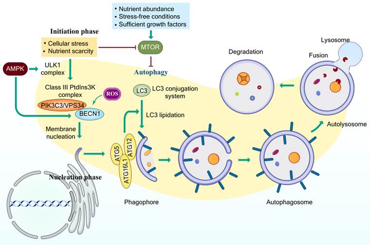
A more detailed, step-by-step schematic of the upstream metabolic cues, membrane nucleation events, and progressive vesicular transitions governing macroautophagy is illustrated in Figure 3.
Molecular mechanisms and sequential stages of the autophagic pathway. Autophagy is tightly regulated upstream by metabolic and environmental cues, wherein conditions such as nutrient abundance, stress-free environments, and sufficient growth factors stimulate MTOR to suppress pathway activity. Conversely, cellular stress and nutrient scarcity alleviate MTOR-mediated repression while activating AMPK, driving initiation via the ULK1 complex. During the subsequent nucleation phase, AMPK, ULK1, and reactive oxygen species (ROS) promote activation of the Class III PtdIns3K complex, comprising PIK3C3/VPS34 and BECN1, to trigger membrane nucleation at endoplasmic reticulum-associated sites. Phagophore elongation and maturation are orchestrated by the ATG12-ATG5-ATG16L1 complex alongside the LC3 conjugation system, which drives LC3 lipidation and incorporation into the growing phagophore membrane to sequester cytoplasmic cargo within a double-membraned autophagosome. Upon completion, the autophagosome undergoes fusion with a lysosome to form an autolysosome, culminating in the degradation of enclosed contents by lysosomal hydrolases to facilitate macromolecular recycling and energy homeostasis.
Necroptosis
Unlike standard apoptotic cell death, necroptosis is a controlled form of cell death that does not rely on caspase enzymes. Instead, it is driven primarily by three key proteins, including receptor-interacting proteins 1 and 3 (RIP1 & RIP3) and mixed lineage kinase domain-like protein (MLKL). Because it bypasses traditional death pathways, it offers a powerful backup plan for destroying treatment-resistant cancer cells while simultaneously initiating a strong immune response against tumors [140].
Necroptosis can be initiated by a wide spectrum of upstream cues; these include members of the tumor necrosis factor receptor (TNFR) superfamily, pattern-recognition receptors (PRRs), T-cell receptors (TCRs), and several cytotoxic chemotherapeutic agents [140, 141]. Environmental stressors such as hypoxia may also activate this pathway, although malignant cells can reportedly evade hypoxia-driven necroptosis through increased glucose uptake and accelerated anaerobic glycolysis [142]. Among these diverse inducers, the TNF-TNFR1 axis remains the prototypical and most thoroughly characterized model [143]. Thus, the fundamental principles of necroptotic initiation are best illustrated through TNF-dependent signaling.
The binding of TNF to TNFR1 induces a conformational rearrangement of TNFR1 homotrimers, promoting the formation of a membrane-associated multiprotein signaling hub termed complex I [144]. This platform recruits several key regulators, including RIPK1, TNFR-associated death domain protein (TRADD), the cellular inhibitors of apoptosis cIAP1/2, and the TNFR-associated factors TRAF2/5. Within this complex, RIPK1 functions as a central determinant of cell fate [145] and undergoes BIRC2/BIRC3-mediated polyubiquitination, a modification that activates canonical NFKB signaling [143]. NFKB then induces the transcription of cytoprotective genes that promote cell survival [144].
Following rapid internalization of the ligand-bound receptor, complex I undergoes compositional and post-translational remodeling [143]. The deubiquitinase CYLD removes ubiquitin chains from RIP1/RIPK1, thereby attenuating sustained NFKB activation and shifting the signaling balance toward cell death [146]. This complex, which is composed of RIPK1, TRADD, CASP8, and FADD, facilitates CASP8 activation [147].
Complex II represents a pivotal checkpoint that dictates whether the cell is undergoing apoptosis or necroptosis. Under physiological conditions, active CASP8 cleaves and inactivates RIPK1 and RIPK3, thereby dismantling the necroptotic machinery and promoting apoptotic execution [148]. CASP8 also supports cell survival by cleaving CYLD, preventing excessive deubiquitination of RIPK1 [149]. However, when CASP8 is genetically absent or pharmacologically inhibited [144], RIP kinase cleavage is blocked, irrevocably diverting the cell toward necroptosis.
Commitment to necroptosis is marked by RIPK1 auto-phosphorylation at serine 161 (S161), which activates its kinase function [150]. Activated RIPK1 then engages RIPK3 through their RIP homotypic interaction motifs (RHIMs), forming an amyloid-like heterodimeric assembly known as the necrosome, the central signaling scaffold for necroptosis [145].
Mitochondrial ROS further potentiate this process. ROS accumulation enhances RIPK1 auto-phosphorylation and accelerates RIPK3 recruitment, whereas RIPK3 itself promotes ROS generation, establishing a self-reinforcing positive feedback loop that ensures robust necroptotic activation [151]. CYLD additionally strengthens necrosome signaling by deubiquitinating RIPK1 after its incorporation into the complex [152]. This pathway can be pharmacologically interrupted via the use of RIPK1 inhibitors such as necrostatin-1 (Nec-1), the MLKL inhibitor necrosulfonamide (NSA), or various RIPK3-targeting small molecules [153].
Within the necrosome, activated RIPK3 phosphorylates its downstream effector, MLKL. Phosphorylated MLKL oligomerizes and translocates to the inner leaflet of the plasma membrane, where it disrupts membrane integrity. This culminates in a lytic form of cell death characterized by cellular swelling, organelle breakdown, and catastrophic membrane rupture [154, 155].
Collectively, these findings illustrate that necroptosis is a tightly regulated, multi-step death program in which upstream receptor signaling, the RIPK1 ubiquitination status, CASP8 activity, and necrosome assembly converge to determine the cellular fate. Once CASP8 inhibition permits RIPK1-RIPK3 complex formation, the necroptotic cascade becomes self-reinforcing through ROS-driven amplification, ultimately culminating in MLKL activation, membrane permeabilization, and lytic cell death. This mechanistic framework underscores necroptosis as a decisive alternative to apoptosis and highlights multiple regulatory nodes, particularly within the RIPK1-RIPK3-MLKL axis, that may be exploited for therapeutic intervention.
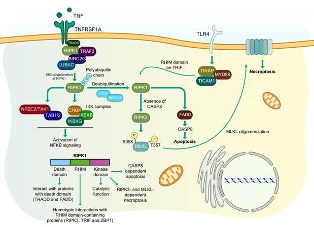
A detailed molecular schematic outlining the specific protein complexes, post-translational modifications, and domain interactions across the necroptotic signaling cascade is provided in Figure 4.
Receptor-mediated signaling networks orchestrating NFKB survival, apoptosis, and necroptosis. Binding of TNF to TNFRSF1A recruits a membrane-proximal complex comprising TRADD, RIPK1, TRAF2, BIRC2/3, and LUBAC, which mediates K63-linked polyubiquitination of RIPK1 to recruit and activate NR2C2/TAK1-TAB1/2 and the IKK complex (CHUK, IKBKB, and IKBKG), thereby driving NFKB survival signaling. Deubiquitination of RIPK1 by the CYLD-SPATA2 complex triggers a transition from survival to cell death pathways, a process also engaged via TLR4 through TIRAP, MYD88, and TICAM1/TRIF via its RHIM domain. In the presence of CASP8, deubiquitinated RIPK1 associates with FADD to activate CASP8, executing apoptosis. Conversely, in the absence or inhibition of CASP8, RIPK1 interacts with RIPK3 to promote downstream phosphorylation of MLKL at residues S358 and T357, driving MLKL oligomerization and plasma membrane translocation to execute necroptosis. Structurally, RIPK1 coordinates these divergent fates through distinct functional modules: its C-terminal death domain mediates interactions with death domain-containing adaptors (TRADD and FADD), the central RHIM domain facilitates homotypic assemblies with RHIM-containing partners (RIPK3, TRIF, and ZBP1), and the N-terminal kinase domain provides catalytic activity critical for both CASP8-dependent apoptosis and RIPK3/MLKL-dependent necroptosis.
Ferroptosis
Ferroptosis is an RCD process driven by lipid peroxidation when the level of ROS exceeds the detoxifying capacity of glutathione (GSH) and glutathione peroxidase 4 (GPX4). Although iron, lipids, and ROS are essential for cellular metabolism, their imbalance, especially under metabolic stress, initiates ferroptosis. This pathway is increasingly recognized as central to malignant biology and is implicated in cancer, neurodegeneration, and inflammatory disorders [156] (see Figure 5 for more details). Cancer cells exhibit metabolic plasticity that can both promote survival and create vulnerabilities to ferroptosis, offering therapeutic opportunities, including synergy with immune-based treatments. However, its clinical translation remains limited [157]. Energy depletion decreases ATP levels and activates AMPK, a key metabolic sensor. AMPK suppresses ACAC1/ACC1 and ACACB/ACC2, reducing malonyl-CoA production and limiting PUFA (polyunsaturated fatty acid) synthesis, thereby decreasing ferroptosis sensitivity [158]. AMPK also phosphorylates BECN1, enabling interaction with SLC7A11 and inhibiting the system Xc⁻ antiporter, a paradoxical pro-ferroptotic effect [159]. These dual roles suggest that tumor cells exploit AMPK signaling to evade ferroptosis, while targeted modulation may enhance therapy.
Metabolic and signaling networks governing ferroptosis sensitivity and resistance. Intracellular and microenvironmental metabolic pathways tightly modulate ferroptotic cell death through lipid remodeling, nutrient sensing, and redox regulation. Glycolysis-derived lactate, transported or signaled via SLC16A1 and HCAR1, depletes ATP and elevates the AMP:ATP ratio, driving phosphorylation and activation of AMPK. Activated AMPK subsequently stimulates the SREBF1-SCD1 axis to convert saturated fatty acids into monounsaturated fatty acids (MUFAs), which potently suppress ferroptosis, while glycolytic flux concurrently inhibits polyunsaturated fatty acid (PUFA) synthesis. Under cellular energy stress, AMPK independently suppresses ACAC (acetyl-CoA carboxylase) activity to prevent the conversion of acetyl-CoA to malonyl-CoA, thereby attenuating PUFA biosynthesis and limiting ferroptotic susceptibility. Concurrently, energy-activated AMPK induces BECN1 phosphorylation, which directly inhibits the cystine/glutamate antiporter SLC7A11 to promote ferroptosis. In parallel, the upstream Hippo kinase LATS1/2 represses MTOR, which otherwise negatively regulates SLC7A11 while phosphorylating SQSTM1/p62. Phosphorylated SQSTM1 complexes with and directs KEAP1 for degradation, promoting the stabilization and accumulation of NFE2L2/NRF2, which upregulates protective MUFA generation to inhibit ferroptosis.
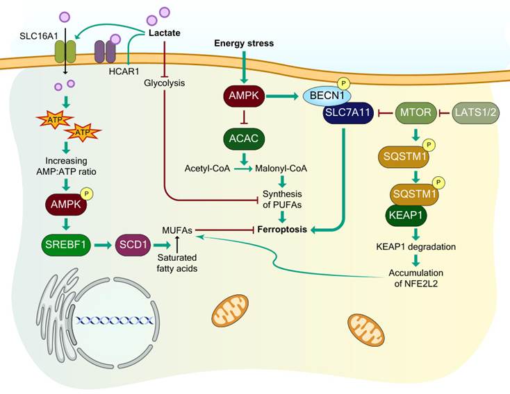
Lactate-loaded HCC cells resist ferroptosis triggered by standard agents like RSL3 and erastin. MCT1-mediated lactate influx fuels ATP generation while blunting AMPK activity, which in turn lifts SREBP1 and its downstream effector SCD1, boosting the supply of monounsaturated fatty acids/MUFAs that shield membranes from ferroptotic peroxidation. Conversely, blocking lactate uptake through HCAR1/MCT1 inhibition re-engages AMPK, suppresses SCD1, and promotes ferroptosis, an effect that may be compounded by concurrent ACSL4 induction, sharply increasing ferroptotic sensitivity [160, 161]. MUFAs resist peroxidation, reducing susceptibility to ferroptosis [162].
Both MTORC1 and MTORC2 (MTOR complexes 1 and 2) regulate ferroptosis; MTORC2 phosphorylates SLC7A11 at Ser26, reducing its transport activity [163]. Work from the Hippo field has uncovered a direct molecular bridge between this growth-suppressive cascade and the nutrient-sensing machinery of MTORC1; the LATS1/2 kinases, which serve as the catalytic core of the Hippo pathway, act on RAPTOR, an obligatory subunit of MTORC1, by phosphorylating its Ser606 residue. This modification restrains MTORC1 activity, and it does so by disrupting the ability of RPTOR to engage RHEB. Genetic evidence reinforces the model; a phosphomimetic knock-in allele, RPTOR-S606D, produces cells of reduced size that divide less readily. Thereby, these findings position the Hippo and MTORC1 pathways in direct communication, providing a mechanism through which organ size can be tuned with precision [164]. The PI3K-AKT-MTOR cascade, which is frequently altered in cancer, enhances MUFA synthesis via SREBF1, promoting ferroptosis resistance [162].
Cancer cells experience increased oxidative stress, which activates NFE2L2/NRF2 (NFE2-like bZIP transcription factor 2), which induces antioxidant response element (ARE)-containing genes [165]. Mutations in KEAP1, CUL3, or NFE2L2 stabilize NFE2L2, increasing its nuclear accumulation in conditions such as non-small cell lung cancer (NSCLC) and lung squamous cell carcinoma (LSCC) [166, 167].
Despite high metabolic demand, dense growth, and oxidative stress, conditions that favor ferroptosis, tumor cells rarely undergo spontaneous ferroptotic death. This reflects robust compensatory mechanisms involving metabolic rewiring, antioxidant activation, and cell-density signaling. Understanding how ferroptosis interfaces with classical oncogenic pathways is essential for developing therapies that selectively activate ferroptosis and overcome current treatment limitations. A comprehensive overview of the metabolic circuits, upstream oncogenic cascades, and lipid-peroxidation checkpoints governing ferroptosis sensitivity is depicted in Figure 5.
Pyroptosis
Pyroptosis is an inflammatory form of RCD mediated by the gasdermin/GSDM family of pore-forming proteins, including GSDMA-GSDME. Upon activation by caspases or granzyme proteases, the amino-terminal GSDM fragment inserts into cellular membranes to form pores, inducing pyroptosis. Although GSDMs have been widely studied in cancer, their overall contribution remains context-dependent, as both pro-tumorigenic and anti-tumorigenic effects have been reported. Pyroptosis can be triggered by pathogen-associated molecular patterns (PAMPs), DAMPs, and various chemotherapeutic agents in GSDM-expressing cells [168].
Inflammasomes are multiprotein complexes activated by intracellular stress or pathogens. Key sensors include MEFV/pyrin, AIM2, and several NLRs (NOD-like receptors), such as NLRP1, NLRP3, and NLRC4 [169]. Among these proteins, NLRP3 is the most extensively characterized.
NLRP3 promotes tumor progression by enhancing the proliferation and motility of A549 lung cancer cells [170], correlates with disease progression in oral squamous cell carcinoma (OSCC) [171], and drives EMT in colon cancer independent of inflammasome activation [172]. It also contributes to chemoresistance in OSCC [173]. Inflammasome/NLRP3-associated secretion of IL18 supports proliferation, immunosuppression, angiogenesis, and metastasis across cancers like lymphoma and gastric cancer [174, 175]. Similarly, NLRP1 enhances melanoma progression by promoting inflammasome activity and inhibiting apoptosis [176].
Conversely, reduced expression of NLRP3, NLRP1, and AIM2 in hepatocellular carcinoma (HCC), colorectal cancer (CRC), and gastric cancer (GC) is correlated with advanced disease, suggesting that tumor-suppressive effects are involved [177-179]. In colitis-associated cancer, NLRP3-driven IL18 protects intestinal epithelial cells from DSS- or AOM-induced injury [180-183]. NLRC4 in tumor-associated macrophages (TAMs) suppresses melanoma growth independently of inflammasome activation [184], whereas NLRP3 in Kupffer cells limits CRC liver metastasis by promoting NK cell maturation and IL18-dependent cytotoxicity [185].
The GSDM family (GSDMA-GSDME) has diverse, tumor-specific functions; GSDMA, expressed mainly in epithelial tissues, is linked to TNF-induced apoptosis [186], and skin inflammation and alopecia [187, 188]. Its frequent silencing in gastric cancer suggests a tumor- suppressive role [189]. GSDMB is often overexpressed in cancers such as breast cancer, where it promotes migration, metastasis, and therapy resistance, particularly in ERBB2/HER2⁺ breast cancer [190, 191]. GSDMC is upregulated in CRC due to TGFBR2 mutation [192] and is associated with poor prognosis in lung adenocarcinoma [193].
GSDMD, a central innate immune mediator, has dual effects: it enhances NSCLC aggressiveness via EGFR-AKT signaling [194] but suppresses gastric cancer progression by regulating the S-to-G2 transition [195]. Several agents, such as α-NETA [196], and polyphyllin VI [197], induce GSDMD-dependent pyroptosis, inhibiting tumor growth. Metformin also triggers pyroptosis in ESCC through the MIR497-PELP1 axis [198]. CARD8-mediated pyroptosis via DPP8/9 inhibition has been proposed as a therapeutic strategy in AML [199], whereas CD38 deficiency reduces inflammasome-driven pyroptosis in HNSCC [200].
GSDME/DFNA5 has highly context-specific roles; however, a novel DFNA5 mutation (IVS8+4 A>G) in the splice donor site of intron 8 has been found to cause late-onset non-syndromic hearing loss in a Chinese family [201, 202]. GSDME is frequently silenced by promoter hypermethylation in breast [203, 204] and colorectal [205-207] cancers, which is correlated with poor prognosis. Restoring GSDME expression suppresses proliferation [206, 208] and increases chemosensitivity, such as in retinoblastoma [209]. However, some studies have reported no association between GSDME methylation and tumorigenesis. Chemotherapy-induced CASP3 cleavage of GSDME triggers pyroptosis [210, 211], enhancing cisplatin sensitivity in some contexts [211], although not universally.
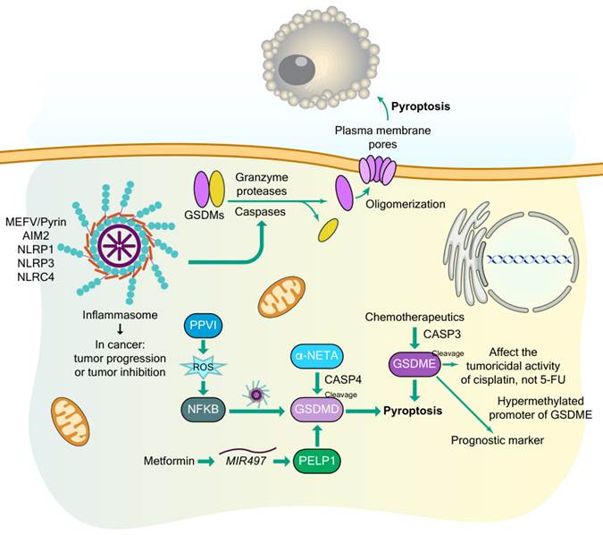
Overall, GSDMs exert heterogeneous, tumor-dependent effects; some (e.g., GSDME) act as suppressors, whereas others (e.g., GSDMB and GSDMC) promote malignancy. Pyroptosis in hypoxic tumor regions may paradoxically sustain inflammation and tumor progression. In addition to pyroptosis, other pyroptosis-independent functions of GSDMs require further study. For a more detailed overview of the proteolytic cascades, gasdermin pore-forming complexes, and drug-induced pyroptotic pathways in cancer, see Figure 6.
Mechanistic pathways, molecular regulators, and clinical implications of pyroptotic cell death in cancer. Canonical and non-canonical pyroptosis execution relies on the proteolytic cleavage of gasdermin family members (GSDMs) by inflammatory caspases or granzyme proteases, releasing active N-terminal domains that oligomerize to form lytic plasma membrane pores. Upstream inflammasome complexes, including MEFV/Pyrin, AIM2, NLRP1, NLRP3, and NLRC4, direct caspase activation to govern contextual tumor progression or suppression. GSDMD-mediated pyroptosis is regulated via multiple pharmacological and signaling axes: polyphyllin VI (PPVI) stimulates reactive oxygen species (ROS) production to drive NFKB signaling and inflammasome assembly, α-NETA engages CASP4-mediated cleavage of GSDMD, and metformin modulates the MIR497-PELP1 axis to promote GSDMD activity. Concurrently, chemotherapeutic agents induce CASP3-dependent cleavage of GSDME to trigger pyroptosis, modulating the tumoricidal efficacy of cisplatin without altering sensitivity to 5-fluorouracil (5-FU). In clinical contexts, hypermethylation of the GSDME promoter serves as an informative prognostic biomarker across malignancies.
Cuproptosis
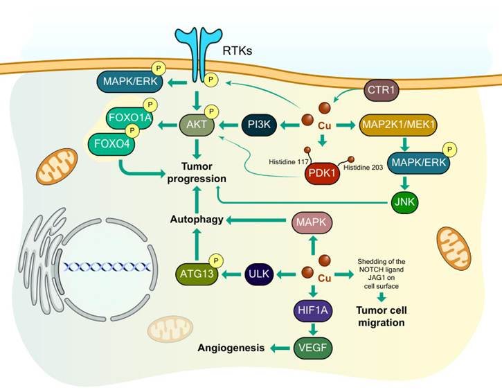
Cuproptosis is a recently described RCD variant that is uniquely reliant on copper ions and executes through a mechanism distinct from all previously established cellular death cascades (see Figure 7 for more details). Although extensive investigations throughout the past decade have examined diverse RCD modalities, the proposition that copper can trigger a completely separate death mechanism remained theoretical until its molecular architecture was definitively characterized [212].
Copper-dependent oncogenic signaling cascades modulating autophagy, tumor progression, angiogenesis, and cell migration. Intracellular uptake of copper via the high-affinity copper transporter CTR1 stimulates multiple oncogenic and metabolic pathways. Cytoplasmic copper engages receptor tyrosine kinases (RTKs) and downstream PI3K signaling while coordinating with PDK1 through histidine residues (His117 and His203) to promote AKT phosphorylation. Activated AKT directly enhances tumor progression and phosphorylates the transcription factors FOXO1A and FOXO4 to drive pro-tumorigenic cascades. In parallel, copper binding stimulates the canonical MAP2K1/MEK1-MAPK/ERK pathway, leading to downstream JNK activation that promotes tumor growth. Copper also orchestrates autophagy via two distinct routes: through activation of intermediate MAPK signaling and by directly binding ULK kinases to induce ATG13 phosphorylation. Concurrently, copper stabilizes HIF1A to upregulate VEGF expression, driving angiogenesis, while simultaneously promoting the cell-surface shedding of the NOTCH ligand JAG1 to stimulate tumor cell migration
Copper performs pivotal functions in multiple intracellular signaling networks within malignant cells, chiefly through its ability to engage and activate critical molecular constituents across various cascades. Copper is extensively integrated into receptor tyrosine kinase (RTK)-facilitated signaling. It can activate RTKs by engaging and phosphorylating them even without their ligands, which then initiates downstream kinases, including MAPK/ERK and AKT, facilitating cellular motility and proliferation [213].
Within the PI3K-AKT axis, copper modulates signaling through two principal mechanisms: (i) Copper directly activates PI3K, resulting in subsequent AKT activation [214]; (ii) Copper engages with histidine residues (H117 and H203) on PDK1 (pyruvate dehydrogenase kinase 1), which also results in AKT activation [215]. Activated AKT then phosphorylates the transcription factors FOXO1/FOXO1A and FOXO4, augmenting tumor cell proliferation and division [216]. Copper is also indispensable for the activation of the MAPK cascade [217]; it directly engages with MAP2K1/MEK1, enabling MAPK1/ERK2 and MAPK3/ERK1 phosphorylation and subsequent MAPK/JNK activation, which contributes to tumor advancement.
Autophagy permits malignant cells to recycle intracellular constituents, supplying energy and increasing survival. Copper regulates this mechanism by engaging with ULK, which activates ATG13 through phosphorylation, launching autophagosome generation and supporting tumor proliferation [218]. In lung adenocarcinoma promoted by the BRAF oncogene, depletion of the copper transporter SLC31A1/CTR1 diminishes intracellular copper, compromising the operation of MAP2K1/MEK1, MAP2K2/MEK2 and ULK1/ULK2, which are essential for both MAPK and autophagy signaling [219].
Although the NOTCH cascade can function as a tumor suppressor, it is also involved in the development of aggressive malignancies [220]. Copper enables the cleavage of the NOTCH ligand JAG1/Jagged1 (jagged canonical Notch ligand 1), augmenting tumor cell migration [221]. Copper is also intimately associated with tumor angiogenesis [222]. It supports HIF1A/HIF-1α (hypoxia inducible factor 1 subunit alpha) engagement with gene promoters, regulating genes, such as HIF1A [223, 224]. Even under normoxic circumstances, copper stabilizes HIF1A, which elevates VEGF expression and facilitates blood vessel generation in tumors [225]. Copper also increases MYC/c-Myc stability by facilitating phosphorylation at threonine 58 and serine 62 [226].
With respect to metabolic regulation, copper modulates both lipid and glucose metabolism. It regulates lipolysis by engaging with cysteine residues on PDE3B, the enzyme responsible for degrading cAMP [227]. Additionally, copper is believed to suppress tumor expansion by downregulating RPS6KB1/S6K1 and its downstream glycolytic targets, including SLC2A1/GLUT1, PKM/PKM2, and LDHA" to "Additionally, disulfiram/copper complexes have been shown to suppress tumor expansion by downregulating RPS6KB1/S6K1, MYC, and downstream glycolytic targets, including SLC2A1/GLUT1, PKM/PKM2, and LDHA [228].
In conclusion, copper has multiple regulatory effects on malignant cell behavior through both direct molecular engagement and indirect modulation of the tumor microenvironment, emphasizing its critical function in oncogenic signaling and the emerging importance of cuproptosis in cancer biology. For an in-depth and more detailed molecular overview of copper-dependent kinase activation cascades, transcriptional reprogramming, and tumor-promoting pathways, see Figure 7.
Other nonapoptotic RCDs
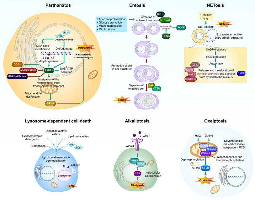
Parthanatos is an RCD modality that is dependent on PARP1 and triggered by DNA damage and chromatin degradation from oxidative stress [229]. Hyperactive PARP1 induces parthanatos via metabolic depletion (NAD⁺, ATP) and mitochondrial dysfunction [230], requiring AIF [231], but not caspases or ENDOG [232]. PARP1 binds AIFM1, causing mitochondrial release and nuclear translocation and inducing chromatinolysis [233]. Negative regulators include ADPRS/ADPRHL2 and RNF146 [234, 235], whereas OGG1 promotes this process [236]. MIF acts as an AIFM1-bound nuclease, generating large DNA fragments [237]. AIFM1-independent parthanatos occurs in some conditions, such as dry macular degeneration [238], and interacts with autophagy-dependent cell death and necroptosis [239, 240].
Entosis is a form of cellular cannibalism in which one cell engulfs another, as observed in epithelial tumor cells under conditions of stress, glucose deprivation, matrix detachment, or mitotic stress [241-243]. The hallmark is cell-in-cell structures [243]. Entosis can promote aneuploidy, nutrient supply, or cancer cell elimination [244-246]. Induction depends on cadherin-mediated adherens junctions (CDH1, CTNNA1), intact actin and microtubules, and RHOA-, ROCK- and myosin-regulated actomyosin contractility [241, 242, 247-250]. Other regulators, including CDC42, AURKA, AMPK, and NUPR1 negatively regulate entosis [241, 242, 251]. Engulfed cells undergo division, release, or lysosomal degradation via LC3-associated phagocytosis and CTSB, independent of caspases or BCL2 proteins [247, 252].
NETosis involves neutrophils releasing neutrophil extracellular traps (NETs) [253] and occurs in other cell types [254-256]. NETs trap pathogens and promote DAMP release, contributing to autoimmunity, ischemia-reperfusion injury, and tumor progression [257-259]. NETosis requires ROS via NOX (NADPH oxidase), granular enzyme translocation, and histone citrullination by PADI4 for chromatin decondensation and nuclear envelope disruption [260-263]. PADI4-independent pathways exist downstream of pyroptosis, necroptosis, or autophagy-dependent cell death. GSDMD can also participate in NETosis [264, 265].
Lysosome-dependent cell death (LCD) occurs via lysosomal membrane permeabilization (LMP), which releases hydrolytic enzymes into the cytosol [266]. As proposed in 1955 and named in 2000, LMP can be induced by detergents, methyl esters, lipid metabolites, and reactive oxygen species (ROS) [266] and can amplify apoptosis, autophagy-dependent cell death, or ferroptosis [267, 268]. Cathepsins are key effectors [269] that can be positively regulated by STAT3 [270] and inhibited by NFKB-induced SERPIN [271]. Furthermore, CTSB cleaves the lysosomal calcium channel MCOLN1/TRPML1 to regulate lysosomal dynamics and autophagy [272], and LMP-mediated iron release contributes to ferroptosis [267, 273, 274].
Alkaliptosis, coined in 2018, is an RCD triggered by intracellular alkalinization [275]. JTC801 induces death in multiple cancer lines through mechanisms distinct from those of other RCDs [275]. Inhibition of alkalinization blocks this death [275]. NFKB-dependent downregulation of CA9, which is controlled by IKBKB, is needed, whereas OPRL1 is dispensable [275].
Oxeiptosis is a type of caspase-independent RCD induced by ROS and is mediated via the KEAP1-PGAM5-AIFM1 pathway [276]. It was introduced in 2018 and is independent of apoptosis, pyroptosis, necroptosis, autophagy, and ferroptosis [276]. Hyperactivated KEAP1 drives H₂O₂-induced oxeiptosis, interacting with PGAM5, which dephosphorylates AIFM1 at Ser116 [276]. Unlike parthanatos, AIFM1 nuclear translocation is unnecessary [276]. pgam5⁻/⁻ mice show increased inflammation after ozone or viral exposure, suggesting that oxeiptosis suppresses inflammation [276].
An in-depth comparative molecular layout detailing the distinct triggering conditions, intermediate enzymes, and terminal structural executions of parthanatos, entosis, NETosis, LCD, alkaliptosis, and oxeiptosis beyond the text is schematically depicted in Figure 8.
Mechanistic pathways and signaling networks underlying non-canonical regulated cell death modalities. Parthanatos is triggered by oxidative stress and severe DNA damage, generating base modifications such as 8-oxo-7,8-dihydroguanine that hyperactivate PARP1, a process counter-regulated by OGG1, ADPRS/ARH3, and RNF146/IDUNA. PARP1 overactivation induces severe NAD⁺/ATP depletion and dissipation of the mitochondrial inner transmembrane potential, driving the release of mitochondrial AIFM1 to translocate to the nucleus and execute parthanatotic chromatinolysis and DNA fragmentation. Entosis is stimulated by triggers including aberrant proliferation, glucose starvation, matrix deadhesion, and mitotic stress, prompting adherens junction formation via CDH1 and CTNNA1 alongside RHOA/ROCK-driven actin-myosin complex contractility; this facilitates internalization into a cell-in-cell structure, followed by lysosome-associated phagocytosis (LAP) and CTSB-mediated degradation of the engulfed cell. NETosis is elicited by infection or tissue injury through NADPH oxidase-dependent ROS generation and autophagy, promoting the release and nuclear translocation of granular enzymes and antimicrobial peptides (including MPO, ELANE, MMPs, elastases, and CAMP) to extrude extracellular net-like DNA-protein structures. Lysosome-dependent cell death (LCD) results from lysosomal membrane permeabilization triggered by cathepsins, lysosomotropic detergents, dipeptide methyl esters, lipid metabolites, and ROS, causing hydrolytic enzyme leakage into the cytosol. Alkaliptosis is driven by GPCR activation via agents such as JTC801, which signals through the IKBKB-NFKB axis to downregulate carbonic anhydrase 9 (CA9), inducing lethal intracellular alkalinization. Oxeiptosis serves as an oxygen radical-induced, caspase-independent cell death cascade activated by hyperoxides or ozone, wherein the KEAP1-PGAM5-NFE2L2 sensory complex prompts the mitochondrial serine-threonine phosphatase PGAM5 to dephosphorylate AIFM1 at Ser116, culminating in mitochondrial dysfunction and oxeiptotic demise.
DRUG RESISTANCE: A MAJOR OBSTACLE IN CANCER TREATMENT
Cancer cells demonstrating treatment unresponsiveness or developing tolerance are subsequently designated drug resistant. This framework originated from infectious disease research, where specific bacterial populations evolved survival mechanisms against antibiotic exposure [292]. Subsequently, analogous resistance mechanisms were characterized in malignant cells, establishing drug resistance as a fundamental weakness in cancer therapeutic approaches [293]. While this phenomenon is well documented, recent scientific progress has focused on its cellular and molecular architecture [294, 295].
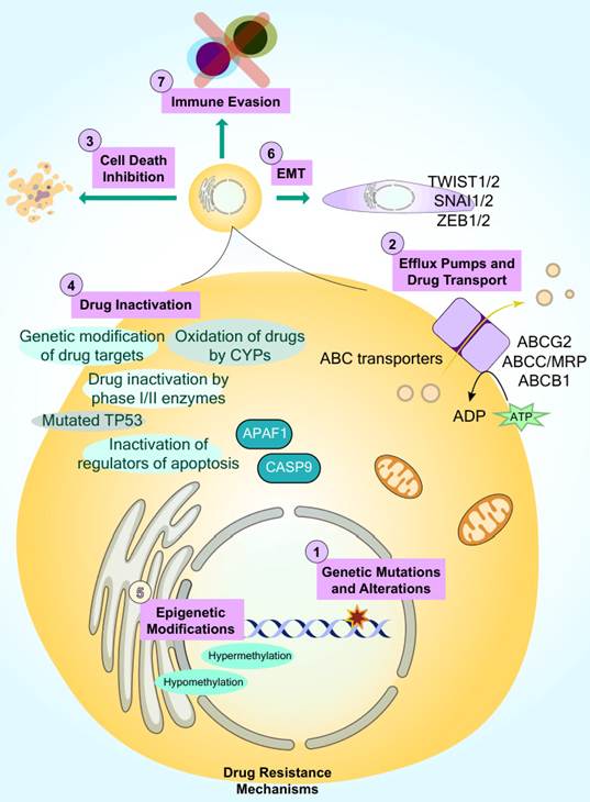
Drug resistance in oncology is primarily stratified into two categories, as shown in Figure 9: (i) Intrinsic (innate or primary) resistance, which refers to the inherent capacity of malignant cells to resist a pharmaceutical agent prior to its administration. The absence of an initial therapeutic response typically indicates this resistance form [11]. (ii) Acquired (extrinsic or secondary) resistance that emerges following treatment initiation, as malignant cells adapt through genetic alterations or protein modifications. Managing acquired resistance frequently presents greater complexity than addressing intrinsic resistance [11]. To provide a more comprehensive framework of drug resistance biology, the following sections review the major molecular mechanisms underlying chemoresistance, which are illustrated in Figure 9 with further details.
Multifactorial mechanisms contributing to cancer drug resistance. Tumor cells acquire therapeutic resistance through diverse intrinsic and extrinsic processes, outlined across seven primary dimensions: (1) genomic instability marked by genetic mutations and alterations; (2) enhanced drug clearance mediated by ATP-dependent efflux pumps and ABC transporters, including ABCG2, ABCC/MRP, and ABCB1; (3) suppression of cell death pathways through the inactivation of key apoptotic regulators, such as mutated TP53, APAF1, and CASP9; (4) metabolic drug inactivation driven by genetic modification of drug targets, oxidation by cytochrome P450 enzymes (CYPs), or biotransformation via phase I/II metabolizing enzymes; (5) epigenetic modifications involving aberrant DNA hypermethylation and hypomethylation patterns; (6) induction of epithelial-mesenchymal transition (EMT) orchestrated by master transcription factors TWIST1/2, SNAI1/2, and ZEB1/2; and (7) immune evasion strategies that shield resistant tumor clones from cytotoxic immune recognition and clearance.
Mechanistic Foundations of Drug Resistance
Multiple cellular and molecular mechanisms contribute to treatment failure, including genomic instability, drug efflux, suppression of cell death pathways, drug metabolism and inactivation, EMT, epigenetic alterations, and immune evasion, as highlighted below:
Genetic modifications and chromosomal instability
Genomic instability is a hallmark of cancer and promotes tumor evolution, heterogeneity, and therapeutic resistance [296]. It includes mutations, microsatellite alterations, and chromosome instability (CIN), all of which can facilitate adaptation to therapeutic stress and are associated with poor clinical outcomes [297, 298]. Studies in multiple cancer types, including non-small cell lung cancer and epithelial ovarian cancer, have demonstrated strong associations between CIN, intratumoral heterogeneity, disease recurrence, and resistance to anticancer therapies [299-301].
Drug efflux transporters
Enhanced drug efflux is one of the most extensively characterized resistance mechanisms. ATP-binding cassette (ABC) transporters actively export chemotherapeutic agents from cancer cells, thereby reducing intracellular drug accumulation and efficacy [302, 303]. Among the most clinically relevant transporters are ABCB1/P-glycoprotein, ABCG2/BCRP, and members of the ABCC/MRP family [302, 304-306]. The overexpression of these transporters has been reported in numerous malignancies and is associated with resistance to different anticancer agents in different cancers, particularly breast cancer [307-309].
Suppression of regulated cell death
Evasion of regulated cell death, particularly apoptosis, represents a central mechanism of cancer drug resistance [14]. Alterations in apoptotic regulators, including BCL2 family proteins, caspases, and inhibitor of apoptosis proteins (IAPs), enable tumor cells to survive despite therapeutic stress [28, 310]. Increased expression of antiapoptotic proteins such as BCL2 and MCL1 has been linked to resistance in several malignancies, while their inhibition can restore treatment sensitivity [311-314]. Given the pivotal role of RCD in therapeutic responses, the relationship between specific RCD modalities and drug resistance is discussed in detail in the next section.
Drug inactivation and metabolic adaptation
Cancer cells can reduce drug efficacy through alterations in drug metabolism, including increased detoxification of active compounds and impaired activation of prodrugs [315]. Enzymes such as glutathione S-transferases (GSTs), cytochrome Ps (e.g., CYP450 and CYP3A4), and UDP-glucuronosyltransferases (UGTs) play major roles in these processes [316-321]. Increased activity or altered expression of these enzymes has been associated with resistance to numerous chemotherapeutic agents.
Epithelial-to-mesenchymal transition
EMT is a reversible cellular program that enhances migration, invasion, metastatic potential, and therapeutic resistance [322-324]. EMT is regulated by transcription factors such as TWIST, ZEB, and SNAI family members and can be activated by signaling pathways, including TGFB, WNT, NOTCH, and Hedgehog [325, 326]. EMT has been implicated in resistance to multiple anticancer agents, through mechanisms involving cancer stem cell maintenance, enhanced survival signaling, and increased drug efflux [327-331].
Epigenetic alterations
Epigenetic modifications such as DNA methylation contribute substantially to cancer drug resistance by altering gene expression without changing the DNA sequence [332-334]. These mechanisms include chromatin remodeling, dysregulated noncoding RNAs (e.g., lncRNA UCA1), and aberrant DNA methylation patterns. These alterations can promote survival pathways, enhance drug detoxification, facilitate EMT, and modulate cellular responses to therapy, thereby contributing to treatment failure [335-338].
Immune evasion
Tumor cells employ diverse strategies to evade immune surveillance, reducing the effectiveness of both conventional therapies and immunotherapies [339, 340]. These mechanisms include impaired antigen presentation, antigen loss, activation of immune checkpoint pathways, remodeling of the tumor microenvironment, epigenetic reprogramming, and increased intratumoral heterogeneity [340-344]. Additional factors, such as antigenic drift, IDO-mediated immunosuppression, aberrant CTNNB1 signaling, and PTEN loss, further contribute to immune escape and therapeutic resistance [345-349].
In conclusion, these mechanisms interact to promote tumor survival under therapeutic pressure. While they provide important context for understanding treatment failure, the following section focuses specifically on how distinct forms of regulated cell death contribute to the development and maintenance of tumor drug resistance. The detailed convergent molecular networks underlying tumor therapeutic failure, detailing the functional coordination between membrane drug-efflux pumps, enzymatic drug neutralization, transcriptional EMT reprogramming, and cell-death evasion, are systematically mapped in Figure 9.
REGULATED CELL DEATH AND CANCER DRUG RESISTANCE: MECHANISTIC AND THERAPEUTIC IMPLICATIONS
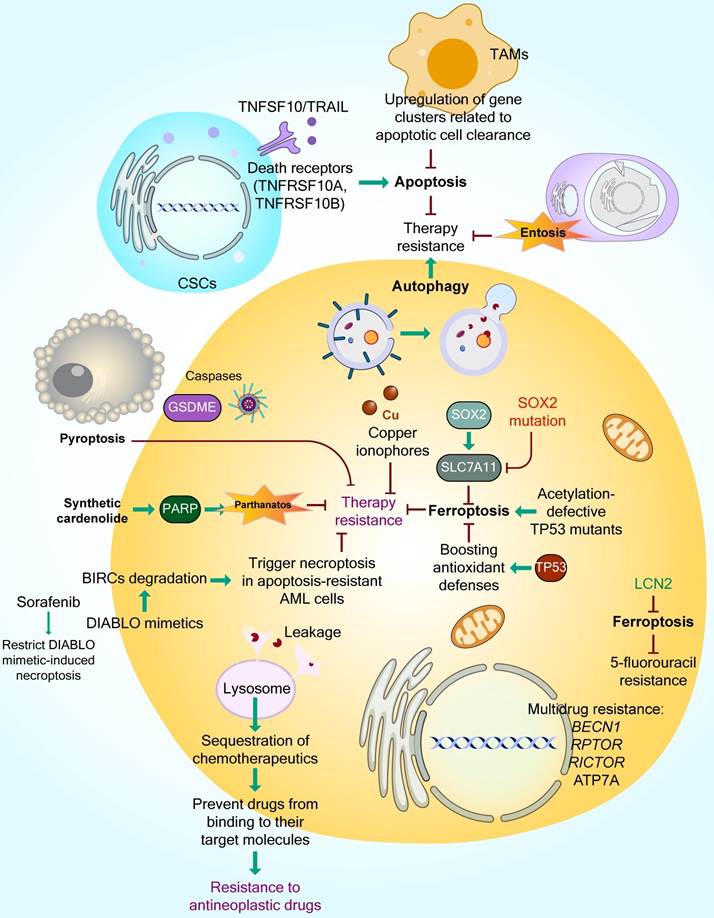
Cancer cells possess a well-documented ability to bypass various cell death pathways, a hallmark of malignancy [17]. One common strategy involves the overproduction of proteins that inhibit apoptosis, preventing the initiation of cell death cascades [16]. Extensive research has clarified how RCD mechanisms contribute to cancer development and therapy resistance across experimental and clinical settings. These insights have helped identify new therapeutic targets, improve prognostic models, and guide strategies for overcoming resistance in refractory cancers. However, the pharmacodynamics of many new agents remain unclear and require further validation through preclinical and clinical studies. Figure 10 provides a detailed and comprehensive beyond-text overview of the interplay between specific regulated cell death pathways and the molecular mechanisms underlying cancer therapy resistance. A central strategy in cancer therapy involves inducing controlled cell death within tumors [350]. By understanding and modulating diverse death pathways, treatments can selectively eliminate cancer cells while sparing normal tissue. This section reviews strategies that target apoptosis, necroptosis, autophagy, ferroptosis, and pyroptosis (Table 2).
Therapeutic Targets and Regulation of Major Cell Death Pathways in Cancer
| TARGET CELL DEATH PATHWAY | THERAPEUTIC STRATEGY/MODULATION | REPRESENTATIVE AGENTS OR TARGETS | MECHANISTIC OR CLINICAL INSIGHTS | REFERENCE(S) |
|---|---|---|---|---|
| Apoptosis | Targeting anti-apoptotic BCL2 family proteins (intrinsic pathway) | Venetoclax (ABT-199), Navitoclax (ABT-263), BH3 mimetics | Venetoclax selectively inhibits BCL2 to restore apoptosis in CLL and AML; combination strategies aim to overcome resistance mediated by MCL1 upregulation. | [356, 447-449] |
| Activating death receptor signaling (extrinsic pathway) | Recombinant TNFSF10/TRAIL, agonistic antibodies against TNFRSF10A, TNFRSF10AB and FASLG | TNFSF10-TNFRSF10A-TNFRSF10B interaction forms DISC and activates CASP8, inducing apoptosis; clinical limitations due to resistance and toxicity in normal cells. | [371, 372, 450, 451] | |
| Autophagy | Inhibiting lysosomal fusion and degradation to block pro-survival autophagy | Chloroquine (CQ), hydroxychloroquine (HCQ) | CQ, HCQ elevate lysosomal pH and inhibit autophagosome-lysosome fusion; enhance chemosensitivity but require monitoring due to retinal toxicity. | [452-454] |
| Necroptosis | Blocking necroptotic mediators to control inflammation or modulate immune response | Necrostatin-1 (Nec-1), GSK2982772 (RIPK1 inhibitor), GSK-872 (RIPK3 inhibitor) | RIPK1 and RIPK3 inhibition prevents necroptosis-related tissue injury; necroptosis induction proposed as an alternative in apoptosis-resistant cancers; potential synergy with immunotherapy. | [141, 279, 455, 456] |
| Ferroptosis | Inhibiting GPX4 to induce iron-dependent lipid peroxidation | RSL3, ML210 | GPX4 inhibition increases ROS and lipid peroxide accumulation, leading to ferroptosis in Ras-driven and drug-resistant cancers. | [457-460] |
| Combination therapies integrating ferroptosis and other modalities | GPX4 inhibitors + ROS inducers or immune checkpoint blockade | Cotargeting oxidative and immune pathways enhances anti-tumor efficacy and overcomes ferroptosis resistance; precision delivery systems under development. | [409, 461, 462] | |
| Pyroptosis | Activating inflammatory caspases to induce immunogenic death | CASP activators; GSDMD agonists | Caspase activation leads to pore formation, cytokine release, and immune stimulation; promising for apoptosis-resistant tumors. | [417, 426, 463] |
Interplay between regulated cell death, microenvironmental interactions, and therapeutic resistance in cancer. Therapy resistance is sustained by interconnected cytoprotective and death-evading programs, wherein enhanced autophagic flux promotes cell survival and entosis dampens anti-tumor efficacy, while tumor-associated macrophages (TAMs) upregulate apoptotic clearance genes to circumvent TNFSF10/TRAIL/death receptor (TNFRSF10A, TNFRSF10B)-mediated apoptosis in cancer stem cells (CSCs). Reactivation of distinct non-apoptotic cell death modes provides pathways to overcome resistance: caspase-dependent cleavage of GSDME induces pyroptosis, synthetic cardenolides activate PARP-driven parthanatos, and copper ionophores elevate intracellular copper levels to circumvent drug refractoriness. Additionally, DIABLO mimetics promote BIRC degradation to trigger necroptosis in apoptosis-resistant acute myeloid leukemia (AML) cells, a process antagonized by sorafenib. Ferroptotic cell death is modulated across multiple regulatory nodes: wild-type TP53 blunts ferroptosis by boosting antioxidant defenses, whereas acetylation-defective TP53 mutants promote ferroptotic susceptibility. Similarly, wild-type SOX2 upregulates the cystine/glutamate antiporter SLC7A11 to suppress ferroptosis, an effect reversed by loss-of-function SOX2 mutations, while LCN2 suppresses ferroptosis to drive 5-fluorouracil resistance. Furthermore, multidrug resistance is sustained transcriptionally by alterations in BECN1, RPTOR, RICTOR, and ATP7A, coupled with lysosomal sequestration and leakage-mediated compartmentalization of chemotherapeutic agents, preventing antineoplastic drugs from reaching their designated intracellular molecular targets.
Apoptosis
Although defects in apoptotic signaling are prevalent in cancer and have led to the development of therapies targeting this pathway, concerns remain about potential resistance to these therapies and unintended toxicity due to widespread apoptosis in healthy cells [14]. Cancer stem cells, which are known for initiating tumors and resisting therapy, often have reduced sensitivity to apoptosis [351-353]. Some CSCs express high levels of death receptors, increasing their susceptibility to TNFSF10/TRAIL-induced apoptosis; for example, SW480 colon cancer CSCs with elevated TNFRSF10A are more responsive to TNFSF10, and AT-3 mammary carcinoma CSC-like cells with high FAS and TNFRSF10B/DR5 levels show increased sensitivity to FASLG and TNFSF10 [354, 355].
BH3 mimetics, such as venetoclax (ABT-199), selectively inhibit BCL2 and have shown efficacy in chronic lymphocytic leukemia with minimal platelet toxicity [356, 357]. Broader-spectrum inhibitors targeting multiple BCL2 family proteins and IAPs are also in development, although concerns about off-target effects persist [358].
Senescent cancer cells, while halting proliferation, can evade apoptosis and contribute to relapse and resistance [359]. TNFSF10, which activates extrinsic apoptosis via TNFRSF10A and TNFRSF10B, is effective against senescent cells; a modified TNFSF10/TRAIL variant with preferential activity toward TNFRSF10B/DR5, induced stronger apoptotic responses in therapy-induced senescent cancer cells than wild-type TNFSF10/TRAIL [360].
Despite its therapeutic potential, apoptosis is a double-edged sword. High apoptosis rates are often observed in aggressive tumors and may promote genomic instability and tumor repopulation [13]. Apoptotic cells can also recruit macrophages and polarize them into M2-like states, which support tumor growth. However, tumor-associated macrophages can also suppress tumor expansion under certain conditions [361].
In a pivotal study, TAMs from “starry-sky” lymphoma showed gene expression profiles favoring apoptotic cell clearance, anti-inflammatory signaling, angiogenesis, and tissue repair, suggesting that phagocytosis of dying cells may enhance tumor-supportive features in the microenvironment [362]. These findings underscore the nuanced role of apoptosis in cancer and the importance of context-specific therapeutic strategies.
Among the agents that pharmacologically affect apoptosis, venetoclax (ABT-199) is a selective BCL2 inhibitor that is approved for CLL and AML patients and is dependent on BCL2 [357, 363]. It effectively triggers apoptosis, but resistance can arise via the upregulation of proteins such as MCL1, which often requires combination with cytotoxic or complementary apoptosis-inducing agents [363].
Venetoclax monotherapy has modest efficacy in AML patients who are unfit for intensive therapy (19% ORR) [364], suggesting the need for combination approaches. Adding venetoclax to HMAs yields substantially higher remission rates, including 61% CR/CRi in untreated elderly AML patients [365], strong activity across molecular subgroups with 10-day decitabine [366], and significantly improved OS and CR rates in VIALE-A patients (14.7 vs. 6.0 months; 36.7% vs. 17.9%) [367].
Compared with LDAC alone, venetoclax plus LDAC has favorable safety, a 54% CR/CRi rate, and an OS benefit, as confirmed in phase 3, with reduced mortality and superior responses [368]. Intensive regimens such as FLAG-Ida plus venetoclax achieve deep remission and improve post-HSCT outcomes [369]. Triplet therapy with venetoclax, decitabine, and an FLT3 inhibitor results in strong responses in patients with FLT3-mutated AML across treatment settings [370].
External apoptosis is triggered via TNF receptor superfamily death receptors (TNFSF10/TRAIL receptors TNFRSF10A and TNFRSF10B, FAS/CD95) [371]. Activators, including recombinant TNFSF10 and agonistic antibodies, stimulate DISC formation and CASP8 activation, which cascades to executioner caspases such as CASP3 [372, 373]. Clinical efficacy is often limited by resistance and toxicity to normal tissues, prompting research into improved delivery and combination strategies.
Overall, apoptosis-promoting agents restore the defective apoptotic machinery, with ongoing studies investigating the specificity, overcoming resistance, and enhancing safety of these agents.
Autophagy
The regulation of autophagy in the context of multidrug resistance has rapidly advanced in cancer research [374]. In an orthotopic mouse model, stable knockdown of the autophagy-related genes Becn1 and Atg5 in HCC cells effectively blocked lung metastasis. Interestingly, while autophagy suppression does not impair cell migration or invasion, it reduces resistance to cell death and hinders successful colonization of lung tissue [375].
In CRC, genes involved in autophagy, such as BECN1, RPTOR, and RICTOR, are associated with disease progression and multidrug resistance [376].
Studies examining oxidative stress and tumor metabolism have revealed a metabolic interplay between autophagy and aerobic glycolysis within the tumor stroma. Cancer cells release hydrogen peroxide, which induces oxidative stress in surrounding fibroblasts. This stress activates autophagy, mitophagy, and glycolysis, reshaping the stromal environment to produce high-energy metabolites such as L-lactate. These nutrients are then utilized by cancer cells to fuel mitochondrial respiration. This metabolic crosstalk may offer new biomarkers and therapeutic targets [377].
Autophagy is increasingly recognized as a survival mechanism that shields cancer cells from apoptosis and contributes to chemotherapy resistance. Inhibiting autophagy has shown potential in sensitizing resistant tumors to anticancer drugs, supporting the strategy of combining autophagy inhibitors with cytotoxic agents [378]. Chloroquine (CQ) and hydroxychloroquine (HCQ), known autophagy inhibitors, have been realized to increase chemotherapy efficacy and overcome resistance in cancers [379, 380]. Cocco et al. found that CQ-mediated inhibition of autophagy could prevent resistance to PI3K and AKT inhibitors and augmented their antitumor effect in combination with paclitaxel in triple negative breast cancer models [381].
Further evidence suggests that silencing ATG14 and the oncogenic transcription factor FOXP1 in ovarian cancer cells increases their sensitivity to cisplatin. This effect is mediated by the overexpression of MIR29C-3p, which plays a key role in tumor progression [382].
With respect to controlling autophagic flux, autophagy recycles damaged organelles and proteins, with dual oncological roles in preventing early tumorigenesis or supporting established tumors. The lysosomotropic agents CQ and HCQ block autophagosome-lysosome fusion by increasing lysosomal pH, increasing the efficacy of cytotoxic and radiation therapy [383, 384]. The role of autophagy is context dependent: early tumor suppression involves reducing oxidative stress and genomic instability, whereas established tumors exploit autophagy for survival under stress [378, 385, 386].
Necroptosis
Cancer cells often evade necroptosis as a survival strategy. In many tumor types, key necroptotic regulators, such as RIPK3, are downregulated, allowing malignant cells to bypass this death pathway [387]. RIPK3 has been shown to suppress tumor growth, particularly in CRC, where its reduced expression is correlated with poorer survival outcomes [388].
Interestingly, sorafenib, a multikinase inhibitor used in AML therapy, interferes with DIABLO mimetic-induced necroptosis by blocking MLKL phosphorylation, thereby limiting cell death in resistant AML cells [389].
Cancer cells may also develop resistance to necroptosis-inducing agents. One mechanism involves adaptation to hypoxic conditions within the TME, which leads to increased reliance on anaerobic glycolysis. This metabolic shift helps neutralize mitochondrial reactive oxygen species via pyruvate, reducing susceptibility to RIPK1/RIP1- and RIPK3/RIP3-mediated necroptosis [142].
Complementary approaches, such as thermal therapy, antihypoxic treatments, or MLKL agonists combined with necroptosis-inducing agents, may further enhance therapeutic efficacy and overcome resistance [140].
Therapeutically, necroptosis can bypass drug resistance via the use of inhibitors targeting RIPK1, RIPK3, and MLKL [390, 391]. RIPK1 antagonists such as necrostatin-1 (Nec-1) prevent RIPK3 interaction [392]. RIPK3 inhibitors block MLKL activation, whereas MLKL antagonists such as necrosulfonamide inhibit membrane translocation [393].
Necroptosis enhances antitumor immunity via DAMP release, supporting immunotherapy combinations, although careful regulation is needed to avoid excessive inflammation or tumor-promoting effects [394]. Clinical translation requires patient selection and biomarker development for susceptibility.
Ferroptosis
Ferroptosis has emerged as a potential mechanism to counteract drug resistance. Ferroptosis susceptibility is influenced by factors other than RAS signaling that were initially linked to RAS-mutant cancers [395]. This pathway is gaining attention for its tumor-suppressive properties and its impact on cancer cell metabolism.
A study revealed that lung cancer stem-like cells resist ferroptosis through the overexpression of the cystine transporter SLC7A11, which is activated by the transcription factor SOX2. Mutation of the SOX2 binding site in the SLC7A11 promoter decreases its expression and increases ferroptosis sensitivity. Tumors with high SOX2 levels are more resistant, and a positive correlation between SOX2 and SLC7A11 is observed in both mouse and human lung cancers. Therefore, targeting SOX2 oxidation may be a novel therapeutic approach [396].
The tumor suppressor TP53 plays a dual role in ferroptosis regulation. In some contexts, acetylation-deficient TP53 mutants can still suppress tumor growth via ferroptosis, even without inducing senescence or apoptosis [397]. In CRC, TP53 acts as a brake on ferroptosis triggered by erastin, and it does so by curbing the enzymatic activity of dipeptidyl peptidase-4 (DPP4) through a route that does not require transcriptional regulation. When TP53 is lost, DPP4 is no longer sequestered in the nucleus and instead accumulates at the plasma membrane, where its activity drives peroxidation of membrane lipids, the terminal event that commits the cell to ferroptotic death [398].
Both preclinical and clinical studies have linked ferroptosis to treatment resistance. High-mesenchymal state cancer cells rely on GPX4 to avoid ferroptosis, and many chemotherapeutics can trigger this pathway. Disruption of ferroptosis signaling often leads to resistance [399, 400]. Moreover, metastatic cells tend to be resistant to ferroptosis, and silencing GPX4 reduces their tumorigenic and metastatic potential [401, 402].
Tumor cells, whether inherently resistant or acquired resistant, often increase their antioxidant defenses to suppress ferroptosis, enabling survival under therapeutic stress. Inducing ferroptosis may reverse this resistance and improve responses to chemotherapy, targeted therapy, and immunotherapy [400]. Recent research has also revealed the role of ferroptosis in immune responses. Jiang et al. reported that the suppression of ferroptosis contributes to resistance to anti-PDCD1-CD274/PD-L1 therapies, suggesting that activating ferroptosis could be key to overcoming immunotherapy resistance [403].
From a therapeutic perspective, ferroptosis induction is promising for drug-resistant tumors [404]. RSL3 has been found to trigger ferroptosis by inactivating GPX4 and ROS generation in CRC [405]. LCN2 (lipocalin 2), which regulates iron metabolism, is elevated in several cancers and contributes to 5-fluorouracil resistance by inhibiting ferroptosis [406].
Iron chelators (e.g., deferoxamine and deferiprone) reduce free iron, modulating ferroptosis, and protecting normal cells [407]. Combining ferroptosis inducers with anti-cancer therapies, particularly immunotherapy, enhances tumor killing [408, 409]. Biomarker-guided delivery systems (e.g., nanoparticles and conjugates) optimize safety and potency [410-412]. Overcoming resistance requires targeting multiple ferroptosis-associated pathways [400].
Pyroptosis
Recent studies have revealed a strong link between pyroptosis and both the onset and spread of various cancers [413-416]. Pyroptosis has the capacity to activate interactions between the innate and adaptive branches of the immune system, reshaping the TME to promote immune activation. This immune-stimulating shift enhances tumor infiltration and supports metastatic progression [285, 417, 418]. Moreover, treatment regimens combining BRAF and MAP2K inhibitors induce pyroptosis, thereby altering the TME [419].
In many cancer cell lines, resistance to chemotherapy arises from defects in apoptotic pathways, making pyroptosis a promising alternative for therapeutic intervention [420]. Pyroptosis also plays a critical role in increasing the efficacy of immunotherapies; for instance, a recent pan-cancer study mapped GSDSM expression across tumor types and linked it to key immune readouts. The standout result was widespread GSDMD overexpression, seen in gastric, colon adenocarcinoma, and breast cancers, which tracked with greater abundance of tumor-infiltrating CD8+ T cells, M1 macrophages, and NK cells, a pattern of potential benefit. By contrast, GSDMB correlated negatively with these parameters in breast cancer [421, 422].
However, compared with normal cells, most tumor cells exhibit reduced GSDME expression, largely due to the methylation of GSDME mRNA [423]. This downregulation limits pyroptosis activation. Therefore, selecting chemotherapeutic agents on the basis of GSDME expression levels may improve treatment outcomes. Increasing GSDME levels in tumor cells can increase their sensitivity to chemotherapy and reduce drug resistance [211].
A recent discovery revealed a pyroptosis-related gene (PRG) signature, including CASP4, CASP9, GSDMC, and IL1A, that enables the classification of glioma patients into high- and low-risk groups. This PRG profile not only aids in diagnosis and prognosis but also provides insights into immune cell infiltration and TME dynamics, offering potential guidance for immunotherapy strategies [424].
Pyroptosis can be triggered by inflammatory caspases (incl. CASP1, CASP4, CASP5, and CASP11) to promote tumor cell death and immune activation [417]. Combining pyroptosis inducers with immunotherapy enhances antitumor efficacy. Precision delivery and prodrug designs improve specificity and safety [420].
CASP1 or CASP4, CASP5 and CASP11 activators induce tumor pyroptosis, releasing cytokines that recruit and activate immune cells, converting the TME to a more immunogenic state [425, 426].
Factors such as DAMPs can enhance pyroptosis activity, resulting in dendritic cell maturation and T-cell responses [417]. Synergistic combinations include immune checkpoint inhibitors and adoptive cell therapies, increasing tumor immunogenicity and therapeutic effectiveness [427, 428]. Targeted delivery and prodrug approaches minimize systemic toxicity, transforming immunologically "cold" tumors into "hot" tumors responsive to immunotherapy [429, 430].
Cuproptosis
Copper ionophores, including elesclomol/ES, are being evaluated in clinical settings for their ability to manipulate intracellular copper and iron levels, aiming to overcome chemotherapy resistance [431, 432]. Disulfiram/DSF, an FDA-approved redox-active molecule, has been demonstrated to enhance the effects of radiation and chemotherapy in NSCLC, particularly under hypoxic conditions. It improves the therapeutic impact of carboplatin and radiation in xenograft models [433]. Additionally, ATP7A, as a key copper transporter, has been found to be associated with tumor immune infiltration and immune checkpoint function in HCC, suggesting its role as a predictive biomarker for immunotherapy and targeted therapy in this malignancy [434]. Despite their promise, copper ionophores often act through multiple pathways, and their specific role in cuproptosis remains underexplored.
Parthanatos
PARP1 plays a vital role in maintaining genomic integrity and orchestrating DNA repair mechanisms. Currently, several clinical trials are investigating PARP inhibition, and several agents, such as niraparib, rucaparib, talazoparib, and olaparib, have received FDA approval [435].
AIFM1/AIF, a key downstream effector of PARP1-mediated cell death, is crucial for the assembly of mitochondrial respiratory complex I and energy production. Experimental models using orthotopically implanted tumors in immunodeficient mice revealed that reduced AIFM1 levels promote EMT, facilitating metastasis. This is partly due to oxidative suppression of the tumor suppressor PTEN [436], which also contributes to drug resistance in NSCLC [437]. Therapeutic strategies aimed at modulating AIFM1 have been explored, with evidence suggesting that AIFM1 activation may be correlated with chemotherapy outcomes in patients with triple negative breast cancer [438].
Interestingly, a synthetic cardenolide, which is structurally similar to digitoxin and digoxin, was found to induce parthanatos by upregulating PARP and poly(ADP-ribose)/PAR in drug-resistant cancer cells overexpressing ABC transporters such as ABCB1 and ABCB5. Notably, this effect occurred independently of ROS [439].
Lysosome-dependent cell death
Lysosomes contribute to chemotherapy resistance by sequestering drugs, preventing them from reaching their intended intracellular targets [440]. Agents such as doxorubicin and TKIs accumulate in lysosomes and stimulate lysosomal biogenesis, further enhancing drug sequestration [441]. Additionally, lysosomal drug buildup can trigger exocytosis, releasing contents outside the cell and reducing drug efficacy through a mechanism distinct from ABC transporter-mediated efflux [441, 442].
Entosis
Entosis, a form of cell-in-cell death, has been observed in multiple cancers and is considered a tumor-suppressive process with potential relevance to therapy resistance [443]. While its exact role remains under investigation, entosis is emerging as a prognostic marker, and compounds that induce entosis are being explored as anticancer agents [444].
However, high levels of entotic structures in tumors have also been linked to disease progression. Clinical studies in head and neck squamous cell carcinoma, lung adenocarcinoma, and pancreatic ductal adenocarcinoma (PDAC) have shown correlations between entosis and poor prognosis or recurrence [444]. The internalized cells within the entotic vacuoles may be shielded from chemotherapy, and their eventual release could lead to treatment failure [247].
In prostate cancer, entosis has been identified as a survival strategy against the TKI nintedanib [445]. This process is particularly relevant in PDAC liver metastases, where the NET1 gene, which is closely related to entosis, is upregulated. NET1 expression is correlated with aggressive tumor behavior and poor outcomes, suggesting that entosis-driven NET1 activation contributes to PDAC progression [446].
CLINICAL TRANSLATIONS
While therapies that activate RCD pathways offer significant potential in oncology, the development of resistance remains a major clinical challenge [464]. This section explores how cancer cells evade cell death-inducing treatments and outlines strategies to counteract this resistance (Table 3). Since therapeutic tolerance is a central obstacle [464], understanding the underlying biological mechanisms is essential for designing effective interventions. Promising approaches include combination regimens, antiapoptotic protein targeting, epigenetic reprogramming, autophagy inhibition, immune system enhancement, and personalized medicine strategies, all aimed at improving treatment outcomes [465-471]. Ongoing research and clinical trials continue to refine these methods and expand the arsenal against resistant cancers.
Major findings on targeting and regulation of cell death pathways in therapeutic resistance
| MECHANISM | THERAPEUTIC TARGET OR APPROACH | MECHANISTIC INSIGHT/REGULATORY FOCUS | REPRESENTATIVE AGENT(S) | THERAPEUTIC IMPLICATION | REFERENCE(S) |
|---|---|---|---|---|---|
| Apoptosis | TP53 | Mutations or altered expression disrupt mitochondrial permeabilization and apoptotic signaling | Venetoclax resistance | Disrupts MYC transcriptional program and overcomes venetoclax resistance in acute myeloid leukemias | [474, 507] |
| MCL1 and BCL2 | Combination Therapy (Preclinical models and a Phase Ib study) | Polatuzumab vedotin and venetoclax | Targeting MCL1 and BCL2 with polatuzumab vedotin and venetoclax overcomes treatment resistance in R/R non-Hodgkin lymphoma | [484] | |
| Cytoprotective and Lysosomal Autophagy | Autophagy-related genes, LC3, and BECN1 | Stress-induced autophagy allows survival during treatment | Chloroquine, hydroxychloroquine | Blocking autophagy enhances cytotoxicity of chemo- and radiotherapy | [477, 508] |
| Epigenetic Reprogramming | m6 methylation, histone acetylation/deacetylation | Silencing of cell death | HDACis, m6 methylating chemo-drugs | Reactivates pro-death genes and sensitizes tumors to therapy | [486, 509] |
| Chemotherapy Integration | DNA damage and replication arrest | Cytotoxic synergy enhances apoptotic cascade | Venetoclax + cytarabine PARP inhibitors + DNA-damaging agents | Improved response in AML and BRCA-mutant cancers | [369, 498] |
Resistance to cell death-inducing treatments often stems from genetic mutations and altered cellular signaling: mutations in BCL2 family proteins such as BAX and BAK1 can prevent mitochondrial membrane permeabilization, a key step in apoptosis [472]; TP53 mutations impair the ability of cells to initiate apoptosis in response to DNA damage and stress signals [91, 102, 473]; overexpression of antiapoptotic proteins such as BCL2, BCL2L1/BCL-XL, and MCL1 can sequester proapoptotic factors, blocking apoptosis and reducing sensitivity to BCL2 inhibitors such as venetoclax [474-476].
Cancer cells also upregulate autophagy in response to stress, including exposure to cell death-inducing agents, enabling survival through cytoprotective autophagy [477].
Epigenetic modifications, such as DNA methylation and histone changes, can silence genes involved in apoptosis, pyroptosis, and ferroptosis further promoting resistance [478-481].
Combination therapies offer a powerful means to enhance efficacy and bypass resistance mechanisms in cancer treatment [482, 483]. Dual-targeting strategies, such as combining BCL2 inhibitors with PI3K inhibitors, can prevent the activation of compensatory survival pathways; moreover, coadministering BCL2 and MCL1 inhibitors addresses multiple nodes in the apoptotic network, effectively restoring cell death. The development of selective MCL1 inhibitors and dual-targeting agents may further overcome resistance [484, 485]. Epigenetic therapies, including DNA methylation inhibitors and histone deacetylase inhibitors, can reactivate silenced proapoptotic genes and promote cell death [486-488].
Enhancing immune responses through immunotherapy, such as combining cell death-targeted treatments with checkpoint inhibitors, can help eliminate resistant cancer cells via immune-mediated mechanisms [489]. Cancer vaccines and adoptive T-cell therapies targeting antigens released during cell death may further strengthen immune responses [490-494]. Precision medicine approaches, including biomarker-driven therapy and comprehensive genomic profiling, also enable the identification of resistance markers and guide the selection of optimal combination strategies tailored to individual patients [495].
Integrating cell death-inducing agents with conventional treatments such as chemotherapy and immunotherapy has shown promise in enhancing therapeutic efficacy and overcoming resistance [25, 496].
Chemotherapeutic agents typically induce cell death by damaging DNA, disrupting replication, or halting cell division. When paired with cell death-targeting drugs, this dual approach can intensify apoptosis. For example, combining venetoclax with cytarabine has demonstrated increased effectiveness in AML [369]. Similarly, PARP inhibitors, which impair DNA repair, can be used alongside DNA-damaging chemotherapy to increase cell death in BRCA-mutated cancers [497, 498].
As we know, immune checkpoint inhibitors (e.g., anti-PDCD1 and anti-CTLA4) enhance the ability of the immune system to recognize and destroy cancer cells [499, 500]. Furthermore, cell death-targeted therapies can increase the release of tumor antigens, increasing immune activation [25, 501]. Thereby, combining these therapies can intensify immune-mediated tumor clearance [502, 503]. Additionally, pairing cell death-inducing agents with adoptive T-cell therapies can improve antigen presentation and enhance T-cell-mediated cytotoxicity [504-506].
LIMITATIONS AND CHALLENGES
The integration of multiple therapeutic approaches has become a strategic method to increase the effectiveness of cancer treatments. These combinations often produce synergistic effects, where the collective impact exceeds the sum of individual therapies [510, 511]. This strategy helps overcome resistance by simultaneously targeting several survival pathways [510]. Utilizing combination regimens may also allow for lower dosages of each drug, thereby minimizing overall toxicity and reducing side effects commonly associated with high-dose monotherapy [512]. Moreover, such approaches can more effectively address tumor heterogeneity by targeting distinct subpopulations of cancer cells within the tumor mass [513]. Treatments that induce cell death can also increase the release of tumor antigens, increasing the efficacy of immunotherapies and stimulating a stronger immune response [514].
Despite their potential, combination therapies also present several complex challenges:
i. Determining the most effective sequence, timing, and dosage of combined therapies to maximize synergy while minimizing toxicity is a complex task [515-517].
ii. Combining multiple therapeutic agents can increase the likelihood of adverse effects, cumulative toxicities, and unexpected pharmacological interactions, necessitating close monitoring and careful management of patient health [518, 519].
iii. The development of reliable biomarkers to predict which patients will benefit from specific therapeutic combinations remains a critical yet demanding endeavor [427, 520]. This requires extensive research and clinical validation.
iv. Even with combination strategies, tumors can evolve new resistance mechanisms [3]; continuous research is essential to understand and counteract these adaptive responses. Tumor plasticity, clonal evolution, and dynamic changes within the tumor microenvironment may enable cancer cells to adapt to simultaneous therapeutic pressures, ultimately reducing long-term treatment efficacy [521, 522].
v. The financial burden associated with multiagent therapies can be substantial, creating barriers for both healthcare systems and patients [523]; this challenge is further amplified when advanced nanotechnology-based delivery systems, personalized treatment approaches, or sophisticated biomarker-guided therapies are incorporated. Ensuring equitable access to these innovative treatments, particularly in low-resource settings, remains a pressing concern.
OUTLOOK AND PERSPECTIVES
Cancer treatment is undergoing rapid transformation, driven by breakthroughs in understanding cell death mechanisms, advanced drug delivery technologies, and personalized therapeutic strategies [524]. Herein, we outline key forward-looking directions and research opportunities that hold promise for improving clinical outcomes in oncology.
Technologies such as high-throughput screening (HTS) and multi-omics platforms are accelerating progress in identifying new therapeutic targets [525]. HTS enables the rapid assessment of thousands of chemical compounds or genetic alterations, facilitating the discovery of agents that can modulate cellular processes such as cell death [526]. HTS applications include screening small molecules and RNA interference (RNAi) tools to uncover novel pathways and targets involved in RCD [527]. For instance, identifying compounds that selectively induce cell death, such as apoptosis, in cancer cells, while sparing healthy tissue can lead to safer and more effective treatments [526, 528].
Omics approaches, encompassing genomics, proteomics, and metabolomics, play a complementary role. Genomic tools such as whole-genome sequencing and transcriptomics reveal mutations and expression changes linked to the regulation of cell death. Proteomics, which uses mass spectrometry, identifies shifts in protein levels, modifications, and interactions within death-related pathways. Metabolomics uncovers metabolic shifts that influence cell death, offering insights into vulnerabilities in cancer metabolism. Integrating these datasets provides a comprehensive view of cell death dynamics and helps pinpoint actionable therapeutic targets [529-531].
Biomarkers are essential for guiding treatment decisions and improving patient outcomes [532]; these kinds of biomarkers are principally categorized into predictive biomarkers that help identify patients likely to benefit from specific therapies, and prognostic biomarkers that offer insights into disease trajectory and help stratify patients by risk, informing treatment intensity [532]. For example, Apoptosis- and autophagy-associated markers can predict survival in patients with neoadjuvant treated esophageal adenocarcinoma [533], and dynamic biomarker assessment can shape therapeutic decisions in HR-positive/ERBB2/HER2-negative breast cancer. Approaches range from tissue and liquid biopsies to metabolic imaging and microbiome profiling, all of which track tumor- or host-derived shifts as they unfold over time [534].
Nanoparticle-based delivery systems offer significant advantages by improving drug accumulation in tumors and minimizing systemic side effects [535, 536]. Liposomal formulations, such as liposomal doxorubicin in breast cancer, increase the drug concentration at the tumor site while reducing toxicity elsewhere [537, 538].
Targeted delivery methods use ligands, antibodies, or peptides to direct drugs specifically to cancer cells. ADCs, which link monoclonal antibodies to cytotoxic agents, exemplify this approach by ensuring selective delivery to tumor cells expressing specific antigens [539, 540].
The TME, characterized by hypoxia, a dense ECM, and aberrant vasculature, presents substantial barriers to effective drug delivery [541]. Strategies to modify the TME, such as the use of agents that normalize blood vessels or degrade ECM components, can improve drug penetration; for instance, combining VEGF inhibitors with chemotherapy enhances vascular function and drug delivery [542]. Resistance mechanisms such as drug efflux and metabolic degradation also reduce drug efficacy; solutions include codelivery of efflux pump inhibitors that ensure selective activation within the tumor [543].
In addition to molecular profiling, personalized oncology will increasingly incorporate patient heterogeneity, including epigenetic characteristics, immune status, metabolic signatures, comorbidities, lifestyle factors, treatment preferences, and psychosocial determinants of health [471, 495]. Integrating these multidimensional datasets through artificial intelligence and systems biology approaches may improve patient stratification, optimize therapeutic selection, and support truly individualized cancer care.
In summary, the future of cancer therapy lies in integrating HTS and omics technologies for target discovery, advanced drug delivery platforms for precise therapeutic delivery, and personalized medicine approaches tailored to each patient's biological and clinical characteristics. Discovering new cell death regulators and developing robust biomarkers will enable more targeted interventions. Advances in nanotechnology and targeted delivery systems will help overcome current limitations and enhance therapeutic impact, whereas personalized therapeutic strategies promise improved efficacy and reduced toxicity through individualized treatment selection. Continued innovation and interdisciplinary research are essential to fully realize these strategies in clinical practice.
Despite these advances, several practical challenges remain. High costs and technological complexity can limit accessibility and scalability, especially in resource-limited settings. Patient variability in treatment response complicates personalization and prediction. Limitations in modeling complex biological systems may lead to unexpected outcomes and reduced effectiveness. Additionally, ethical and regulatory concerns surrounding genetically engineered therapies and potential off-target effects must be addressed to ensure safe and equitable implementation. These challenges underscore the need for ongoing research, technological refinement, and policy development to support the responsible integration of emerging cancer therapies.
Conclusion and remarks
The complex control of RCD serves as a vital factor in determining the success of cancer interventions. The capacity of cancer cells to evade apoptotic and nonapoptotic death processes is the core of the ongoing issue of drug resistance, tumor reemergence, and spread. Progress in clarifying the molecular foundations of RCD, encompassing the equilibrium between pro- and antiapoptotic elements, alternative forms of cell death, and their connections to inflammation and immune reactions, presents encouraging opportunities for therapeutic action. Addressing elements of these routes together with tolerance processes such as autophagy, necroptosis, and efflux carriers delivers a multidimensional tactic to heighten tumor vulnerability and prevent intervention shortcomings. Prospective translational studies focused on accurately adjusting RCD and surmounting tumor diversity will prove essential in refining anticancer interventions. This thorough insight promotes the development of advanced therapeutics that could markedly improve patients' ability to combat tolerant cancers.
Abbreviations
ABC: ATP-binding cassette; ADR: adriamycin; ALL: acute lymphoblastic leukemia; AML: acute myeloid leukemia; AMPK: AMP-activated protein kinase; BCSCs: breast cancer stem cells; CIN: chromosome instability; CLL: chronic lymphocytic leukemia; CQ: chloroquine; CRC: colorectal cancer; CS: chromosomal instability score; CSCs: cancer stem cells; ctDNA: circulating tumor DNA; CTLs: cytotoxic T lymphocytes; DAMPs: danger/damage-associated molecular patterns; DCs: dendritic cells; DISC: death-inducing signaling complex; ECM: extracellular matrix; EMT: epithelial-mesenchymal transition; EOC: epithelial ovarian cancer; ER: endoplasmic reticulum; HCC: hepatocellular carcinoma; HCQ: hydroxychloroquine; HGSOC: high-grade serous ovarian carcinoma; NSCLC: non-small cell lung cancer; HTS: high-throughput screening; IAPs: inhibitors of apoptosis; ICD: immunogenic cell death; ITH: intratumor heterogeneity; LCD: lysosome-dependent cell death; LMP: lysosomal membrane permeabilization; lncRNA: long noncoding RNA; LPS: lipopolysaccharide; MUFA: monounsaturated fatty acid; NBD: nucleotide-binding domain; NK: natural killer; NSCLC: non-small cell lung cancer; PCD: programmed cell death; PDAC: pancreatic ductal adenocarcinoma; PI3K: class I phosphoinositide 3-kinase.
Acknowledgements
No generative AI was used to write any part of this manuscript. OpenAI ChatGPT (GPT-5.6), Google AI Pro (5 TB), and Microsoft Copilot were used solely for language polishing, grammar editing, and sentence arrangement. All content was reviewed and verified by the authors, who take full responsibility for the work.
Funding
A.P.K. was supported by the NUHS Seed Grant, National University of Singapore (NUHSRO/2023/039/RO5+6/Seed-Mar/04).
Author contributions
Conceptualization: OV, KH, and APK. Literature search & Data collection: OV, DJK, JR, MA, WJO, RJR, WCC, KSA, and KH. Writing-original draft: OV, DJK, JR, MA, WJO, RJR, WCC, KSA, and KH. Writing-review & editing: OV, DJK, JR, MA, WJO, RJR, WCC, KSA, KH, and APK. Supervision & Project administration: KH and APK. All the authors read and approved the final manuscript.
Competing Interests
The authors have declared that no competing interest exists.
References
1. Nikolaou M, Pavlopoulou A, Georgakilas AG, Kyrodimos E. The challenge of drug resistance in cancer treatment: a current overview. Clin Exp Metastasis. 2018;35:309-18
2. Saha M, Sarkar A. Review on Multiple Facets of Drug Resistance: A Rising Challenge in the 21st Century. J Xenobiot. 2021;11:197-214
3. Boumahdi S, de Sauvage FJ. The great escape: tumour cell plasticity in resistance to targeted therapy. Nat Rev Drug Discov. 2020;19:39-56
4. Michaelis M, Wass MN, Cinatl J. Drug-adapted cancer cell lines as preclinical models of acquired resistance. Cancer Drug Resist. 2019;2:447-56
5. Aramini B, Masciale V, Grisendi G, Bertolini F, Maur M, Guaitoli G. et al. Dissecting Tumor Growth: The Role of Cancer Stem Cells in Drug Resistance and Recurrence. Cancers (Basel). 2022 14
6. Masoudi M, Gopalan V. Roles of cancer stem cells in therapy resistance and disease recurrence. Cancer Stem Cells: Basic Concept and Therapeutic Implications: Springer. 2023 p. 149-65
7. Zhang Z, Zhou L, Xie N, Nice EC, Zhang T, Cui Y, Huang C. Overcoming cancer therapeutic bottleneck by drug repurposing. Signal Transduct Target Ther. 2020;5:113
8. DeRidder L, Rubinson DA, Langer R, Traverso G. The past, present, and future of chemotherapy with a focus on individualization of drug dosing. J Control Release. 2022;352:840-60
9. Lei ZN, Tian Q, Teng QX, Wurpel JND, Zeng L, Pan Y, Chen ZS. Understanding and targeting resistance mechanisms in cancer. MedComm (2020). 2023;4:e265
10. Mansoori B, Mohammadi A, Davudian S, Shirjang S, Baradaran B. The Different Mechanisms of Cancer Drug Resistance: A Brief Review. Adv Pharm Bull. 2017;7:339-48
11. Wang X, Zhang H, Chen X. Drug resistance and combating drug resistance in cancer. Cancer Drug Resist. 2019;2:141-60
12. Gong L, Huang D, Shi Y, Liang Z, Bu H. Regulated cell death in cancer: from pathogenesis to treatment. Chin Med J (Engl). 2023;136:653-65
13. Morana O, Wood W, Gregory CD. The Apoptosis Paradox in Cancer. Int J Mol Sci. 2022 23
14. Pistritto G, Trisciuoglio D, Ceci C, Garufi A, D'Orazi G. Apoptosis as anticancer mechanism: function and dysfunction of its modulators and targeted therapeutic strategies. Aging (Albany NY). 2016;8:603-19
15. Hanggi K, Ruffell B. Cell death, therapeutics, and the immune response in cancer. Trends Cancer. 2023;9:381-96
16. Mohammad RM, Muqbil I, Lowe L, Yedjou C, Hsu HY, Lin LT. et al. Broad targeting of resistance to apoptosis in cancer. Semin Cancer Biol. 2015;35(Suppl):S78-S103
17. Zhu Y, Yang R, Law JH, Khan M, Yip KW, Sun Q. Editorial: Hallmark of cancer: Resisting cell death. Front Oncol. 2022;12:1069947
18. Meng Y, Chen Q, Zhou Z, Li M. Regulated cell death in cancer: Mechanisms, crosstalk, and opportunities for therapy. Cancer Lett. 2025;635:218077
19. Zhou J, Huang R, Aimaiti M, Zhou Q, Wu X, Zhu J. et al. Mechanism of RCD and the Role of Different Death Signaling Pathways in Cancer. Biomedicines. 2025 13
20. Krysko O, Aaes TL, Kagan VE, D'Herde K, Bachert C, Leybaert L. et al. Necroptotic cell death in anti-cancer therapy. Immunol Rev. 2017;280:207-19
21. Murao A, Aziz M, Wang H, Brenner M, Wang P. Release mechanisms of major DAMPs. Apoptosis. 2021;26:152-62
22. Sun C, Zhan J, Li Y, Zhou C, Huang S, Zhu X, Huang K. Non-apoptotic regulated cell death mediates reprogramming of the tumour immune microenvironment by macrophages. J Cell Mol Med. 2024;28:e18348
23. Wu Y, Li H, Yue K, Jing C, Duan Y. Ferroptosis in cancer: metabolism, mechanisms and therapeutic prospects. Mol Cancer. 2025;24:303
24. Lei K, Chen J, Deng Y, Peng Y, Zhai X, Ren X. et al. Cracking the code of cancer immunotherapy resistance: emerging roles of pyroptosis and necroptosis. J Exp Clin Cancer Res. 2025;44:308
25. Zhang H, Li S, Liu S, Liao Y, Liu H, Yang M, Chen P. Therapeutic targeting of cell death-immune crosstalk in cancer to rewire the tumor immune microenvironment. Mol Cancer. 2025;24:277
26. Kerr JF, Wyllie AH, Currie AR. Apoptosis: a basic biological phenomenon with wide-ranging implications in tissue kinetics. Br J Cancer. 1972;26:239-57
27. Tang D, Kang R, Berghe TV, Vandenabeele P, Kroemer G. The molecular machinery of regulated cell death. Cell Res. 2019;29:347-64
28. Elmore S. Apoptosis: a review of programmed cell death. Toxicol Pathol. 2007;35:495-516
29. Baig S, Seevasant I, Mohamad J, Mukheem A, Huri HZ, Kamarul T. Potential of apoptotic pathway-targeted cancer therapeutic research: Where do we stand? Cell Death Dis. 2016;7:e2058
30. Jan R, Chaudhry GE. Understanding Apoptosis and Apoptotic Pathways Targeted Cancer Therapeutics. Adv Pharm Bull. 2019;9:205-18
31. Martinou JC, Youle RJ. Mitochondria in apoptosis: Bcl-2 family members and mitochondrial dynamics. Dev Cell. 2011;21:92-101
32. Green DR. The Death Receptor Pathway of Apoptosis. Cold Spring Harb Perspect Biol. 2022 14
33. Le Gallo M, Poissonnier A, Blanco P, Legembre P. CD95/Fas, Non-Apoptotic Signaling Pathways, and Kinases. Front Immunol. 2017;8:1216
34. Walczak H. Death receptor-ligand systems in cancer, cell death, and inflammation. Cold Spring Harb Perspect Biol. 2013;5:a008698
35. Devin A, Lin Y, Liu ZG. The role of the death-domain kinase RIP in tumour-necrosis-factor-induced activation of mitogen-activated protein kinases. EMBO Rep. 2003;4:623-7
36. Guicciardi ME, Gores GJ. Life and death by death receptors. FASEB J. 2009;23:1625-37
37. Pobezinskaya YL, Liu Z. The role of TRADD in death receptor signaling. Cell Cycle. 2012;11:871-6
38. Kantari C, Lafont E, Walczak H. Death Receptor-Induced Apoptotic and Nonapoptotic Signaling. Pathobiology of Human Disease: A Dynamic Encyclopedia of Disease Mechanisms. Academic Press. 2014:131-44
39. Kim JW, Choi EJ, Joe CO. Activation of death-inducing signaling complex (DISC) by pro-apoptotic C-terminal fragment of RIP. Oncogene. 2000;19:4491-9
40. Park HH, Lo YC, Lin SC, Wang L, Yang JK, Wu H. The death domain superfamily in intracellular signaling of apoptosis and inflammation. Annu Rev Immunol. 2007;25:561-86
41. Yang JK. Death effecter domain for the assembly of death-inducing signaling complex. Apoptosis. 2015;20:235-9
42. Morishima N, Nakanishi K, Takenouchi H, Shibata T, Yasuhiko Y. An endoplasmic reticulum stress-specific caspase cascade in apoptosis. Cytochrome c-independent activation of caspase-9 by caspase-12. J Biol Chem. 2002;277:34287-94
43. Trapani JA, Smyth MJ. Functional significance of the perforin/granzyme cell death pathway. Nat Rev Immunol. 2002;2:735-47
44. Nadendla EK, Tweedell RE, Kasof G, Kanneganti TD. Caspases: structural and molecular mechanisms and functions in cell death, innate immunity, and disease. Cell Discov. 2025;11:42
45. Velotti F, Barchetta I, Cimini FA, Cavallo MG. Granzyme B in Inflammatory Diseases: Apoptosis, Inflammation, Extracellular Matrix Remodeling, Epithelial-to-Mesenchymal Transition and Fibrosis. Front Immunol. 2020;11:587581
46. Morrison DK. MAP kinase pathways. Cold Spring Harb Perspect Biol. 2012 4
47. Dhanasekaran DN, Reddy EP. JNK signaling in apoptosis. Oncogene. 2008;27:6245-51
48. Yue J, Lopez JM. Understanding MAPK Signaling Pathways in Apoptosis. Int J Mol Sci. 2020 21
49. Xia Z, Dickens M, Raingeaud J, Davis RJ, Greenberg ME. Opposing effects of ERK and JNK-p38 MAP kinases on apoptosis. Science. 1995;270:1326-31
50. Kondoh K, Nishida E. Regulation of MAP kinases by MAP kinase phosphatases. Biochim Biophys Acta. 2007;1773:1227-37
51. Van Opdenbosch N, Lamkanfi M. Caspases in Cell Death, Inflammation, and Disease. Immunity. 2019;50:1352-64
52. Deming P, Kurokawa M. Dismantling the Apoptotic Cell by Caspases. eLS. 2017:1-10
53. Puccini J, Kumar S. Caspases. Encyclopedia of cell biology: Elsevier. 2016 p. 364-73
54. Nair P, Lu M, Petersen S, Ashkenazi A. Apoptosis initiation through the cell-extrinsic pathway. Methods Enzymol. 2014;544:99-128
55. Tummers B, Green DR. Caspase-8: regulating life and death. Immunol Rev. 2017;277:76-89
56. Dewson G, Kluck RM. Mechanisms by which Bak and Bax permeabilise mitochondria during apoptosis. J Cell Sci. 2009;122:2801-8
57. Kale J, Osterlund EJ, Andrews DW. BCL-2 family proteins: changing partners in the dance towards death. Cell Death Differ. 2018;25:65-80
58. Shamas-Din A, Kale J, Leber B, Andrews DW. Mechanisms of action of Bcl-2 family proteins. Cold Spring Harb Perspect Biol. 2013;5:a008714
59. Zhou L, Chang DC. Dynamics and structure of the Bax-Bak complex responsible for releasing mitochondrial proteins during apoptosis. J Cell Sci. 2008;121:2186-96
60. Hu Q, Wu D, Chen W, Yan Z, Yan C, He T. et al. Molecular determinants of caspase-9 activation by the Apaf-1 apoptosome. Proc Natl Acad Sci U S A. 2014;111:16254-61
61. Asadi M, Taghizadeh S, Kaviani E, Vakili O, Taheri-Anganeh M, Tahamtan M, Savardashtaki A. Caspase-3: Structure, function, and biotechnological aspects. Biotechnol Appl Biochem. 2022;69:1633-45
62. Deveraux QL, Takahashi R, Salvesen GS, Reed JC. X-linked IAP is a direct inhibitor of cell-death proteases. Nature. 1997;388:300-4
63. Du C, Fang M, Li Y, Li L, Wang X. Smac, a mitochondrial protein that promotes cytochrome c-dependent caspase activation by eliminating IAP inhibition. Cell. 2000;102:33-42
64. Verhagen AM, Ekert PG, Pakusch M, Silke J, Connolly LM, Reid GE. et al. Identification of DIABLO, a mammalian protein that promotes apoptosis by binding to and antagonizing IAP proteins. Cell. 2000;102:43-53
65. Suzuki Y, Takahashi-Niki K, Akagi T, Hashikawa T, Takahashi R. Mitochondrial protease Omi/HtrA2 enhances caspase activation through multiple pathways. Cell Death Differ. 2004;11:208-16
66. Wischhof L, Scifo E, Ehninger D, Bano D. AIFM1 beyond cell death: An overview of this OXPHOS-inducing factor in mitochondrial diseases. EBioMedicine. 2022;83:104231
67. Gerschenson LE, Rotello RJ. Apoptosis: a different type of cell death. FASEB J. 1992;6:2450-5
68. Pereira WO, Amarante-Mendes GP. Apoptosis: a programme of cell death or cell disposal? Scand J Immunol. 2011;73:401-7
69. Mustafa M, Ahmad R, Tantry IQ, Ahmad W, Siddiqui S, Alam M. et al. Apoptosis: A Comprehensive Overview of Signaling Pathways, Morphological Changes, and Physiological Significance and Therapeutic Implications. Cells. 2024 13
70. Ow YP, Green DR, Hao Z, Mak TW. Cytochrome c: functions beyond respiration. Nat Rev Mol Cell Biol. 2008;9:532-42
71. Sevrioukova IF. Apoptosis-inducing factor: structure, function, and redox regulation. Antioxid Redox Signal. 2011;14:2545-79
72. Cossu F, Milani M, Mastrangelo E, Lecis D. Targeting the BIR Domains of Inhibitor of Apoptosis (IAP) Proteins in Cancer Treatment. Comput Struct Biotechnol J. 2019;17:142-50
73. Zhao XY, Wang XY, Wei QY, Xu YM, Lau ATY. Potency and Selectivity of SMAC/DIABLO Mimetics in Solid Tumor Therapy. Cells. 2020 9
74. Galluzzi L, Joza N, Tasdemir E, Maiuri MC, Hengartner M, Abrams JM. et al. No death without life: vital functions of apoptotic effectors. Cell Death Differ. 2008;15:1113-23
75. Kelly PN, Strasser A. The role of Bcl-2 and its pro-survival relatives in tumourigenesis and cancer therapy. Cell Death Differ. 2011;18:1414-24
76. Eleftheriadis T, Pissas G, Liakopoulos V, Stefanidis I. Cytochrome c as a Potentially Clinical Useful Marker of Mitochondrial and Cellular Damage. Front Immunol. 2016;7:279
77. Garrido C, Galluzzi L, Brunet M, Puig PE, Didelot C, Kroemer G. Mechanisms of cytochrome c release from mitochondria. Cell Death Differ. 2006;13:1423-33
78. Lanneau D, Brunet M, Frisan E, Solary E, Fontenay M, Garrido C. Heat shock proteins: essential proteins for apoptosis regulation. J Cell Mol Med. 2008;12:743-61
79. Adrain C, Creagh EM, Martin SJ. Apoptosis-associated release of Smac/DIABLO from mitochondria requires active caspases and is blocked by Bcl-2. EMBO J. 2001;20:6627-36
80. Silke J, Meier P. Inhibitor of apoptosis (IAP) proteins-modulators of cell death and inflammation. Cold Spring Harb Perspect Biol. 2013 5
81. Yang QH, Church-Hajduk R, Ren J, Newton ML, Du C. Omi/HtrA2 catalytic cleavage of inhibitor of apoptosis (IAP) irreversibly inactivates IAPs and facilitates caspase activity in apoptosis. Genes Dev. 2003;17:1487-96
82. Yuan S, Akey CW. Apoptosome structure, assembly, and procaspase activation. Structure. 2013;21:501-15
83. Cain K, Bratton SB, Langlais C, Walker G, Brown DG, Sun XM, Cohen GM. Apaf-1 oligomerizes into biologically active approximately 700-kDa and inactive approximately 1.4-MDa apoptosome complexes. J Biol Chem. 2000;275:6067-70
84. Martinou JC, Desagher S, Antonsson B. Cytochrome c release from mitochondria: all or nothing. Nat Cell Biol. 2000;2:E41-3
85. Purring-Koch C, McLendon G. Cytochrome c binding to Apaf-1: the effects of dATP and ionic strength. Proc Natl Acad Sci U S A. 2000;97:11928-31
86. Reubold TF, Eschenburg S. A molecular view on signal transduction by the apoptosome. Cell Signal. 2012;24:1420-5
87. Wu CC, Lee S, Malladi S, Chen MD, Mastrandrea NJ, Zhang Z, Bratton SB. The Apaf-1 apoptosome induces formation of caspase-9 homo- and heterodimers with distinct activities. Nat Commun. 2016;7:13565
88. Inoue S, Browne G, Melino G, Cohen GM. Ordering of caspases in cells undergoing apoptosis by the intrinsic pathway. Cell Death Differ. 2009;16:1053-61
89. Wang Y, Tjandra N. Structural insights of tBid, the caspase-8-activated Bid, and its BH3 domain. J Biol Chem. 2013;288:35840-51
90. Feoktistova M, Makarov R, Yazdi AS, Panayotova-Dimitrova D. RIPK1 and TRADD Regulate TNF-Induced Signaling and Ripoptosome Formation. Int J Mol Sci. 2021 22
91. Aubrey BJ, Kelly GL, Janic A, Herold MJ, Strasser A. How does p53 induce apoptosis and how does this relate to p53-mediated tumour suppression? Cell Death Differ. 2018;25:104-13
92. Blandino G, Valenti F, Sacconi A, Di Agostino S. Wild type- and mutant p53 proteins in mitochondrial dysfunction: emerging insights in cancer disease. Semin Cell Dev Biol. 2020;98:105-17
93. Chen J. The Cell-Cycle Arrest and Apoptotic Functions of p53 in Tumor Initiation and Progression. Cold Spring Harb Perspect Med. 2016;6:a026104
94. D'Orazi G. p53 Function and Dysfunction in Human Health and Diseases. Biomolecules. 2023 13
95. Eischen CM. Genome Stability Requires p53. Cold Spring Harb Perspect Med. 2016 6
96. Engeland K. Cell cycle regulation: p53-p21-RB signaling. Cell Death Differ. 2022;29:946-60
97. Kamada R, Toguchi Y, Nomura T, Imagawa T, Sakaguchi K. Tetramer formation of tumor suppressor protein p53: Structure, function, and applications. Biopolymers. 2016;106:598-612
98. Kruse JP, Gu W. Modes of p53 regulation. Cell. 2009;137:609-22
99. Lain S, Xirodimas D, Lane DP. Accumulating active p53 in the nucleus by inhibition of nuclear export: a novel strategy to promote the p53 tumor suppressor function. Exp Cell Res. 1999;253:315-24
100. Lavin MF, Gueven N. The complexity of p53 stabilization and activation. Cell Death Differ. 2006;13:941-50
101. Ljungman M. Dial 9-1-1 for p53: mechanisms of p53 activation by cellular stress. Neoplasia. 2000;2:208-25
102. Marei HE, Althani A, Afifi N, Hasan A, Caceci T, Pozzoli G. et al. p53 signaling in cancer progression and therapy. Cancer Cell Int. 2021;21:703
103. Ozaki T, Nakagawara A. Role of p53 in Cell Death and Human Cancers. Cancers (Basel). 2011;3:994-1013
104. Zawacka-Pankau JE. The Role of p53 Family in Cancer. Cancers (Basel). 2022 14
105. Zilfou JT, Lowe SW. Tumor suppressive functions of p53. Cold Spring Harb Perspect Biol. 2009;1:a001883
106. Nag S, Qin J, Srivenugopal KS, Wang M, Zhang R. The MDM2-p53 pathway revisited. J Biomed Res. 2013;27:254-71
107. Levine AJ, Oren M. The first 30 years of p53: growing ever more complex. Nat Rev Cancer. 2009;9:749-58
108. Rivlin N, Brosh R, Oren M, Rotter V. Mutations in the p53 Tumor Suppressor Gene: Important Milestones at the Various Steps of Tumorigenesis. Genes Cancer. 2011;2:466-74
109. Johnson DE. Noncaspase proteases in apoptosis. Leukemia. 2000;14:1695-703
110. Sorimachi H, Ishiura S, Suzuki K. Structure and physiological function of calpains. Biochem J. 1997;328( Pt 3):721-32
111. Zatz M, Starling A. Calpains and disease. N Engl J Med. 2005;352:2413-23
112. Vakili O, Klionsky DJ, Reiter RJ, Ren J, Deshpande A, Ahn KS. et al. Autophagy as a therapeutic linchpin in metabolic diseases and obesity-associated diabetes. Autophagy. 2026;22:1444-74
113. Kaushik S, Singh R, Cuervo AM. Autophagic pathways and metabolic stress. Diabetes Obes Metab. 2010;12(Suppl 2):4-14
114. Mizushima N, Komatsu M. Autophagy: renovation of cells and tissues. Cell. 2011;147:728-41
115. Kaushik S, Cuervo AM. The coming of age of chaperone-mediated autophagy. Nat Rev Mol Cell Biol. 2018;19:365-81
116. Mijaljica D, Prescott M, Devenish RJ. Microautophagy in mammalian cells: revisiting a 40-year-old conundrum. Autophagy. 2011;7:673-82
117. Amaravadi RK, Lippincott-Schwartz J, Yin XM, Weiss WA, Takebe N, Timmer W. et al. Principles and current strategies for targeting autophagy for cancer treatment. Clin Cancer Res. 2011;17:654-66
118. Mizushima N. Autophagy: process and function. Genes Dev. 2007;21:2861-73
119. Mercer CA, Kaliappan A, Dennis PB. A novel, human Atg13 binding protein, Atg101, interacts with ULK1 and is essential for macroautophagy. Autophagy. 2009;5:649-62
120. Russell RC, Tian Y, Yuan H, Park HW, Chang YY, Kim J. et al. ULK1 induces autophagy by phosphorylating Beclin-1 and activating VPS34 lipid kinase. Nat Cell Biol. 2013;15:741-50
121. Mizushima N, Noda T, Yoshimori T, Tanaka Y, Ishii T, George MD. et al. A protein conjugation system essential for autophagy. Nature. 1998;395:395-8
122. Eskelinen EL. Maturation of autophagic vacuoles in Mammalian cells. Autophagy. 2005;1:1-10
123. He C, Klionsky DJ. Regulation mechanisms and signaling pathways of autophagy. Annu Rev Genet. 2009;43:67-93
124. Jung CH, Ro SH, Cao J, Otto NM, Kim DH. mTOR regulation of autophagy. FEBS Lett. 2010;584:1287-95
125. Kim J, Guan KL. mTOR as a central hub of nutrient signalling and cell growth. Nat Cell Biol. 2019;21:63-71
126. Laplante M, Sabatini DM. mTOR Signaling. Cold Spring Harb Perspect Biol. 2012 4
127. Hardie DG, Schaffer BE, Brunet A. AMPK: An Energy-Sensing Pathway with Multiple Inputs and Outputs. Trends Cell Biol. 2016;26:190-201
128. Herzig S, Shaw RJ. AMPK: guardian of metabolism and mitochondrial homeostasis. Nat Rev Mol Cell Biol. 2018;19:121-35
129. Wang S, Li H, Yuan M, Fan H, Cai Z. Role of AMPK in autophagy. Front Physiol. 2022;13:1015500
130. Kim J, Kundu M, Viollet B, Guan KL. AMPK and mTOR regulate autophagy through direct phosphorylation of Ulk1. Nat Cell Biol. 2011;13:132-41
131. Zhang D, Wang W, Sun X, Xu D, Wang C, Zhang Q. et al. AMPK regulates autophagy by phosphorylating BECN1 at threonine 388. Autophagy. 2016;12:1447-59
132. Eisenberg-Lerner A, Kimchi A. The paradox of autophagy and its implication in cancer etiology and therapy. Apoptosis. 2009;14:376-91
133. Thorburn A, Thamm DH, Gustafson DL. Autophagy and cancer therapy. Mol Pharmacol. 2014;85:830-8
134. Denton D, Kumar S. Autophagy-dependent cell death. Cell Death Differ. 2019;26:605-16
135. Fulda S, Kogel D. Cell death by autophagy: emerging molecular mechanisms and implications for cancer therapy. Oncogene. 2015;34:5105-13
136. Nagelkerke A, Bussink J, Geurts-Moespot A, Sweep FC, Span PN. Therapeutic targeting of autophagy in cancer. Part II: pharmacological modulation of treatment-induced autophagy. Semin Cancer Biol. 2015;31:99-105
137. White E. Deconvoluting the context-dependent role for autophagy in cancer. Nat Rev Cancer. 2012;12:401-10
138. Yang ZJ, Chee CE, Huang S, Sinicrope FA. The role of autophagy in cancer: therapeutic implications. Mol Cancer Ther. 2011;10:1533-41
139. Salimi-Jeda A, Ghabeshi S, Gol Mohammad Pour Z, Jazaeri EO, Araiinejad M, Sheikholeslami F. et al. Autophagy Modulation and Cancer Combination Therapy: A Smart Approach in Cancer Therapy. Cancer Treat Res Commun. 2022;30:100512
140. Gong Y, Fan Z, Luo G, Yang C, Huang Q, Fan K. et al. The role of necroptosis in cancer biology and therapy. Mol Cancer. 2019;18:100
141. Zhang C, Wang X, Miao Z, Shi T. Unleashing necroptosis: Transforming the tumor immune microenvironment for cancer therapy. Biomed Pharmacother. 2026;195:118988
142. Huang CY, Kuo WT, Huang YC, Lee TC, Yu LC. Resistance to hypoxia-induced necroptosis is conferred by glycolytic pyruvate scavenging of mitochondrial superoxide in colorectal cancer cells. Cell Death Dis. 2013;4:e622
143. Fulda S. The mechanism of necroptosis in normal and cancer cells. Cancer Biol Ther. 2013;14:999-1004
144. Vandenabeele P, Galluzzi L, Vanden Berghe T, Kroemer G. Molecular mechanisms of necroptosis: an ordered cellular explosion. Nat Rev Mol Cell Biol. 2010;11:700-14
145. Li J, McQuade T, Siemer AB, Napetschnig J, Moriwaki K, Hsiao YS. et al. The RIP1/RIP3 necrosome forms a functional amyloid signaling complex required for programmed necrosis. Cell. 2012;150:339-50
146. Wright A, Reiley WW, Chang M, Jin W, Lee AJ, Zhang M, Sun SC. Regulation of early wave of germ cell apoptosis and spermatogenesis by deubiquitinating enzyme CYLD. Dev Cell. 2007;13:705-16
147. Hitomi J, Christofferson DE, Ng A, Yao J, Degterev A, Xavier RJ, Yuan J. Identification of a molecular signaling network that regulates a cellular necrotic cell death pathway. Cell. 2008;135:1311-23
148. Wu W, Liu P, Li J. Necroptosis: an emerging form of programmed cell death. Crit Rev Oncol Hematol. 2012;82:249-58
149. O'Donnell MA, Perez-Jimenez E, Oberst A, Ng A, Massoumi R, Xavier R. et al. Caspase 8 inhibits programmed necrosis by processing CYLD. Nat Cell Biol. 2011;13:1437-42
150. Degterev A, Hitomi J, Germscheid M, Ch'en IL, Korkina O, Teng X. et al. Identification of RIP1 kinase as a specific cellular target of necrostatins. Nat Chem Biol. 2008;4:313-21
151. Zhang Y, Su SS, Zhao S, Yang Z, Zhong CQ, Chen X. et al. RIP1 autophosphorylation is promoted by mitochondrial ROS and is essential for RIP3 recruitment into necrosome. Nat Commun. 2017;8:14329
152. Moquin DM, McQuade T, Chan FK. CYLD deubiquitinates RIP1 in the TNFalpha-induced necrosome to facilitate kinase activation and programmed necrosis. PLoS One. 2013;8:e76841
153. Zhang J, Yang Y, He W, Sun L. Necrosome core machinery: MLKL. Cell Mol Life Sci. 2016;73:2153-63
154. Sun L, Wang H, Wang Z, He S, Chen S, Liao D. et al. Mixed lineage kinase domain-like protein mediates necrosis signaling downstream of RIP3 kinase. Cell. 2012;148:213-27
155. Murphy JM, Czabotar PE, Hildebrand JM, Lucet IS, Zhang JG, Alvarez-Diaz S. et al. The pseudokinase MLKL mediates necroptosis via a molecular switch mechanism. Immunity. 2013;39:443-53
156. Tang D, Chen X, Kang R, Kroemer G. Ferroptosis: molecular mechanisms and health implications. Cell Res. 2021;31:107-25
157. Jiang X, Stockwell BR, Conrad M. Ferroptosis: mechanisms, biology and role in disease. Nat Rev Mol Cell Biol. 2021;22:266-82
158. Lee H, Zandkarimi F, Zhang Y, Meena JK, Kim J, Zhuang L. et al. Energy-stress-mediated AMPK activation inhibits ferroptosis. Nat Cell Biol. 2020;22:225-34
159. Song X, Zhu S, Chen P, Hou W, Wen Q, Liu J. et al. AMPK-Mediated BECN1 Phosphorylation Promotes Ferroptosis by Directly Blocking System X(c)(-) Activity. Curr Biol. 2018;28:2388-99 e5
160. Bertolio R, Napoletano F, Mano M, Maurer-Stroh S, Fantuz M, Zannini A. et al. Sterol regulatory element binding protein 1 couples mechanical cues and lipid metabolism. Nat Commun. 2019;10:1326
161. Zhao Y, Li M, Yao X, Fei Y, Lin Z, Li Z. et al. HCAR1/MCT1 Regulates Tumor Ferroptosis through the Lactate-Mediated AMPK-SCD1 Activity and Its Therapeutic Implications. Cell Rep. 2020;33:108487
162. Yi J, Zhu J, Wu J, Thompson CB, Jiang X. Oncogenic activation of PI3K-AKT-mTOR signaling suppresses ferroptosis via SREBP-mediated lipogenesis. Proc Natl Acad Sci U S A. 2020;117:31189-97
163. Gu Y, Albuquerque CP, Braas D, Zhang W, Villa GR, Bi J. et al. mTORC2 Regulates Amino Acid Metabolism in Cancer by Phosphorylation of the Cystine-Glutamate Antiporter xCT. Mol Cell. 2017;67:128-38 e7
164. Gan W, Dai X, Dai X, Xie J, Yin S, Zhu J. et al. LATS suppresses mTORC1 activity to directly coordinate Hippo and mTORC1 pathways in growth control. Nat Cell Biol. 2020;22:246-56
165. Sporn MB, Liby KT. NRF2 and cancer: the good, the bad and the importance of context. Nat Rev Cancer. 2012;12:564-71
166. Jeong Y, Hellyer JA, Stehr H, Hoang NT, Niu X, Das M. et al. Role of KEAP1/NFE2L2 Mutations in the Chemotherapeutic Response of Patients with Non-Small Cell Lung Cancer. Clin Cancer Res. 2020;26:274-81
167. Zhang Y, Fan H, Fang S, Wang L, Chen L, Jin Y. et al. Mutations and expression of the NFE2L2/KEAP1/CUL3 pathway in Chinese patients with lung squamous cell carcinoma. J Thorac Dis. 2016;8:1639-44
168. Lu X, Guo T, Zhang X. Pyroptosis in Cancer: Friend or Foe? Cancers (Basel). 2021 13
169. Wang L, Sharif H, Vora SM, Zheng Y, Wu H. Structures and functions of the inflammasome engine. J Allergy Clin Immunol. 2021;147:2021-9
170. Wang Y, Kong H, Zeng X, Liu W, Wang Z, Yan X. et al. Activation of NLRP3 inflammasome enhances the proliferation and migration of A549 lung cancer cells. Oncol Rep. 2016;35:2053-64
171. Wang H, Luo Q, Feng X, Zhang R, Li J, Chen F. NLRP3 promotes tumor growth and metastasis in human oral squamous cell carcinoma. BMC Cancer. 2018;18:500
172. Wang H, Wang Y, Du Q, Lu P, Fan H, Lu J, Hu R. Inflammasome-independent NLRP3 is required for epithelial-mesenchymal transition in colon cancer cells. Exp Cell Res. 2016;342:184-92
173. Feng X, Luo Q, Zhang H, Wang H, Chen W, Meng G, Chen F. The role of NLRP3 inflammasome in 5-fluorouracil resistance of oral squamous cell carcinoma. J Exp Clin Cancer Res. 2017;36:81
174. Zhao X, Zhang C, Hua M, Wang R, Zhong C, Yu J. et al. NLRP3 inflammasome activation plays a carcinogenic role through effector cytokine IL-18 in lymphoma. Oncotarget. 2017;8:108571-83
175. Terme M, Ullrich E, Aymeric L, Meinhardt K, Desbois M, Delahaye N. et al. IL-18 induces PD-1-dependent immunosuppression in cancer. Cancer Res. 2011;71:5393-9
176. Zhai Z, Liu W, Kaur M, Luo Y, Domenico J, Samson JM. et al. NLRP1 promotes tumor growth by enhancing inflammasome activation and suppressing apoptosis in metastatic melanoma. Oncogene. 2017;36:3820-30
177. Wei Q, Mu K, Li T, Zhang Y, Yang Z, Jia X. et al. Deregulation of the NLRP3 inflammasome in hepatic parenchymal cells during liver cancer progression. Lab Invest. 2014;94:52-62
178. Williams TM, Leeth RA, Rothschild DE, Coutermarsh-Ott SL, McDaniel DK, Simmons AE. et al. The NLRP1 inflammasome attenuates colitis and colitis-associated tumorigenesis. J Immunol. 2015;194:3369-80
179. Dihlmann S, Tao S, Echterdiek F, Herpel E, Jansen L, Chang-Claude J. et al. Lack of Absent in Melanoma 2 (AIM2) expression in tumor cells is closely associated with poor survival in colorectal cancer patients. Int J Cancer. 2014;135:2387-96
180. Nowarski R, Jackson R, Gagliani N, de Zoete MR, Palm NW, Bailis W. et al. Epithelial IL-18 Equilibrium Controls Barrier Function in Colitis. Cell. 2015;163:1444-56
181. Zaki MH, Vogel P, Body-Malapel M, Lamkanfi M, Kanneganti TD. IL-18 production downstream of the Nlrp3 inflammasome confers protection against colorectal tumor formation. J Immunol. 2010;185:4912-20
182. Allen IC, TeKippe EM, Woodford RM, Uronis JM, Holl EK, Rogers AB. et al. The NLRP3 inflammasome functions as a negative regulator of tumorigenesis during colitis-associated cancer. J Exp Med. 2010;207:1045-56
183. Song-Zhao GX, Srinivasan N, Pott J, Baban D, Frankel G, Maloy KJ. Nlrp3 activation in the intestinal epithelium protects against a mucosal pathogen. Mucosal Immunol. 2014;7:763-74
184. Janowski AM, Colegio OR, Hornick EE, McNiff JM, Martin MD, Badovinac VP. et al. NLRC4 suppresses melanoma tumor progression independently of inflammasome activation. J Clin Invest. 2016;126:3917-28
185. Dupaul-Chicoine J, Arabzadeh A, Dagenais M, Douglas T, Champagne C, Morizot A. et al. The Nlrp3 Inflammasome Suppresses Colorectal Cancer Metastatic Growth in the Liver by Promoting Natural Killer Cell Tumoricidal Activity. Immunity. 2015;43:751-63
186. Lei M, Bai X, Yang T, Lai X, Qiu W, Yang L, Lian X. Gsdma3 is a new factor needed for TNF-α-mediated apoptosis signal pathway in mouse skin keratinocytes. Histochemistry and cell biology. 2012;138:385-96
187. Runkel F, Marquardt A, Stoeger C, Kochmann E, Simon D, Kohnke B. et al. The dominant alopecia phenotypes Bareskin, Rex-denuded, and Reduced Coat 2 are caused by mutations in gasdermin 3. Genomics. 2004;84:824-35
188. Zhou Y, Jiang X, Gu P, Chen W, Zeng X, Gao X. Gsdma3 mutation causes bulge stem cell depletion and alopecia mediated by skin inflammation. Am J Pathol. 2012;180:763-74
189. Saeki N, Kim DH, Usui T, Aoyagi K, Tatsuta T, Aoki K. et al. GASDERMIN, suppressed frequently in gastric cancer, is a target of LMO1 in TGF-beta-dependent apoptotic signalling. Oncogene. 2007;26:6488-98
190. Hergueta-Redondo M, Sarrio D, Molina-Crespo A, Vicario R, Bernado-Morales C, Martinez L. et al. Gasdermin B expression predicts poor clinical outcome in HER2-positive breast cancer. Oncotarget. 2016;7:56295-308
191. Molina-Crespo A, Cadete A, Sarrio D, Gamez-Chiachio M, Martinez L, Chao K. et al. Intracellular Delivery of an Antibody Targeting Gasdermin-B Reduces HER2 Breast Cancer Aggressiveness. Clin Cancer Res. 2019;25:4846-58
192. Miguchi M, Hinoi T, Shimomura M, Adachi T, Saito Y, Niitsu H. et al. Gasdermin C Is Upregulated by Inactivation of Transforming Growth Factor beta Receptor Type II in the Presence of Mutated Apc, Promoting Colorectal Cancer Proliferation. PLoS One. 2016;11:e0166422
193. Wei J, Xu Z, Chen X, Wang X, Zeng S, Qian L. et al. Overexpression of GSDMC is a prognostic factor for predicting a poor outcome in lung adenocarcinoma. Mol Med Rep. 2020;21:360-70
194. Gao J, Qiu X, Xi G, Liu H, Zhang F, Lv T, Song Y. Downregulation of GSDMD attenuates tumor proliferation via the intrinsic mitochondrial apoptotic pathway and inhibition of EGFR/Akt signaling and predicts a good prognosis in non-small cell lung cancer. Oncol Rep. 2018;40:1971-84
195. Wang WJ, Chen D, Jiang MZ, Xu B, Li XW, Chu Y. et al. Downregulation of gasdermin D promotes gastric cancer proliferation by regulating cell cycle-related proteins. J Dig Dis. 2018;19:74-83
196. Qiao L, Wu X, Zhang J, Liu L, Sui X, Zhang R. et al. alpha-NETA induces pyroptosis of epithelial ovarian cancer cells through the GSDMD/caspase-4 pathway. FASEB J. 2019;33:12760-7
197. Teng JF, Mei QB, Zhou XG, Tang Y, Xiong R, Qiu WQ. et al. Polyphyllin VI Induces Caspase-1-Mediated Pyroptosis via the Induction of ROS/NF-kappaB/NLRP3/GSDMD Signal Axis in Non-Small Cell Lung Cancer. Cancers (Basel). 2020 12
198. Wang L, Li K, Lin X, Yao Z, Wang S, Xiong X. et al. Metformin induces human esophageal carcinoma cell pyroptosis by targeting the miR-497/PELP1 axis. Cancer Lett. 2019;450:22-31
199. Johnson DC, Taabazuing CY, Okondo MC, Chui AJ, Rao SD, Brown FC. et al. DPP8/DPP9 inhibitor-induced pyroptosis for treatment of acute myeloid leukemia. Nat Med. 2018;24:1151-6
200. Zhang MJ, Gao W, Liu S, Siu SP, Yin M, Ng JC. et al. CD38 triggers inflammasome-mediated pyroptotic cell death in head and neck squamous cell carcinoma. Am J Cancer Res. 2020;10:2895-908
201. Cheng J, Han DY, Dai P, Sun HJ, Tao R, Sun Q. et al. A novel DFNA5 mutation, IVS8+4 A>G, in the splice donor site of intron 8 causes late-onset non-syndromic hearing loss in a Chinese family. Clin Genet. 2007;72:471-7
202. Laer LV, Huizing EH, Verstreken M, Zuijlen Dv, Wauters JG, Bossuyt PJ. et al. Nonsyndromic hearing impairment is associated with a mutation in DFNA5. Nature genetics. 1998;20:194-7
203. Croes L, Beyens M, Fransen E, Ibrahim J, Vanden Berghe W, Suls A. et al. Large-scale analysis of DFNA5 methylation reveals its potential as biomarker for breast cancer. Clin Epigenetics. 2018;10:51
204. Kim MS, Lebron C, Nagpal JK, Chae YK, Chang X, Huang Y. et al. Methylation of the DFNA5 increases risk of lymph node metastasis in human breast cancer. Biochem Biophys Res Commun. 2008;370:38-43
205. Ibrahim J, Op de Beeck K, Fransen E, Croes L, Beyens M, Suls A. et al. Methylation analysis of Gasdermin E shows great promise as a biomarker for colorectal cancer. Cancer medicine. 2019;8:2133-45
206. Kim MS, Chang X, Yamashita K, Nagpal JK, Baek JH, Wu G. et al. Aberrant promoter methylation and tumor suppressive activity of the DFNA5 gene in colorectal carcinoma. Oncogene. 2008;27:3624-34
207. Yokomizo K, Harada Y, Kijima K, Shinmura K, Sakata M, Sakuraba K. et al. Methylation of the DFNA5 gene is frequently detected in colorectal cancer. Anticancer research. 2012;32:1319-22
208. Wang CJ, Tang L, Shen DW, Wang C, Yuan QY, Gao W. et al. The expression and regulation of DFNA5 in human hepatocellular carcinoma DFNA5 in hepatocellular carcinoma. Mol Biol Rep. 2013;40:6525-31
209. Li F, Xia Q, Ren L, Nie Y, Ren H, Guo X. et al. GSDME Increases Chemotherapeutic Drug Sensitivity by Inducing Pyroptosis in Retinoblastoma Cells. Oxid Med Cell Longev. 2022;2022:2371807
210. Yu P, Wang H-y, Tian M, Li A-x, Chen X-s, Wang X-l. et al. Eukaryotic elongation factor-2 kinase regulates the cross-talk between autophagy and pyroptosis in doxorubicin-treated human melanoma cells in vitro. Acta Pharmacologica Sinica. 2019;40:1237-44
211. Zhang CC, Li CG, Wang YF, Xu LH, He XH, Zeng QZ. et al. Chemotherapeutic paclitaxel and cisplatin differentially induce pyroptosis in A549 lung cancer cells via caspase-3/GSDME activation. Apoptosis. 2019;24:312-25
212. Xie J, Yang Y, Gao Y, He J. Cuproptosis: mechanisms and links with cancers. Mol Cancer. 2023;22:46
213. He F, Chang C, Liu B, Li Z, Li H, Cai N, Wang HH. Copper (II) Ions Activate Ligand-Independent Receptor Tyrosine Kinase (RTK) Signaling Pathway. Biomed Res Int. 2019;2019:4158415
214. Ostrakhovitch EA, Lordnejad MR, Schliess F, Sies H, Klotz LO. Copper ions strongly activate the phosphoinositide-3-kinase/Akt pathway independent of the generation of reactive oxygen species. Arch Biochem Biophys. 2002;397:232-9
215. Guo J, Cheng J, Zheng N, Zhang X, Dai X, Zhang L. et al. Copper Promotes Tumorigenesis by Activating the PDK1-AKT Oncogenic Pathway in a Copper Transporter 1 Dependent Manner. Adv Sci (Weinh). 2021;8:e2004303
216. Walter PL, Kampkotter A, Eckers A, Barthel A, Schmoll D, Sies H, Klotz LO. Modulation of FoxO signaling in human hepatoma cells by exposure to copper or zinc ions. Arch Biochem Biophys. 2006;454:107-13
217. Turski ML, Brady DC, Kim HJ, Kim BE, Nose Y, Counter CM. et al. A novel role for copper in Ras/mitogen-activated protein kinase signaling. Mol Cell Biol. 2012;32:1284-95
218. Tsang T, Posimo JM, Gudiel AA, Cicchini M, Feldser DM, Brady DC. Copper is an essential regulator of the autophagic kinases ULK1/2 to drive lung adenocarcinoma. Nature cell biology. 2020;22:412-24
219. Tsang T, Gu X, Davis CI, Posimo JM, Miller ZA, Brady DC. BRAFV600E-Driven Lung Adenocarcinoma Requires Copper to Sustain Autophagic Signaling and Processing. Mol Cancer Res. 2022;20:1096-107
220. Nowell CS, Radtke F. Notch as a tumour suppressor. Nat Rev Cancer. 2017;17:145-59
221. Parr-Sturgess CA, Tinker CL, Hart CA, Brown MD, Clarke NW, Parkin ET. Copper modulates zinc metalloproteinase-dependent ectodomain shedding of key signaling and adhesion proteins and promotes the invasion of prostate cancer epithelial cells. Mol Cancer Res. 2012;10:1282-93
222. Onuma T, Mizutani T, Fujita Y, Yamada S, Yoshida Y. Copper content in ascitic fluid is associated with angiogenesis and progression in ovarian cancer. J Trace Elem Med Biol. 2021;68:126865
223. Feng W, Ye F, Xue W, Zhou Z, Kang YJ. Copper regulation of hypoxia-inducible factor-1 activity. Mol Pharmacol. 2009;75:174-82
224. Wu Z, Zhang W, Kang YJ. Copper affects the binding of HIF-1alpha to the critical motifs of its target genes. Metallomics. 2019;11:429-38
225. Martin F, Linden T, Katschinski DM, Oehme F, Flamme I, Mukhopadhyay CK. et al. Copper-dependent activation of hypoxia-inducible factor (HIF)-1: implications for ceruloplasmin regulation. Blood. 2005;105:4613-9
226. Chen GS, Chen SY, Liu ST, Hsieh CC, Lee SP, Huang SM. Stabilization of the c-Myc Protein via the Modulation of Threonine 58 and Serine 62 Phosphorylation by the Disulfiram/Copper Complex in Oral Cancer Cells. Int J Mol Sci. 2022 23
227. Krishnamoorthy L, Cotruvo JA Jr, Chan J, Kaluarachchi H, Muchenditsi A, Pendyala VS. et al. Copper regulates cyclic-AMP-dependent lipolysis. Nat Chem Biol. 2016;12:586-92
228. Du C, Guan X, Liu Y, Xu Z, Du X, Li B. et al. Disulfiram/copper induces antitumor activity against gastric cancer cells in vitro and in vivo by inhibiting S6K1 and c-Myc. Cancer Chemother Pharmacol. 2022;89:451-8
229. David KK, Andrabi SA, Dawson TM, Dawson VL. Parthanatos, a messenger of death. Front Biosci (Landmark Ed). 2009;14:1116-28
230. Andrabi SA, Dawson TM, Dawson VL. Mitochondrial and nuclear cross talk in cell death: parthanatos. Ann N Y Acad Sci. 2008;1147:233-41
231. Huang P, Chen G, Jin W, Mao K, Wan H, He Y. Molecular Mechanisms of Parthanatos and Its Role in Diverse Diseases. Int J Mol Sci. 2022 23
232. Yu SW, Wang H, Poitras MF, Coombs C, Bowers WJ, Federoff HJ. et al. Mediation of poly(ADP-ribose) polymerase-1-dependent cell death by apoptosis-inducing factor. Science. 2002;297:259-63
233. Wang Y, Kim NS, Haince JF, Kang HC, David KK, Andrabi SA. et al. Poly(ADP-ribose) (PAR) binding to apoptosis-inducing factor is critical for PAR polymerase-1-dependent cell death (parthanatos). Sci Signal. 2011;4:ra20
234. Andrabi SA, Kang HC, Haince JF, Lee YI, Zhang J, Chi Z. et al. Iduna protects the brain from glutamate excitotoxicity and stroke by interfering with poly(ADP-ribose) polymer-induced cell death. Nat Med. 2011;17:692-9
235. Mashimo M, Kato J, Moss J. ADP-ribosyl-acceptor hydrolase 3 regulates poly (ADP-ribose) degradation and cell death during oxidative stress. Proc Natl Acad Sci U S A. 2013;110:18964-9
236. Wang R, Li C, Qiao P, Xue Y, Zheng X, Chen H. et al. OGG1-initiated base excision repair exacerbates oxidative stress-induced parthanatos. Cell Death Dis. 2018;9:628
237. Wang Y, An R, Umanah GK, Park H, Nambiar K, Eacker SM. et al. A nuclease that mediates cell death induced by DNA damage and poly(ADP-ribose) polymerase-1. Science. 2016 354
238. Jang KH, Do YJ, Son D, Son E, Choi JS, Kim E. AIF-independent parthanatos in the pathogenesis of dry age-related macular degeneration. Cell Death Dis. 2017;8:e2526
239. Rodriguez-Vargas JM, Ruiz-Magana MJ, Ruiz-Ruiz C, Majuelos-Melguizo J, Peralta-Leal A, Rodriguez MI. et al. ROS-induced DNA damage and PARP-1 are required for optimal induction of starvation-induced autophagy. Cell Res. 2012;22:1181-98
240. Xu X, Chua CC, Zhang M, Geng D, Liu CF, Hamdy RC, Chua BH. The role of PARP activation in glutamate-induced necroptosis in HT-22 cells. Brain Res. 2010;1343:206-12
241. Durgan J, Tseng Y-Y, Hamann JC, Domart M-C, Collinson L, Hall A. et al. Mitosis can drive cell cannibalism through entosis. Elife. 2017;6:e27134
242. Hamann JC, Surcel A, Chen R, Teragawa C, Albeck JG, Robinson DN, Overholtzer M. Entosis Is Induced by Glucose Starvation. Cell Rep. 2017;20:201-10
243. Overholtzer M, Mailleux AA, Mouneimne G, Normand G, Schnitt SJ, King RW. et al. A nonapoptotic cell death process, entosis, that occurs by cell-in-cell invasion. Cell. 2007;131:966-79
244. Krajcovic M, Johnson NB, Sun Q, Normand G, Hoover N, Yao E. et al. A non-genetic route to aneuploidy in human cancers. Nat Cell Biol. 2011;13:324-30
245. Hamann JC, Overholtzer M. Entosis enables a population response to starvation. Oncotarget. 2017;8:57934-5
246. Krishna S, Overholtzer M. Mechanisms and consequences of entosis. Cell Mol Life Sci. 2016Jun;73(11-12):2379-2386
247. Durgan J, Florey O. Cancer cell cannibalism: Multiple triggers emerge for entosis. Biochim Biophys Acta Mol Cell Res. 2018;1865:831-41
248. Martins I, Raza SQ, Voisin L, Dakhli H, Law F, De Jong D. et al. Entosis: The emerging face of non-cell-autonomous type IV programmed death. Biomed J. 2017;40:133-40
249. Sun Q, Cibas ES, Huang H, Hodgson L, Overholtzer M. Induction of entosis by epithelial cadherin expression. Cell Res. 2014;24:1288-98
250. Sun Q, Luo T, Ren Y, Florey O, Shirasawa S, Sasazuki T. et al. Competition between human cells by entosis. Cell Res. 2014;24:1299-310
251. Santagostino SF, Assenmacher CA, Tarrant JC, Adedeji AO, Radaelli E. Mechanisms of Regulated Cell Death: Current Perspectives. Vet Pathol. 2021;58:596-623
252. Chen Y, Li X, Yang M, Liu SB. Research progress on morphology and mechanism of programmed cell death. Cell Death Dis. 2024;15:327
253. Brinkmann V, Reichard U, Goosmann C, Fauler B, Uhlemann Y, Weiss DS. et al. Neutrophil extracellular traps kill bacteria. Science. 2004;303:1532-5
254. Arazna M, Pruchniak MP, Demkow U. Reactive Oxygen Species, Granulocytes, and NETosis. Adv Exp Med Biol. 2015;836:1-7
255. Kazzaz NM, Sule G, Knight JS. Intercellular Interactions as Regulators of NETosis. Front Immunol. 2016;7:453
256. Remijsen Q, Kuijpers TW, Wirawan E, Lippens S, Vandenabeele P, Vanden Berghe T. Dying for a cause: NETosis, mechanisms behind an antimicrobial cell death modality. Cell Death Differ. 2011;18:581-8
257. Shahzad A, Ni Y, Yang Y, Liu W, Teng Z, Bai H. et al. Neutrophil Extracellular Traps (NETs) in health and disease. Mol Biomed. 2025;6:130
258. Wang H, Kim SJ, Lei Y, Wang S, Wang H, Huang H. et al. Neutrophil extracellular traps in homeostasis and disease. Signal Transduct Target Ther. 2024;9:235
259. Bhatia N, George B, Masih D, Khan MMU, Malik P. Mechanistic insights into PAMP and DAMP driven activation of NETosis in autoimmune disorders. Int Immunopharmacol. 2025;162:115149
260. Li P, Li M, Lindberg MR, Kennett MJ, Xiong N, Wang Y. PAD4 is essential for antibacterial innate immunity mediated by neutrophil extracellular traps. J Exp Med. 2010;207:1853-62
261. Remijsen Q, Vanden Berghe T, Wirawan E, Asselbergh B, Parthoens E, De Rycke R. et al. Neutrophil extracellular trap cell death requires both autophagy and superoxide generation. Cell Res. 2011;21:290-304
262. Skendros P, Mitroulis I, Ritis K. Autophagy in Neutrophils: From Granulopoiesis to Neutrophil Extracellular Traps. Front Cell Dev Biol. 2018;6:109
263. Yipp BG, Petri B, Salina D, Jenne CN, Scott BN, Zbytnuik LD. et al. Infection-induced NETosis is a dynamic process involving neutrophil multitasking in vivo. Nat Med. 2012;18:1386-93
264. Chen KW, Monteleone M, Boucher D, Sollberger G, Ramnath D, Condon ND. et al. Noncanonical inflammasome signaling elicits gasdermin D-dependent neutrophil extracellular traps. Sci Immunol. 2018 3
265. Sollberger G, Choidas A, Burn GL, Habenberger P, Di Lucrezia R, Kordes S. et al. Gasdermin D plays a vital role in the generation of neutrophil extracellular traps. Sci Immunol. 2018 3
266. Aits S, Jaattela M. Lysosomal cell death at a glance. J Cell Sci. 2013;126:1905-12
267. Gao H, Bai Y, Jia Y, Zhao Y, Kang R, Tang D, Dai E. Ferroptosis is a lysosomal cell death process. Biochem Biophys Res Commun. 2018;503:1550-6
268. Kroemer G, Jaattela M. Lysosomes and autophagy in cell death control. Nat Rev Cancer. 2005;5:886-97
269. Repnik U, Stoka V, Turk V, Turk B. Lysosomes and lysosomal cathepsins in cell death. Biochim Biophys Acta. 2012;1824:22-33
270. Kreuzaler PA, Staniszewska AD, Li W, Omidvar N, Kedjouar B, Turkson J. et al. Stat3 controls lysosomal-mediated cell death in vivo. Nat Cell Biol. 2011;13:303-9
271. Liu N, Raja SM, Zazzeroni F, Metkar SS, Shah R, Zhang M. et al. NF-kappaB protects from the lysosomal pathway of cell death. EMBO J. 2003;22:5313-22
272. Man SM, Kanneganti TD. Regulation of lysosomal dynamics and autophagy by CTSB/cathepsin B. Autophagy. 2016;12:2504-5
273. Terman A, Kurz T. Lysosomal iron, iron chelation, and cell death. Antioxid Redox Signal. 2013;18:888-98
274. Torii S, Shintoku R, Kubota C, Yaegashi M, Torii R, Sasaki M. et al. An essential role for functional lysosomes in ferroptosis of cancer cells. Biochem J. 2016;473:769-77
275. Song X, Zhu S, Xie Y, Liu J, Sun L, Zeng D. et al. JTC801 Induces pH-dependent Death Specifically in Cancer Cells and Slows Growth of Tumors in Mice. Gastroenterology. 2018;154:1480-93
276. Holze C, Michaudel C, Mackowiak C, Haas DA, Benda C, Hubel P. et al. Oxeiptosis, a ROS-induced caspase-independent apoptosis-like cell-death pathway. Nat Immunol. 2018;19:130-40
277. Galluzzi L, Bravo-San Pedro JM, Levine B, Green DR, Kroemer G. Pharmacological modulation of autophagy: therapeutic potential and persisting obstacles. Nat Rev Drug Discov. 2017;16:487-511
278. You H, Wang L, Meng H, Li J, Fang G. Autophagy: Shedding Light on the Mechanisms and Multifaceted Roles in Cancers. Biomolecules. 2025 15
279. Liang J, Tan C, Li X, Fan J, Liu B. Programmed cell death in cancer: targeting necroptosis to kill tumor cell. Cell Death Discov. 2026 12
280. Xiao H, Han Z, Xu M, Gao X, Qiu S, Ren N. et al. The Role of Post-Translational Modifications in Necroptosis. Biomolecules. 2025 15
281. Turska-Skrodzka W, Ostrowska A, Kretowski AJ, Niemira M. The functional role of ferroptosis in the development and progression of common cancers. Exp Mol Pathol. 2026;146:105050
282. Yang Y, Yu S, Liu W, Zhuo Y, Qu C, Zeng Y. Ferroptosis-related signaling pathways in cancer drug resistance. Cancer Drug Resist. 2025;8:1
283. Mou Y, Wang J, Wu J, He D, Zhang C, Duan C, Li B. Ferroptosis, a new form of cell death: opportunities and challenges in cancer. J Hematol Oncol. 2019;12:34
284. Ouyang X, Zhou J, Lin L, Zhang Z, Luo S, Hu D. Pyroptosis, inflammasome, and gasdermins in tumor immunity. Innate Immun. 2023;29:3-13
285. Alishvandi A, Aram C, Shahrivar FF, Kesharwani P, Sahebkar A. Pyroptosis in cancer therapy: a double-edged sword for immune activation and tumor progression. Mol Cancer. 2025;24:297
286. Gaidt MM, Hornung V. Pore formation by GSDMD is the effector mechanism of pyroptosis. EMBO J. 2016;35:2167-9
287. Huang XY, Shen JY, Huang K, Wang L, Sethi G, Ma Z. Cuproptosis in cancers: Function and implications from bench to bedside. Biomed Pharmacother. 2024;176:116874
288. Hao Q, Gan Y, Zhou X. Tackling cuproptosis: from metabolic rewiring to therapeutic exploitation in cancer. Cell Mol Immunol. 2026;23:239-60
289. Jiang C, Xin H, Liu Y, Han Y. Cuproptosis as a therapeutic target in cancer: a Systematic Review and bibliometric analysis of the research landscape. Front Oncol. 2025;15:1566986
290. Springer C, Humayun D, Skouta R. Cuproptosis: Unraveling the Mechanisms of Copper-Induced Cell Death and Its Implication in Cancer Therapy. Cancers (Basel). 2024 16
291. Susin SA, Lorenzo HK, Zamzami N, Marzo I, Snow BE, Brothers GM. et al. Molecular characterization of mitochondrial apoptosis-inducing factor. Nature. 1999;397:441-6
292. Davies J, Davies D. Origins and evolution of antibiotic resistance. Microbiol Mol Biol Rev. 2010;74:417-33
293. Szakacs G, Hall MD, Gottesman MM, Boumendjel A, Kachadourian R, Day BJ. et al. Targeting the Achilles heel of multidrug-resistant cancer by exploiting the fitness cost of resistance. Chem Rev. 2014;114:5753-74
294. Cheng X, Xu M, Wei J, Cao C. Cancer drug response and resistance: molecular mechanisms and combating strategies. Signal Transduct Target Ther. 2026 11
295. Li J, Hu J, Yang Y, Zhang H, Liu Y, Fang Y. et al. Drug resistance in cancer: molecular mechanisms and emerging treatment strategies. Mol Biomed. 2025;6:111
296. Yao Y, Dai W. Genomic Instability and Cancer. J Carcinog Mutagen. 2014 5
297. Lukow DA, Sheltzer JM. Chromosomal instability and aneuploidy as causes of cancer drug resistance. Trends Cancer. 2022;8:43-53
298. Lee AJ, Endesfelder D, Rowan AJ, Walther A, Birkbak NJ, Futreal PA. et al. Chromosomal instability confers intrinsic multidrug resistance. Cancer Res. 2011;71:1858-70
299. Replogle JM, Zhou W, Amaro AE, McFarland JM, Villalobos-Ortiz M, Ryan J. et al. Aneuploidy increases resistance to chemotherapeutics by antagonizing cell division. Proc Natl Acad Sci U S A. 2020;117:30566-76
300. Jamal-Hanjani M, Wilson GA, McGranahan N, Birkbak NJ, Watkins TBK, Veeriah S. et al. Tracking the Evolution of Non-Small-Cell Lung Cancer. N Engl J Med. 2017;376:2109-21
301. Penner-Goeke S, Lichtensztejn Z, Neufeld M, Ali JL, Altman AD, Nachtigal MW, McManus KJ. The temporal dynamics of chromosome instability in ovarian cancer cell lines and primary patient samples. PLoS Genet. 2017;13:e1006707
302. Vasiliou V, Vasiliou K, Nebert DW. Human ATP-binding cassette (ABC) transporter family. Hum Genomics. 2009;3:281-90
303. Ramos A, Sadeghi S, Tabatabaeian H. Battling Chemoresistance in Cancer: Root Causes and Strategies to Uproot Them. Int J Mol Sci. 2021 22
304. Wang JQ, Wu ZX, Yang Y, Teng QX, Li YD, Lei ZN. et al. ATP-binding cassette (ABC) transporters in cancer: A review of recent updates. J Evid Based Med. 2021;14:232-56
305. Wilkens S. Structure and mechanism of ABC transporters. F1000Prime Rep. 2015;7:14
306. Xiao H, Zheng Y, Ma L, Tian L, Sun Q. Clinically-Relevant ABC Transporter for Anti-Cancer Drug Resistance. Front Pharmacol. 2021;12:648407
307. Nakanishi T, Ross DD. Breast cancer resistance protein (BCRP/ABCG2): its role in multidrug resistance and regulation of its gene expression. Chin J Cancer. 2012;31:73-99
308. Natarajan K, Xie Y, Baer MR, Ross DD. Role of breast cancer resistance protein (BCRP/ABCG2) in cancer drug resistance. Biochem Pharmacol. 2012;83:1084-103
309. Yamada A, Ishikawa T, Ota I, Kimura M, Shimizu D, Tanabe M. et al. High expression of ATP-binding cassette transporter ABCC11 in breast tumors is associated with aggressive subtypes and low disease-free survival. Breast Cancer Res Treat. 2013;137:773-82
310. Neophytou CM, Trougakos IP, Erin N, Papageorgis P. Apoptosis Deregulation and the Development of Cancer Multi-Drug Resistance. Cancers (Basel). 2021 13
311. Chen KF, Lin JP, Shiau CW, Tai WT, Liu CY, Yu HC. et al. Inhibition of Bcl-2 improves effect of LCL161, a SMAC mimetic, in hepatocellular carcinoma cells. Biochem Pharmacol. 2012;84:268-77
312. Placzek WJ, Wei J, Kitada S, Zhai D, Reed JC, Pellecchia M. A survey of the anti-apoptotic Bcl-2 subfamily expression in cancer types provides a platform to predict the efficacy of Bcl-2 antagonists in cancer therapy. Cell Death Dis. 2010;1:e40
313. Wei SH, Dong K, Lin F, Wang X, Li B, Shen JJ. et al. Inducing apoptosis and enhancing chemosensitivity to gemcitabine via RNA interference targeting Mcl-1 gene in pancreatic carcinoma cell. Cancer Chemother Pharmacol. 2008;62:1055-64
314. Hussain SR, Cheney CM, Johnson AJ, Lin TS, Grever MR, Caligiuri MA. et al. Mcl-1 is a relevant therapeutic target in acute and chronic lymphoid malignancies: down-regulation enhances rituximab-mediated apoptosis and complement-dependent cytotoxicity. Clin Cancer Res. 2007;13:2144-50
315. Pan ST, Li ZL, He ZX, Qiu JX, Zhou SF. Molecular mechanisms for tumour resistance to chemotherapy. Clin Exp Pharmacol Physiol. 2016;43:723-37
316. Townsend DM, Tew KD. The role of glutathione-S-transferase in anti-cancer drug resistance. Oncogene. 2003;22:7369-75
317. Rodriguez-Antona C, Ingelman-Sundberg M. Cytochrome P450 pharmacogenetics and cancer. Oncogene. 2006;25:1679-91
318. Alzahrani AM, Rajendran P. The Multifarious Link between Cytochrome P450s and Cancer. Oxid Med Cell Longev. 2020;2020:3028387
319. Wang F, Zhang X, Wang Y, Chen Y, Lu H, Meng X. et al. Activation/Inactivation of Anticancer Drugs by CYP3A4: Influencing Factors for Personalized Cancer Therapy. Drug Metab Dispos. 2023;51:543-59
320. Esteves F, Rueff J, Kranendonk M. The Central Role of Cytochrome P450 in Xenobiotic Metabolism-A Brief Review on a Fascinating Enzyme Family. J Xenobiot. 2021;11:94-114
321. Jarrar Y, Lee SJ. The Functionality of UDP-Glucuronosyltransferase Genetic Variants and their Association with Drug Responses and Human Diseases. J Pers Med. 2021 11
322. Chen T, You Y, Jiang H, Wang ZZ. Epithelial-mesenchymal transition (EMT): A biological process in the development, stem cell differentiation, and tumorigenesis. J Cell Physiol. 2017;232:3261-72
323. Huang Y, Hong W, Wei X. The molecular mechanisms and therapeutic strategies of EMT in tumor progression and metastasis. J Hematol Oncol. 2022;15:129
324. Heerboth S, Housman G, Leary M, Longacre M, Byler S, Lapinska K. et al. EMT and tumor metastasis. Clin Transl Med. 2015;4:6
325. Debnath P, Huirem RS, Dutta P, Palchaudhuri S. Epithelial-mesenchymal transition and its transcription factors. Biosci Rep. 2022 42
326. Kumar V, Vashishta M, Kong L, Wu X, Lu JJ, Guha C, Dwarakanath BS. The Role of Notch, Hedgehog, and Wnt Signaling Pathways in the Resistance of Tumors to Anticancer Therapies. Front Cell Dev Biol. 2021;9:650772
327. Gooding AJ, Schiemann WP. Epithelial-Mesenchymal Transition Programs and Cancer Stem Cell Phenotypes: Mediators of Breast Cancer Therapy Resistance. Mol Cancer Res. 2020;18:1257-70
328. Li QQ, Xu JD, Wang WJ, Cao XX, Chen Q, Tang F. et al. Twist1-mediated adriamycin-induced epithelial-mesenchymal transition relates to multidrug resistance and invasive potential in breast cancer cells. Clin Cancer Res. 2009;15:2657-65
329. De Las Rivas J, Brozovic A, Izraely S, Casas-Pais A, Witz IP, Figueroa A. Cancer drug resistance induced by EMT: novel therapeutic strategies. Arch Toxicol. 2021;95:2279-97
330. Jiang M, Wang J, Li Y, Zhang K, Wang T, Bo Z. et al. EMT and cancer stem cells: Drivers of therapy resistance and promising therapeutic targets. Drug Resist Updat. 2025;83:101276
331. Shibue T, Weinberg RA. EMT, CSCs, and drug resistance: the mechanistic link and clinical implications. Nat Rev Clin Oncol. 2017;14:611-29
332. Yang J, Zhang M, Zhong Y, Sun C, Zhuang J. Epigenetic modifications in cancer drug resistance: molecular mechanisms and therapeutic interventions. Mol Biomed. 2026 7
333. Sadida HQ, Abdulla A, Marzooqi SA, Hashem S, Macha MA, Akil ASA, Bhat AA. Epigenetic modifications: Key players in cancer heterogeneity and drug resistance. Transl Oncol. 2024;39:101821
334. Romero-Garcia S, Prado-Garcia H, Carlos-Reyes A. Role of DNA Methylation in the Resistance to Therapy in Solid Tumors. Front Oncol. 2020;10:1152
335. Swayden M, Chhouri H, Anouar Y, Grumolato L. Tolerant/Persister Cancer Cells and the Path to Resistance to Targeted Therapy. Cells. 2020 9
336. Wang Z, Hausmann S, Lyu R, Li TM, Lofgren SM, Flores NM. et al. SETD5-Coordinated Chromatin Reprogramming Regulates Adaptive Resistance to Targeted Pancreatic Cancer Therapy. Cancer Cell. 2020;37:834-49 e13
337. Ivanova T, Zouridis H, Wu Y, Cheng LL, Tan IB, Gopalakrishnan V. et al. Integrated epigenomics identifies BMP4 as a modulator of cisplatin sensitivity in gastric cancer. Gut. 2013;62:22-33
338. Fan Y, Shen B, Tan M, Mu X, Qin Y, Zhang F, Liu Y. Long non-coding RNA UCA1 increases chemoresistance of bladder cancer cells by regulating Wnt signaling. FEBS J. 2014;281:1750-8
339. Zhang M, Liu C, Tu J, Tang M, Ashrafizadeh M, Nabavi N. et al. Advances in cancer immunotherapy: historical perspectives, current developments, and future directions. Mol Cancer. 2025;24:136
340. Tufail M, Jiang CH, Li N. Immune evasion in cancer: mechanisms and cutting-edge therapeutic approaches. Signal Transduct Target Ther. 2025;10:227
341. Taylor BC, Balko JM. Mechanisms of MHC-I Downregulation and Role in Immunotherapy Response. Front Immunol. 2022;13:844866
342. Kim SK, Cho SW. The Evasion Mechanisms of Cancer Immunity and Drug Intervention in the Tumor Microenvironment. Front Pharmacol. 2022;13:868695
343. Li YR, Halladay T, Yang L. Immune evasion in cell-based immunotherapy: unraveling challenges and novel strategies. J Biomed Sci. 2024;31:5
344. Czajka-Francuz P, Prendes MJ, Mankan A, Quintana A, Pabla S, Ramkissoon S. et al. Mechanisms of immune modulation in the tumor microenvironment and implications for targeted therapy. Front Oncol. 2023;13:1200646
345. Bai XF, Liu J, Li O, Zheng P, Liu Y. Antigenic drift as a mechanism for tumor evasion of destruction by cytolytic T lymphocytes. J Clin Invest. 2003;111:1487-96
346. Spranger S, Spaapen RM, Zha Y, Williams J, Meng Y, Ha TT, Gajewski TF. Up-regulation of PD-L1, IDO, and T(regs) in the melanoma tumor microenvironment is driven by CD8(+) T cells. Sci Transl Med. 2013;5:200ra116
347. Beatty GL, Gladney WL. Immune escape mechanisms as a guide for cancer immunotherapy. Clin Cancer Res. 2015;21:687-92
348. Peng W, Chen JQ, Liu C, Malu S, Creasy C, Tetzlaff MT. et al. Loss of PTEN Promotes Resistance to T Cell-Mediated Immunotherapy. Cancer Discov. 2016;6:202-16
349. Ruiz de Galarreta M, Bresnahan E, Molina-Sanchez P, Lindblad KE, Maier B, Sia D. et al. beta-Catenin Activation Promotes Immune Escape and Resistance to Anti-PD-1 Therapy in Hepatocellular Carcinoma. Cancer Discov. 2019;9:1124-41
350. Kalkavan H, Ruhl S, Shaw JJP, Green DR. Non-lethal outcomes of engaging regulated cell death pathways in cancer. Nat Cancer. 2023;4:795-806
351. Lee H, Kim B, Park J, Park S, Yoo G, Yum S. et al. Cancer stem cells: landscape, challenges and emerging therapeutic innovations. Signal Transduct Target Ther. 2025;10:248
352. He YC, Zhou FL, Shen Y, Liao DF, Cao D. Apoptotic death of cancer stem cells for cancer therapy. Int J Mol Sci. 2014;15:8335-51
353. Safa AR. Drug and apoptosis resistance in cancer stem cells: a puzzle with many pieces. Cancer Drug Resist. 2022;5:850-72
354. Sussman RT, Ricci MS, Hart LS, Sun SY, El-Deiry WS. Chemotherapy-resistant side-population of colon cancer cells has a higher sensitivity to TRAIL than the non-SP, a higher expression of c-Myc and TRAIL-receptor DR4. Cancer Biol Ther. 2007;6:1490-5
355. Li M, Knight DA, Smyth MJ, Stewart TJ. Sensitivity of a novel model of mammary cancer stem cell-like cells to TNF-related death pathways. Cancer Immunol Immunother. 2012;61:1255-68
356. Souers AJ, Leverson JD, Boghaert ER, Ackler SL, Catron ND, Chen J. et al. ABT-199, a potent and selective BCL-2 inhibitor, achieves antitumor activity while sparing platelets. Nat Med. 2013;19:202-8
357. Itchaki G, Brown JR. The potential of venetoclax (ABT-199) in chronic lymphocytic leukemia. Ther Adv Hematol. 2016;7:270-87
358. Perez-Galan P, Roue G, Villamor N, Campo E, Colomer D. The BH3-mimetic GX15-070 synergizes with bortezomib in mantle cell lymphoma by enhancing Noxa-mediated activation of Bak. Blood. 2007;109:4441-9
359. Song KX, Wang JX, Huang D. Therapy-induced senescent tumor cells in cancer relapse. J Natl Cancer Cent. 2023;3:273-8
360. Soto-Gamez A, Wang Y, Zhou X, Seras L, Quax W, Demaria M. Enhanced extrinsic apoptosis of therapy-induced senescent cancer cells using a death receptor 5 (DR5) selective agonist. Cancer Lett. 2022;525:67-75
361. Mantovani A, Sica A. Macrophages, innate immunity and cancer: balance, tolerance, and diversity. Curr Opin Immunol. 2010;22:231-7
362. Ford CA, Petrova S, Pound JD, Voss JJ, Melville L, Paterson M. et al. Oncogenic properties of apoptotic tumor cells in aggressive B cell lymphoma. Curr Biol. 2015;25:577-88
363. Cang S, Iragavarapu C, Savooji J, Song Y, Liu D. ABT-199 (venetoclax) and BCL-2 inhibitors in clinical development. J Hematol Oncol. 2015;8:129
364. Konopleva M, Pollyea DA, Potluri J, Chyla B, Hogdal L, Busman T. et al. Efficacy and Biological Correlates of Response in a Phase II Study of Venetoclax Monotherapy in Patients with Acute Myelogenous Leukemia. Cancer Discov. 2016;6:1106-17
365. DiNardo CD, Pratz KW, Letai A, Jonas BA, Wei AH, Thirman M. et al. Safety and preliminary efficacy of venetoclax with decitabine or azacitidine in elderly patients with previously untreated acute myeloid leukaemia: a non-randomised, open-label, phase 1b study. Lancet Oncol. 2018;19:216-28
366. DiNardo CD, Maiti A, Rausch CR, Pemmaraju N, Naqvi K, Daver NG. et al. 10-day decitabine with venetoclax for newly diagnosed intensive chemotherapy ineligible, and relapsed or refractory acute myeloid leukaemia: a single-centre, phase 2 trial. Lancet Haematol. 2020;7:e724-e36
367. DiNardo CD, Jonas BA, Pullarkat V, Thirman MJ, Garcia JS, Wei AH. et al. Azacitidine and Venetoclax in Previously Untreated Acute Myeloid Leukemia. N Engl J Med. 2020;383:617-29
368. Wei AH, Montesinos P, Ivanov V, DiNardo CD, Novak J, Laribi K. et al. Venetoclax plus LDAC for newly diagnosed AML ineligible for intensive chemotherapy: a phase 3 randomized placebo-controlled trial. Blood. 2020;135:2137-45
369. DiNardo CD, Lachowiez CA, Takahashi K, Loghavi S, Xiao L, Kadia T. et al. Venetoclax Combined With FLAG-IDA Induction and Consolidation in Newly Diagnosed and Relapsed or Refractory Acute Myeloid Leukemia. J Clin Oncol. 2021;39:2768-78
370. Maiti A, DiNardo CD, Daver NG, Rausch CR, Ravandi F, Kadia TM. et al. Triplet therapy with venetoclax, FLT3 inhibitor and decitabine for FLT3-mutated acute myeloid leukemia. Blood Cancer J. 2021;11:25
371. Gampa SC, Garimella SV, Pandrangi S. Nano-TRAIL: a promising path to cancer therapy. Cancer Drug Resist. 2023;6:78-102
372. Di Cristofano F, George A, Tajiknia V, Ghandali M, Wu L, Zhang Y. et al. Therapeutic targeting of TRAIL death receptors. Biochem Soc Trans. 2023;51:57-70
373. Pimentel JM, Zhou JY, Wu GS. The Role of TRAIL in Apoptosis and Immunosurveillance in Cancer. Cancers (Basel). 2023 15
374. Zhu J, Lv L, Yan Y, Wang S, Lu X, Ma X. et al. Role of autophagy in tumorigenesis and drug resistance: molecular mechanisms and therapeutic targets. Mol Biomed. 2026 7
375. Peng YF, Shi YH, Ding ZB, Ke AW, Gu CY, Hui B. et al. Autophagy inhibition suppresses pulmonary metastasis of HCC in mice via impairing anoikis resistance and colonization of HCC cells. Autophagy. 2013;9:2056-68
376. Shuhua W, Chenbo S, Yangyang L, Xiangqian G, Shuang H, Tangyue L, Dong T. Autophagy-related genes Raptor, Rictor, and Beclin1 expression and relationship with multidrug resistance in colorectal carcinoma. Hum Pathol. 2015;46:1752-9
377. Pavlides S, Vera I, Gandara R, Sneddon S, Pestell RG, Mercier I. et al. Warburg meets autophagy: cancer-associated fibroblasts accelerate tumor growth and metastasis via oxidative stress, mitophagy, and aerobic glycolysis. Antioxid Redox Signal. 2012;16:1264-84
378. Yun CW, Jeon J, Go G, Lee JH, Lee SH. The Dual Role of Autophagy in Cancer Development and a Therapeutic Strategy for Cancer by Targeting Autophagy. Int J Mol Sci. 2020 22
379. Ferreira PMP, Sousa RWR, Ferreira JRO, Militao GCG, Bezerra DP. Chloroquine and hydroxychloroquine in antitumor therapies based on autophagy-related mechanisms. Pharmacol Res. 2021;168:105582
380. Verbaanderd C, Maes H, Schaaf MB, Sukhatme VP, Pantziarka P, Sukhatme V. et al. Repurposing Drugs in Oncology (ReDO)-chloroquine and hydroxychloroquine as anti-cancer agents. Ecancermedicalscience. 2017;11:781
381. Cocco S, Leone A, Roca MS, Lombardi R, Piezzo M, Caputo R. et al. Inhibition of autophagy by chloroquine prevents resistance to PI3K/AKT inhibitors and potentiates their antitumor effect in combination with paclitaxel in triple negative breast cancer models. J Transl Med. 2022;20:290
382. Hu Z, Cai M, Zhang Y, Tao L, Guo R. miR-29c-3p inhibits autophagy and cisplatin resistance in ovarian cancer by regulating FOXP1/ATG14 pathway. Cell Cycle. 2020;19:193-206
383. Chen JL, Wu X, Yin D, Jia XH, Chen X, Gu ZY, Zhu XM. Autophagy inhibitors for cancer therapy: Small molecules and nanomedicines. Pharmacol Ther. 2023;249:108485
384. Mudaliar P, Nalawade A, Devarajan S, Aich J. Therapeutic potential of autophagy activators and inhibitors in lung and breast cancer- a review. Mol Biol Rep. 2022;49:10783-95
385. Kwantwi LB. The dual role of autophagy in the regulation of cancer treatment. Amino Acids. 2024;56:7
386. Verma AK, Bharti PS, Rafat S, Bhatt D, Goyal Y, Pandey KK. et al. Autophagy Paradox of Cancer: Role, Regulation, and Duality. Oxid Med Cell Longev. 2021;2021:8832541
387. Koo GB, Morgan MJ, Lee DG, Kim WJ, Yoon JH, Koo JS. et al. Methylation-dependent loss of RIP3 expression in cancer represses programmed necrosis in response to chemotherapeutics. Cell Res. 2015;25:707-25
388. Feng X, Song Q, Yu A, Tang H, Peng Z, Wang X. Receptor-interacting protein kinase 3 is a predictor of survival and plays a tumor suppressive role in colorectal cancer. Neoplasma. 2015;62:592-601
389. Feldmann F, Schenk B, Martens S, Vandenabeele P, Fulda S. Sorafenib inhibits therapeutic induction of necroptosis in acute leukemia cells. Oncotarget. 2017;8:68208-20
390. Huang X, Xiao F, Li Y, Qian W, Ding W, Ye X. Bypassing drug resistance by triggering necroptosis: recent advances in mechanisms and its therapeutic exploitation in leukemia. J Exp Clin Cancer Res. 2018;37:310
391. Niu L, Liu F, Zhao Y, Li J, Wang H. Regulatory complexity and therapeutic targeting of the necroptosis network. Front Immunol. 2026;17:1824460
392. Yao K, Shi Z, Zhao F, Tan C, Zhang Y, Fan H. et al. RIPK1 in necroptosis and recent progress in related pharmaceutics. Front Immunol. 2025;16:1480027
393. Yang W, Tao K, Wang Y, Huang Y, Duan C, Wang T. et al. Necrosulfonamide ameliorates intestinal inflammation via inhibiting GSDMD-medicated pyroptosis and MLKL-mediated necroptosis. Biochem Pharmacol. 2022;206:115338
394. Zhang Z, Zhang F, Xie W, Niu Y, Wang H, Li G. et al. Induced Necroptosis and Its Role in Cancer Immunotherapy. Int J Mol Sci. 2024 25
395. Xu T, Ding W, Ji X, Ao X, Liu Y, Yu W, Wang J. Molecular mechanisms of ferroptosis and its role in cancer therapy. J Cell Mol Med. 2019;23:4900-12
396. Wang X, Chen Y, Wang X, Tian H, Wang Y, Jin J. et al. Stem Cell Factor SOX2 Confers Ferroptosis Resistance in Lung Cancer via Upregulation of SLC7A11. Cancer Res. 2021;81:5217-29
397. Wang SJ, Li D, Ou Y, Jiang L, Chen Y, Zhao Y, Gu W. Acetylation Is Crucial for p53-Mediated Ferroptosis and Tumor Suppression. Cell Rep. 2016;17:366-73
398. Xie Y, Zhu S, Song X, Sun X, Fan Y, Liu J. et al. The Tumor Suppressor p53 Limits Ferroptosis by Blocking DPP4 Activity. Cell Rep. 2017;20:1692-704
399. Viswanathan VS, Ryan MJ, Dhruv HD, Gill S, Eichhoff OM, Seashore-Ludlow B. et al. Dependency of a therapy-resistant state of cancer cells on a lipid peroxidase pathway. Nature. 2017;547:453-7
400. Zhang C, Liu X, Jin S, Chen Y, Guo R. Ferroptosis in cancer therapy: a novel approach to reversing drug resistance. Mol Cancer. 2022;21:47
401. Liu W, Chakraborty B, Safi R, Kazmin D, Chang CY, McDonnell DP. Dysregulated cholesterol homeostasis results in resistance to ferroptosis increasing tumorigenicity and metastasis in cancer. Nat Commun. 2021;12:5103
402. Safari MH, Rahimzadeh P, Alaei E, Alimohammadi M, Esfandiari N, Daneshi S. et al. Targeting ferroptosis in gastrointestinal tumors: Interplay of iron-dependent cell death and autophagy. Mol Cell Probes. 2025;79:102013
403. Jiang Z, Lim SO, Yan M, Hsu JL, Yao J, Wei Y. et al. TYRO3 induces anti-PD-1/PD-L1 therapy resistance by limiting innate immunity and tumoral ferroptosis. J Clin Invest. 2021 131
404. Ma W, Hu N, Xu W, Zhao L, Tian C, Kamei KI. Ferroptosis inducers: A new frontier in cancer therapy. Bioorg Chem. 2024;146:107331
405. Sui X, Zhang R, Liu S, Duan T, Zhai L, Zhang M. et al. RSL3 Drives Ferroptosis Through GPX4 Inactivation and ROS Production in Colorectal Cancer. Front Pharmacol. 2018;9:1371
406. Chaudhary N, Choudhary BS, Shah SG, Khapare N, Dwivedi N, Gaikwad A. et al. Lipocalin 2 expression promotes tumor progression and therapy resistance by inhibiting ferroptosis in colorectal cancer. Int J Cancer. 2021;149:1495-511
407. Kourti M, Kontoghiorghes GJ. Linking Iron Metabolism, Ferroptosis, and Cancer: New Targets and Prospects for Effective Anticancer Therapeutic Interventions. Cancers (Basel). 2026 18
408. Cai H, Ren Y, Chen S, Wang Y, Chu L. Ferroptosis and tumor immunotherapy: A promising combination therapy for tumors. Front Oncol. 2023;13:1119369
409. Gan L, Lin X, Zhong Z, Zheng Y, Chen X, Chen J. et al. Ferroptosis meets cancer immunotherapy: Overcoming the crosstalk challenges through advanced drug delivery strategies. Acta Pharm Sin B. 2025;15:6307-41
410. Li B, Chen X, Qiu W, Zhao R, Duan J, Zhang S. et al. Synchronous Disintegration of Ferroptosis Defense Axis via Engineered Exosome-Conjugated Magnetic Nanoparticles for Glioblastoma Therapy. Adv Sci (Weinh). 2022;9:e2105451
411. Szwed M, Poczta-Krawczyk A, Bukowski K, Marczak A. Nanoparticle-Mediated Ferroptosis for Cancer Therapy: Mechanisms and Therapeutic Strategies. Nanotechnol Sci Appl. 2025;18:445-70
412. Sheikh A, Kesharwani P, Almalki WH, Almujri SS, Dai L, Chen ZS. et al. Understanding the Novel Approach of Nanoferroptosis for Cancer Therapy. Nanomicro Lett. 2024;16:188
413. Liu Z, Xu S, Chen L, Gong J, Wang M. The role of pyroptosis in cancer: key components and therapeutic potential. Cell Commun Signal. 2024;22:548
414. Liu SW, Song WJ, Ma GK, Wang H, Yang L. Pyroptosis and its role in cancer. World J Clin Cases. 2023;11:2386-95
415. Xia X, Wang X, Cheng Z, Qin W, Lei L, Jiang J, Hu J. The role of pyroptosis in cancer: pro-cancer or pro-"host"? Cell Death Dis. 2019;10:650
416. Huang C, Li J, Zhang C. What role does pyroptosis play in cancer? Mol Metab. 2022;65:101587
417. Chen G, Zhang Z, Chong W, Qiu G, Li Z, Yang S. et al. The molecular mechanisms of pyroptosis and its implications in tumor immunotherapy. Mol Cancer. 2026 25
418. Du T, Gao J, Li P, Wang Y, Qi Q, Liu X. et al. Pyroptosis, metabolism, and tumor immune microenvironment. Clin Transl Med. 2021;11:e492
419. Erkes DA, Cai W, Sanchez IM, Purwin TJ, Rogers C, Field CO. et al. Mutant BRAF and MEK Inhibitors Regulate the Tumor Immune Microenvironment via Pyroptosis. Cancer Discov. 2020;10:254-69
420. Wang L, Qin X, Liang J, Ge P. Induction of Pyroptosis: A Promising Strategy for Cancer Treatment. Front Oncol. 2021;11:635774
421. Wang YY, Shi LY, Xu MH, Jing Y, Sun CC, Yang JH. et al. A pan-cancer analysis of the expression of gasdermin genes in tumors and their relationship with the immune microenvironment. Transl Cancer Res. 2021;10:4125-47
422. Magnani L, Colantuoni M, Mortellaro A. Gasdermins: New Therapeutic Targets in Host Defense, Inflammatory Diseases, and Cancer. Front Immunol. 2022;13:898298
423. Gong W, Fang P, Leng M, Shi Y. Promoting GSDME expression through DNA demethylation to increase chemosensitivity of breast cancer MCF-7 / Taxol cells. PLoS One. 2023;18:e0282244
424. Zhang Y, Zhang C, Yang Y, Wang G, Wang Z, Liu J. et al. Pyroptosis-Related Gene Signature Predicts Prognosis and Indicates Immune Microenvironment Infiltration in Glioma. Front Cell Dev Biol. 2022;10:862493
425. Hou J, Hsu JM, Hung MC. Molecular mechanisms and functions of pyroptosis in inflammation and antitumor immunity. Mol Cell. 2021;81:4579-90
426. Wu H, Qian D, Bai X, Sun S. Targeted Pyroptosis Is a Potential Therapeutic Strategy for Cancer. J Oncol. 2022;2022:2515525
427. Butterfield LH, Najjar YG. Immunotherapy combination approaches: mechanisms, biomarkers and clinical observations. Nat Rev Immunol. 2024;24:399-416
428. Seliger B. Combinatorial Approaches With Checkpoint Inhibitors to Enhance Anti-tumor Immunity. Front Immunol. 2019;10:999
429. Ding B, Chen H, Tan J, Meng Q, Zheng P, Ma P, Lin J. ZIF-8 Nanoparticles Evoke Pyroptosis for High-Efficiency Cancer Immunotherapy. Angew Chem Int Ed Engl. 2023;62:e202215307
430. Luo QW, Yao L, Li L, Yang Z, Zhao MM, Zheng YZ. et al. Inherent Capability of Self-Assembling Nanostructures in Specific Proteasome Activation for Cancer Cell Pyroptosis. Small. 2023;19:e2205531
431. Yao L, Jiang B, Xu D. Strategies to combat cancer drug resistance: focus on copper metabolism and cuproptosis. Cancer Drug Resist. 2025;8:15
432. Zheng P, Zhou C, Lu L, Liu B, Ding Y. Elesclomol: a copper ionophore targeting mitochondrial metabolism for cancer therapy. J Exp Clin Cancer Res. 2022;41:271
433. Falls-Hubert KC, Butler AL, Gui K, Anderson M, Li M, Stolwijk JM. et al. Disulfiram causes selective hypoxic cancer cell toxicity and radio-chemo-sensitization via redox cycling of copper. Free Radic Biol Med. 2020;150:1-11
434. Shao K, Shen H, Chen X, Shao Z, Liu Y, Wang Y. et al. Copper transporter gene ATP7A: A predictive biomarker for immunotherapy and targeted therapy in hepatocellular carcinoma. Int Immunopharmacol. 2023;114:109518
435. Kim D, Nam HJ. PARP Inhibitors: Clinical Limitations and Recent Attempts to Overcome Them. Int J Mol Sci. 2022 23
436. Shen SM, Guo M, Xiong Z, Yu Y, Zhao XY, Zhang FF, Chen GQ. AIF inhibits tumor metastasis by protecting PTEN from oxidation. EMBO Rep. 2015;16:1563-80
437. Gallego MA, Joseph B, Hemstrom TH, Tamiji S, Mortier L, Kroemer G. et al. Apoptosis-inducing factor determines the chemoresistance of non-small-cell lung carcinomas. Oncogene. 2004;23:6282-91
438. Torok M, Nagy A, Cserni G, Karancsi Z, Gregus B, Nagy DH. et al. Prognostic Potential of Apoptosis-Related Biomarker Expression in Triple-Negative Breast Cancers. Int J Mol Sci. 2025 26
439. Zhou M, Boulos JC, Klauck SM, Efferth T. The cardiac glycoside ZINC253504760 induces parthanatos-type cell death and G2/M arrest via downregulation of MEK1/2 phosphorylation in leukemia cells. Cell Biol Toxicol. 2023;39:2971-97
440. Hrabeta J, Belhajova M, Subrtova H, Merlos Rodrigo MA, Heger Z, Eckschlager T. Drug Sequestration in Lysosomes as One of the Mechanisms of Chemoresistance of Cancer Cells and the Possibilities of Its Inhibition. Int J Mol Sci. 2020 21
441. Domagala A, Fidyt K, Bobrowicz M, Stachura J, Szczygiel K, Firczuk M. Typical and Atypical Inducers of Lysosomal Cell Death: A Promising Anticancer Strategy. Int J Mol Sci. 2018 19
442. Zhitomirsky B, Assaraf YG. Lysosomal accumulation of anticancer drugs triggers lysosomal exocytosis. Oncotarget. 2017;8:45117-32
443. Kianfar M, Balcerak A, Chmielarczyk M, Tarnowski L, Grzybowska EA. Cell Death by Entosis: Triggers, Molecular Mechanisms and Clinical Significance. Int J Mol Sci. 2022 23
444. Mlynarczuk-Bialy I, Dziuba I, Sarnecka A, Platos E, Kowalczyk M, Pels KK. et al. Entosis: From Cell Biology to Clinical Cancer Pathology. Cancers (Basel). 2020 12
445. Liu J, Wang L, Zhang Y, Li S, Sun F, Wang G. et al. Induction of entosis in prostate cancer cells by nintedanib and its therapeutic implications. Oncol Lett. 2019;17:3151-62
446. Song J, Xu R, Zhang H, Xue X, Ruze R, Chen Y. et al. Cell-in-Cell-Mediated Entosis Reveals a Progressive Mechanism in Pancreatic Cancer. Gastroenterology. 2023;165:1505-21 e20
447. DiNardo CD, Pratz K, Pullarkat V, Jonas BA, Arellano M, Becker PS. et al. Venetoclax combined with decitabine or azacitidine in treatment-naive, elderly patients with acute myeloid leukemia. Blood. 2019;133:7-17
448. Xu J, Dong X, Huang DCS, Xu P, Zhao Q, Chen B. Current Advances and Future Strategies for BCL-2 Inhibitors: Potent Weapons against Cancers. Cancers (Basel). 2023 15
449. Pan R, Hogdal LJ, Benito JM, Bucci D, Han L, Borthakur G. et al. Selective BCL-2 inhibition by ABT-199 causes on-target cell death in acute myeloid leukemia. Cancer Discov. 2014;4:362-75
450. Snajdauf M, Havlova K, Vachtenheim J Jr, Ozaniak A, Lischke R, Bartunkova J. et al. The TRAIL in the Treatment of Human Cancer: An Update on Clinical Trials. Front Mol Biosci. 2021;8:628332
451. Amarante-Mendes GP, Griffith TS. Therapeutic applications of TRAIL receptor agonists in cancer and beyond. Pharmacol Ther. 2015;155:117-31
452. Halcrow PW, Geiger JD, Chen X. Overcoming Chemoresistance: Altering pH of Cellular Compartments by Chloroquine and Hydroxychloroquine. Front Cell Dev Biol. 2021;9:627639
453. Chude CI, Amaravadi RK. Targeting Autophagy in Cancer: Update on Clinical Trials and Novel Inhibitors. Int J Mol Sci. 2017 18
454. Marmor MF, Kellner U, Lai TY, Lyons JS, Mieler WF, American Academy of O. Revised recommendations on screening for chloroquine and hydroxychloroquine retinopathy. Ophthalmology. 2011;118:415-22
455. Cao L, Mu W. Necrostatin-1 and necroptosis inhibition: Pathophysiology and therapeutic implications. Pharmacol Res. 2021;163:105297
456. Fernando W, Clucas J, Rizzo A, Singer R, Goode E, Tiu C. et al. Necroptosis in both tumour and stromal compartments determines responsiveness to immunogenic cell death-based immunotherapy. Nat Commun. 2026 17
457. Wang L, Chen X, Yan C. Ferroptosis: An emerging therapeutic opportunity for cancer. Genes Dis. 2022;9:334-46
458. Wang J, Guo D, Jiang S, Wu W, Gao X. Targeting ferroptosis in cancer: from mechanistic insights to therapeutic approaches. Mol Biomed. 2026 7
459. Diao J, Jia Y, Dai E, Liu J, Kang R, Tang D. et al. Ferroptotic therapy in cancer: benefits, side effects, and risks. Mol Cancer. 2024;23:89
460. Zhang JJ, Du J, Kong N, Zhang GY, Liu MZ, Liu C. Mechanisms and pharmacological applications of ferroptosis: a narrative review. Ann Transl Med. 2021;9:1503
461. Ailun G, Gu H, Amadi PU, Chen X, Abbas M, Tuo Y, Zhang DW. Ferroptosis and immunity: rewiring the tumor microenvironment for therapy. RSC Adv. 2026;16:10261-83
462. Lv Z, Liu Z, Nie Z, Li N, Kong L, Shen F. et al. Ferroptosis reshapes the tumor immune microenvironment: molecular mechanisms, immune regulation, and therapeutic synergistic strategies. Front Immunol. 2026;17:1772259
463. Liu J, Chen T, Liu X, Li Z, Zhang Y. Engineering materials for pyroptosis induction in cancer treatment. Bioact Mater. 2024;33:30-45
464. Sazonova EV, Yapryntseva MA, Pervushin NV, Tsvetcov RI, Zhivotovsky B, Kopeina GS. Cancer Drug Resistance: Targeting Proliferation or Programmed Cell Death. Cells. 2024 13
465. Aldea M, Andre F, Marabelle A, Dogan S, Barlesi F, Soria JC. Overcoming Resistance to Tumor-Targeted and Immune-Targeted Therapies. Cancer Discov. 2021;11:874-99
466. Drago JZ, Modi S, Chandarlapaty S. Unlocking the potential of antibody-drug conjugates for cancer therapy. Nat Rev Clin Oncol. 2021;18:327-44
467. Dhiman VK, Kumari M, Singh D. Chemoresistance: The hidden barrier in cancer treatment. Cancer Pathog Ther. 2026;4:98-109
468. Shahar N, Larisch S. Inhibiting the inhibitors: Targeting anti-apoptotic proteins in cancer and therapy resistance. Drug Resist Updat. 2020;52:100712
469. Costa P, Sales SLA, Pinheiro DP, Pontes LQ, Maranhao SS, Pessoa CDO. et al. Epigenetic reprogramming in cancer: From diagnosis to treatment. Front Cell Dev Biol. 2023;11:1116805
470. Chang H, Zou Z. Targeting autophagy to overcome drug resistance: further developments. J Hematol Oncol. 2020;13:159
471. He W, McCoy MD, Riggins RB, Beckman RA, Yeang CH. Personalized cancer treatment strategies incorporating irreversible and reversible drug resistance mechanisms. NPJ Syst Biol Appl. 2025;11:70
472. Vogler M, Braun Y, Smith VM, Westhoff MA, Pereira RS, Pieper NM. et al. The BCL2 family: from apoptosis mechanisms to new advances in targeted therapy. Signal Transduct Target Ther. 2025;10:91
473. Thomas AF, Kelly GL, Strasser A. Of the many cellular responses activated by TP53, which ones are critical for tumour suppression? Cell Death Differ. 2022;29:961-71
474. Nechiporuk T, Kurtz SE, Nikolova O, Liu T, Jones CL, D'Alessandro A. et al. The TP53 Apoptotic Network Is a Primary Mediator of Resistance to BCL2 Inhibition in AML Cells. Cancer Discov. 2019;9:910-25
475. Kamihara Y, Kikuchi S, Ishikawa N, Shinomiya N, Kunimoto K, Horaguchi R. et al. Antiapoptotic BCL2 family proteins BCL-XL and MCL1 as factors predicting resistance against venetoclax plus azacitidine for patients with newly diagnosed acute myelogenous leukemia. PLoS One. 2026;21:e0341461
476. Roberts AW, Wei AH, Huang DCS. BCL2 and MCL1 inhibitors for hematologic malignancies. Blood. 2021;138:1120-36
477. Zhang L, Zhu Y, Zhang J, Zhang L, Chen L. Inhibiting Cytoprotective Autophagy in Cancer Therapy: An Update on Pharmacological Small-Molecule Compounds. Front Pharmacol. 2022;13:966012
478. Ketkar M, Dutt S. Epigenetic Regulation Towards Acquired Drug Resistance in Cancer. Subcell Biochem. 2022;100:473-502
479. Zhou S, Liu J, Wan A, Zhang Y, Qi X. Epigenetic regulation of diverse cell death modalities in cancer: a focus on pyroptosis, ferroptosis, cuproptosis, and disulfidptosis. J Hematol Oncol. 2024;17:22
480. Zhang RN, Jing ZQ, Zhang L, Sun ZJ. Epigenetic regulation of pyroptosis in cancer: Molecular pathogenesis and targeting strategies. Cancer Lett. 2023;575:216413
481. Wang Y, Hu J, Wu S, Fleishman JS, Li Y, Xu Y. et al. Targeting epigenetic and posttranslational modifications regulating ferroptosis for the treatment of diseases. Signal Transduct Target Ther. 2023;8:449
482. Yi M, Zheng X, Niu M, Zhu S, Ge H, Wu K. Combination strategies with PD-1/PD-L1 blockade: current advances and future directions. Mol Cancer. 2022;21:28
483. Zhu S, Zhang T, Zheng L, Liu H, Song W, Liu D. et al. Combination strategies to maximize the benefits of cancer immunotherapy. J Hematol Oncol. 2021;14:156
484. Lasater EA, Amin DN, Bannerji R, Mali RS, Barrett K, Rys RN. et al. Targeting MCL-1 and BCL-2 with polatuzumab vedotin and venetoclax overcomes treatment resistance in R/R non-Hodgkin lymphoma: Results from preclinical models and a Phase Ib study. Am J Hematol. 2023;98:449-63
485. Zhao T, He Q, Xie S, Zhan H, Jiang C, Lin S. et al. A novel Mcl-1 inhibitor synergizes with venetoclax to induce apoptosis in cancer cells. Mol Med. 2023;29:10
486. Zhou M, Yuan M, Zhang M, Lei C, Aras O, Zhang X, An F. Combining histone deacetylase inhibitors (HDACis) with other therapies for cancer therapy. Eur J Med Chem. 2021;226:113825
487. Suraweera A, O'Byrne KJ, Richard DJ. Epigenetic drugs in cancer therapy. Cancer Metastasis Rev. 2025;44:37
488. Ma C, Cheng J, Gu J, Wang Q. Epigenetic drugs in cancer therapy: mechanisms, immune modulation, and therapeutic applications. Mol Biomed. 2025;6:132
489. Shi F, Huang X, Hong Z, Lu N, Huang X, Liu L. et al. Improvement strategy for immune checkpoint blockade: A focus on the combination with immunogenic cell death inducers. Cancer Lett. 2023;562:216167
490. Liu J, Fu M, Wang M, Wan D, Wei Y, Wei X. Cancer vaccines as promising immuno-therapeutics: platforms and current progress. J Hematol Oncol. 2022;15:28
491. Chen W, Baig J, Choi H, Son S. Combinatorial in situ cancer vaccines: unlocking broad and enhanced antitumor responses. Signal Transduct Target Ther. 2026 11
492. Leon E, Ranganathan R, Savoldo B. Adoptive T cell therapy: Boosting the immune system to fight cancer. Semin Immunol. 2020;49:101437
493. Wang Z, Cao YJ. Adoptive Cell Therapy Targeting Neoantigens: A Frontier for Cancer Research. Front Immunol. 2020;11:176
494. Rota M, Torresan I, Palmerio S, Tasselli E, Rossi A, Zivi A, Zacchi F. The role of therapeutic cancer vaccines in the modern immunotherapy era: State of the art with recent progress and future challenges. Crit Rev Oncol Hematol. 2026;217:105068
495. Tsimberidou AM, Fountzilas E, Nikanjam M, Kurzrock R. Review of precision cancer medicine: Evolution of the treatment paradigm. Cancer Treat Rev. 2020;86:102019
496. Li C, Qi X, Yan M. Chemotherapy-induced immunogenic cell death in combination with ICIs: a brief review of mechanisms, clinical insights, and therapeutic implications. Front Pharmacol. 2025;16:1572195
497. Abbotts R, Dellomo AJ, Rassool FV. Pharmacologic Induction of BRCAness in BRCA-Proficient Cancers: Expanding PARP Inhibitor Use. Cancers (Basel). 2022 14
498. Sun W, Wu Y, Ma F, Fan J, Qiao Y. Efficacy of PARP Inhibitor, Platinum, and Immunotherapy in BRCA-Mutated HER2-Negative Breast Cancer Patients: A Systematic Review and Network Meta-Analysis. J Clin Med. 2023 12
499. Seidel JA, Otsuka A, Kabashima K. Anti-PD-1 and Anti-CTLA-4 Therapies in Cancer: Mechanisms of Action, Efficacy, and Limitations. Front Oncol. 2018;8:86
500. Shiravand Y, Khodadadi F, Kashani SMA, Hosseini-Fard SR, Hosseini S, Sadeghirad H. et al. Immune Checkpoint Inhibitors in Cancer Therapy. Curr Oncol. 2022;29:3044-60
501. Wang W, Li T, Wu K. Cell death in tumor microenvironment: an insight for exploiting novel therapeutic approaches. Cell Death Discov. 2025;11:93
502. Li J, Al Faruque H, Cortes-Sanchez E, VanBrocklin M, Hu-Lieskovan S, Cina M. et al. Polymer-based chemo-immunotherapy: Combining immunogenic cell death induction and PD-L1 blockade enhances antitumor immunity in melanoma. J Control Release. 2025;387:114193
503. Niu Y, Zhu S, Fu K, Lai Y, Pan C, Yang Y. et al. Immunogenic cell death as a cornerstone for combination therapies with immune checkpoint blockade. Mol Cancer. 2026 25
504. Apetoh L, Ladoire S, Coukos G, Ghiringhelli F. Combining immunotherapy and anticancer agents: the right path to achieve cancer cure? Ann Oncol. 2015;26:1813-23
505. Yi R, Zhang Z, Yang Y, Zhu H. Combining Small-Molecular Compounds With CAR T-Cell Therapy: Novel Strategies for Enhanced Cancer Immunotherapy. Cancer Innov. 2026;5:e70053
506. Al-Haideri M, Tondok SB, Safa SH, Maleki AH, Rostami S, Jalil AT. et al. CAR-T cell combination therapy: the next revolution in cancer treatment. Cancer Cell Int. 2022;22:365
507. Nishida Y, Ishizawa J, Ayoub E, Montoya RH, Ostermann LB, Muftuoglu M. et al. Enhanced TP53 reactivation disrupts MYC transcriptional program and overcomes venetoclax resistance in acute myeloid leukemias. Sci Adv. 2023;9:eadh1436
508. Xu J, Elshazly AM, Gewirtz DA. The Cytoprotective, Cytotoxic and Nonprotective Functional Forms of Autophagy Induced by Microtubule Poisons in Tumor Cells-Implications for Autophagy Modulation as a Therapeutic Strategy. Biomedicines. 2022 10
509. Zhuang H, Yu B, Tao D, Xu X, Xu Y, Wang J. et al. The role of m6A methylation in therapy resistance in cancer. Mol Cancer. 2023;22:91
510. Bayat Mokhtari R, Homayouni TS, Baluch N, Morgatskaya E, Kumar S, Das B, Yeger H. Combination therapy in combating cancer. Oncotarget. 2017;8:38022-43
511. Qin SY, Cheng YJ, Lei Q, Zhang AQ, Zhang XZ. Combinational strategy for high-performance cancer chemotherapy. Biomaterials. 2018;171:178-97
512. Duarte D, Vale N. Evaluation of synergism in drug combinations and reference models for future orientations in oncology. Curr Res Pharmacol Drug Discov. 2022;3:100110
513. Aissa AF, Islam A, Ariss MM, Go CC, Rader AE, Conrardy RD. et al. Single-cell transcriptional changes associated with drug tolerance and response to combination therapies in cancer. Nat Commun. 2021;12:1628
514. Hadian K, Stockwell BR. The therapeutic potential of targeting regulated non-apoptotic cell death. Nat Rev Drug Discov. 2023;22:723-42
515. Adhikarla V, Awuah D, Caserta E, Minnix M, Kuznetsov M, Krishnan A. et al. Designing combination therapies for cancer treatment: application of a mathematical framework combining CAR T-cell immunotherapy and targeted radionuclide therapy. Front Immunol. 2024;15:1358478
516. Wei Q, Li P, Yang T, Zhu J, Sun L, Zhang Z. et al. The promise and challenges of combination therapies with antibody-drug conjugates in solid tumors. J Hematol Oncol. 2024;17:1
517. Lopez JS, Banerji U. Combine and conquer: challenges for targeted therapy combinations in early phase trials. Nat Rev Clin Oncol. 2017;14:57-66
518. Park SR, Davis M, Doroshow JH, Kummar S. Safety and feasibility of targeted agent combinations in solid tumours. Nat Rev Clin Oncol. 2013;10:154-68
519. Rached L, Laparra A, Sakkal M, Danlos FX, Barlesi F, Carbonnel F. et al. Toxicity of immunotherapy combinations with chemotherapy across tumor indications: Current knowledge and practical recommendations. Cancer Treat Rev. 2024;127:102751
520. Batis N, Brooks JM, Payne K, Sharma N, Nankivell P, Mehanna H. Lack of predictive tools for conventional and targeted cancer therapy: Barriers to biomarker development and clinical translation. Adv Drug Deliv Rev. 2021;176:113854
521. Dagar G, Dagar M, Gupta A, Surendranath A, Das SK, Rehmani MU. et al. Tumor Plasticity and Microenvironmental Crosstalk as Drivers of Metastasis and Therapy Resistance. MedComm (2020). 2026;7:e70836
522. Li X, Dai S, Cao D, Hou W. Dynamic tumor microenvironment remodeling in cancer therapy resistance: molecular mechanisms and translational opportunities. Front Cell Dev Biol. 2026;14:1887065
523. Smith GL, Lopez-Olivo MA, Advani PG, Ning MS, Geng Y, Giordano SH, Volk RJ. Financial Burdens of Cancer Treatment: A Systematic Review of Risk Factors and Outcomes. J Natl Compr Canc Netw. 2019;17:1184-92
524. Eskandar K. Therapeutic breakthroughs in oncology: Enhancing treatment and management. Can Oncol Nurs J. 2025;35:590-605
525. Fatima I, Rehman A, Ding Y, Wang P, Meng Y, Rehman HU. et al. Breakthroughs in AI and multi-omics for cancer drug discovery: A review. Eur J Med Chem. 2024;280:116925
526. Szymanski P, Markowicz M, Mikiciuk-Olasik E. Adaptation of high-throughput screening in drug discovery-toxicological screening tests. Int J Mol Sci. 2012;13:427-52
527. Gao S, Yang C, Jiang S, Xu XN, Lu X, He YW. et al. Applications of RNA interference high-throughput screening technology in cancer biology and virology. Protein Cell. 2014;5:805-15
528. Carneiro BA, El-Deiry WS. Targeting apoptosis in cancer therapy. Nat Rev Clin Oncol. 2020;17:395-417
529. Chakraborty S, Sharma G, Karmakar S, Banerjee S. Multi-OMICS approaches in cancer biology: New era in cancer therapy. Biochim Biophys Acta Mol Basis Dis. 2024;1870:167120
530. Jendoubi T. Approaches to Integrating Metabolomics and Multi-Omics Data: A Primer. Metabolites. 2021 11
531. Menyhart O, Gyorffy B. Multi-omics approaches in cancer research with applications in tumor subtyping, prognosis, and diagnosis. Comput Struct Biotechnol J. 2021;19:949-60
532. Bossuyt PM, Parvin T. Evaluating Biomarkers for Guiding Treatment Decisions. EJIFCC. 2015;26:63-70
533. El Mashed S, O'Donovan TR, Kay E, O'Grady A, McManus D, Turkington RC, McKenna SL. Apoptosis and autophagy markers predict survival in neoadjuvant treated oesophageal adenocarcinoma patients. BMC Cancer. 2022;22:908
534. Di Grazia G, Sanchez-Bayona R, Casals-Pascual C, Pascual T, Generali D, Gennari A. et al. Dynamic biomarkers in hormone receptor-positive/HER2-negative breast cancer trials: a new hope for precision oncology. NPJ Breast Cancer. 2026 12
535. Yao Y, Zhou Y, Liu L, Xu Y, Chen Q, Wang Y. et al. Nanoparticle-Based Drug Delivery in Cancer Therapy and Its Role in Overcoming Drug Resistance. Front Mol Biosci. 2020;7:193
536. Firouzpour H, Najafi F, Mohammadgholi A, Alizadegan D, Beram FM, Akhtari N. et al. Nanoparticle-based drug delivery systems for effective cancer treatment: Mechanisms and applications. Next Nanotechnology. 2026;9:100372
537. Ghahremanloo A, Erfani B, Asgharzadeh F, Mansoori S, Gheybi F, Hashemy SI. Reducing toxicity and enhancing efficacy of doxorubicin by liposomal doxorubicin and aprepitant in breast cancer. Sci Rep. 2025;15:9798
538. Izadiyan Z, Misran M, Kalantari K, Webster TJ, Kia P, Basrowi NA. et al. Advancements in Liposomal Nanomedicines: Innovative Formulations, Therapeutic Applications, and Future Directions in Precision Medicine. Int J Nanomedicine. 2025;20:1213-62
539. Desai A, Abdayem P, Adjei AA, Planchard D. Antibody-drug conjugates: A promising novel therapeutic approach in lung cancer. Lung Cancer. 2022;163:96-106
540. Passaro A, Janne PA, Peters S. Antibody-Drug Conjugates in Lung Cancer: Recent Advances and Implementing Strategies. J Clin Oncol. 2023;41:3747-61
541. Zhou Y, Chen X, Cao J, Gao H. Overcoming the biological barriers in the tumor microenvironment for improving drug delivery and efficacy. J Mater Chem B. 2020;8:6765-81
542. Fuso Nerini I, Cesca M, Bizzaro F, Giavazzi R. Combination therapy in cancer: effects of angiogenesis inhibitors on drug pharmacokinetics and pharmacodynamics. Chin J Cancer. 2016;35:61
543. Chen Y, Chen H, Shi J. Inorganic nanoparticle-based drug codelivery nanosystems to overcome the multidrug resistance of cancer cells. Mol Pharm. 2014;11:2495-510
Author contact
Corresponding author: Kiavash Hushmandi: houshmandi.kia7ac.ir. Alan Prem Kumar: apkumaredu.sg.