Impact Factor ISSN: 1449-2288




Int J Biol Sci 2026; 22(13):7003-7017. doi:10.7150/ijbs.131639 This issue Cite
Research Paper
Calcitriol Modulates Age-dependent Drug Response in Paired Patient-derived Normal and Tumor Colorectal Organoids
Asunción Fernández-Barral1,2,3,4,#,  , Antonio Barbáchano1,2,3,#,†, Silvia Rodríguez-Marrero1,2,3, Luis del Peso1,3,4,5, Alfonso Herreros-Cabello1, Estrella Arroyo-Gascón1,3,4, Nuria Rodríguez-Salas2,3,6, Isabel Prieto2,3,7, Aurora Burgos2,3,8, Miguel León-Arellano9, Damián García-Olmo9, Tomás Olleros10, José Manuel González-Sancho1,2,3,4, María Jesús Larriba1,2,3, , Alberto Muñoz1,2,3
1. Instituto de Investigaciones Biomédicas Sols-Morreale (IIBM), Consejo Superior de Investigaciones Científicas-Universidad Autónoma de Madrid (CSIC-UAM), 28029 Madrid, Spain.
2. Centro de Investigación Biomédica en Red de Cáncer (CIBERONC), 28029 Madrid, Spain.
3. Instituto de Investigación Sanitaria Hospital Universitario La Paz (IdiPAZ), 28046 Madrid, Spain.
4. Departamento de Bioquímica, Facultad de Medicina, Universidad Autónoma de Madrid, 28029 Madrid, Spain.
5. Centro de Investigación Biomédica en Red de Enfermedades Respiratorias (CIBERES), 28029 Madrid, Spain.
6. Servicio de Oncología Médica, Hospital Universitario La Paz, 28046 Madrid, Spain.
7. Servicio de Cirugía General, Hospital Universitario La Paz, 28046 Madrid, Spain.
8. Servicio de Digestivo, Hospital Universitario La Paz, 28046 Madrid, Spain.
9. Servicio de Cirugía, Hospital Universitario Fundación Jiménez Díaz, 28040 Madrid, Spain.
10. Grupo Farmasierra, San Sebastián de los Reyes, 28706 Madrid, Spain.
# Indicates equal contribution.
† Present address: National Centre for Biotechnology (CNB-CSIC), 28049 Madrid, Spain.
Received 2026-1-17; Accepted 2026-6-22; Published 2026-7-30
Abstract
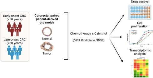
The incidence of early-onset colorectal cancer (EO-CRC, <50 years) is rising worldwide, highlighting the need to better understand the underlying causes of this age-related divergence. We used paired patient-derived normal and tumor colorectal organoids to investigate the therapeutic window of chemotherapeutic agents and its modulation by calcitriol (the most active vitamin D metabolite). Dose-response analyses revealed marked interpatient variability, with SN38 being more potent than 5-fluorouracil (5-FU) and oxaliplatin. Normal organoids were more resistant than paired tumor counterparts to 5-FU and oxaliplatin in both EO-CRC and late-onset CRC (LO-CRC, >50 years), indicating selective tumor cytotoxicity. In contrast, the therapeutic window for SN38 was preserved in LO-CRC but not in EO-CRC organoids, revealing age-dependent differences in drug sensitivity. Moreover, calcitriol reduced the cytotoxicity of 5-FU and SN38 in normal organoids regardless of patient age, while in tumor organoids this protective effect was restricted to EO-CRC. As a consequence, calcitriol treatment selectively expanded the therapeutic window for 5-FU and SN38 in LO-CRC organoids. Mechanistically, these effects correlated with calcitriol-induced antiproliferative action and transcriptional regulation of drug metabolism-related pathways. Overall, our findings identify age-dependent differences in chemotherapy response and support the importance of maintaining adequate vitamin D status to reduce chemotherapy-associated toxicity.
Keywords: colorectal cancer, patient-derived organoids, early-onset colorectal cancer, drug assays, therapeutic window, vitamin D
Introduction
Colorectal cancer (CRC) is a leading neoplasia in terms of incidence and mortality. Paradoxically, the incidence of CRC in patients less than 50 years of age (early-onset CRC, EO-CRC) is increasing for not well understood causes, while incidence in patients aged ≥50 years (late-onset CRC, LO-CRC) is declining, probably due to implementation of screening programs targeting preferentially aged populations and improved treatments [1-4]. EO-CRC is usually diagnosed at advanced stages and is characterized by distinctive genetic, molecular and microbiome profiles, as well as more aggressive clinical behavior that may be due to delay in diagnosis. However, there are no age-specific treatment protocols for CRC patients [2, 5-10].
Organoids are considered an advantageous preclinical model for cancer modeling and precision therapy [11-14], in particular to assay the activity of antitumor drugs, with respect to classically used immortal cell lines and animal models [15]. Aiming at a personalized anticancer therapy, organoids may provide functional data to enrich the genomic information obtained from mutational tumor analysis. As for CRC, organoids are being used in drug discovery and toxicity studies, and for the personalized treatment of CRC patients, although methodology needs to be improved [16-20]. Of note, tumor organoids derived from EO-CRC patients have revealed a striking diversity of molecular phenotypes and heterogeneous genetic background [21].
A large number of epidemiological studies indicate that vitamin D deficiency is a risk factor for developing and dying from CRC [22, 23], and many supplementation studies and clinical trials support a protective action of vitamin D against CRC [24-27]. Interestingly, high total vitamin D intake is associated with decreased risk of EO-CRC in a cohort of young women [28]. Concordantly, the active vitamin D metabolite 1α,25-dihydroxyvitamin D3 (calcitriol) displays a battery of anti-CRC mechanisms in experimental systems [29-33]. Among these mechanisms, calcitriol has been proposed to potentiate cancer therapy and to reverse drug-resistance in bladder, gastric, breast, glioblastoma, liver, ovarian, pancreatic and other cancer cell types [34-38]. Furthermore, calcitriol promotes cell sensitivity to 5-fluorouracil (5-FU) in human colon carcinoma cells by downregulating thymidylate synthase (5-FU molecular target), and the anti-apoptotic protein survivin [39].
Importantly, most reported studies on the action of calcitriol on CRC were performed using immortal long-term cultured carcinoma cell lines, which do not properly reproduce the in vivo situation. Tumors are thought to originate from genetically and epigenetically altered tissue resident stem cells (known as cancer stem cells, CSC) which are also proposed as responsible for chemotherapy resistance and tumor relapse [40]. Unlike 2D monolayers, 3D patient-derived organoids (PDOs) better recapitulate tissue architecture and function. Therefore, to gain more relevant information we have now studied the response to three anticancer drugs currently used in the clinic against CRC in patient-derived tumor organoids generated from CSC. Moreover, we used paired organoids from normal tissue from the same CRC patient to examine the therapeutic window of drug activity. This strategy allows the evaluation of both the efficacy and toxicity of anticancer treatments within the same genetic background, something that is not possible with other preclinical models. In addition, to investigate putative different age-dependent drug sensitivities we have separately analyzed the response to drugs in organoids from EO-CRC and LO-CRC patients. Lastly, we examined the effect of calcitriol on drug activity in normal and tumor PDOs across age groups.
Methods
Human samples
The collection of paired normal and tumor CRC patient tissue samples for the establishment of organoids was approved by the Ethics Committee of Hospital Universitario La Paz (HULP-PI-1425, HULP-PI-3196, HULP-PI-4462, HULP-PI-6274). Fresh human tissue biopsies were obtained from CRC patients undergoing surgery or diagnostic colonoscopy between 2014 and 2023 and provided by IdiPAZ Biobank (PT23/00028), integrated into the Spanish Biobank Network (https://www.isciiibiobanksbiomodels.es/). All patients provided written informed consent.
Generation and culture of patient-derived organoids
Colorectal normal and tumor PDOs were generated as previously described [41]. Briefly, human colon normal biopsies, collected from areas distant to the tumor, were incubated in rotation with an antibiotic mixture (Primocin [Invivogen, CA, USA], gentamicin and fungizone [Thermo Fisher Scientific, MA, USA]) for 1 h at room temperature (RT). Biopsies were then cut into small fragments and incubated under slow rotation twice for 5 min with 10 mM dithiothreitol (DTT [Tocris, Bristol, UK]) at RT followed by incubation with 8 mM EDTA for 5 min at RT and 60 min at 4°C. After several PBS washes to remove EDTA, samples were gently shaken in fresh PBS to release isolated crypts. Pelleted crypts were washed in washing buffer (Advanced DMEM/F12, 10 mM HEPES, and 10 mM Glutamax [Thermo Fisher Scientific]), embedded in Matrigel (Corning, NY, USA) and seeded as drops onto pre-warmed 6-well plates. Following Matrigel polymerization, normal culture medium (Table S1) was added. To establish patient-derived tumor organoids, tumor tissues were incubated with the same antibiotic mixture as normal samples. Biopsies were enzymatically digested with 1 mg/mL collagenase type IV (Sigma-Aldrich, MA, USA) in PBS for 30 min under rotation at 37ºC. Cell suspension was passed through an 18 G syringe and subsequently filtered through a 100 μm mesh filter. Erythrocytes were lysed by incubating the cell suspension with 157 mM NH4Cl for 5 min. Single cells were then embedded in Matrigel and plated onto pre-warmed 12-well dishes. Following Matrigel polymerization, tumor culture medium (normal medium lacking L-WNR conditioned medium, RSPO1 and nicotinamide) (Table S1) was added. Culture medium for both normal and tumor organoids was refreshed every other day.
PDOs were passaged according to a previously described protocol [42]. Briefly, organoids were incubated for 30 min with 1 mg/mL dispase (Thermo Fisher Scientific) at 37°C. Matrigel-embedded organoids were collected with a scraper and centrifuged at 269 x g for 5 min. Pelleted organoids were resuspended in a 1:4 TrypLE Express (Gibco, NY, USA) in PBS solution for 5 min at 37°C, followed by mechanical disaggregation through a 21 G syringe. Cells were pelleted after two washes in washing buffer, resuspended in fresh Matrigel and replated into new culture dishes. Single cell viability requires the addition of Y-27632 in both normal and tumor media until they generate complete organoids. Colorectal PDOs have been reported to preserve the genetic and phenotypic characteristics of the tissue of origin during long-term culture [43-45]. The passage number of normal and tumor organoids used in each experimental analysis is indicated in Table S2.
Drug activity assays
Culture conditions for cell viability assays of normal PDOs were optimized from a protocol previously described by our group for tumor PDOs [46]. Modifications were introduced to obtain a proportional relationship between luminescence signal and organoid number, through the adjustment of organoid size and seeding density. For this characterization prior to the assay, normal organoids were released from Matrigel with 1 mg/mL dispase and sequentially filtered through 70 μm, 40 μm and 20 μm strainers (Pluriselect, Leipzig, Germany) to obtain two size-selected populations: 20-40 μm and 40-70 μm. Both fractions were resuspended in 1 mL culture medium, and organoid concentration was determined. Eight conditions (20-40 μm: 125, 250, 500, and 1000 organoids; 40-70 μm: 50, 100, 200, and 400 organoids) were tested in triplicate by seeding organoids into 96-well dishes containing 2% Matrigel. After four days of treatment, cell viability was measured as cellular ATP level using CellTiter-Glo 3D Luminescent Cell Viability Assay (Promega, WI, USA) following the manufacturer's instructions. The optimal seeding density for normal PDOs was defined as 500 organoids of 20-40 μm per well, which ensures a proportional luminescence signal relative to the number of organoids seeded. Tumor PDOs were previously optimized (250 organoids of 20-40 μm per well) using an equivalent protocol [46].
Following this pre-assay characterization, full-grown paired normal and tumor PDOs were collected after removing Matrigel with 1 mg/mL dispase treatment. Pelleted organoids were resuspended in washing buffer and filtered through 20 μm and 40 μm cell strainers. Size-selected organoids were centrifuged and resuspended in 500 μL of the corresponding culture medium. Organoid density was determined and adjusted to seed 250 tumor and 500 normal organoids per well, distributed in four 5 μL drops within a 24-well dish. For each drug, two complete 24-well dishes were seeded: one treated with calcitriol (100 nM) and the other with vehicle (ethanol). On the following day, organoids were treated in triplicate with seven drug concentrations plus a control (vehicle: water for oxaliplatin, and DMSO for SN38 and 5-FU). We used 5-FU, oxaliplatin and SN38 (active metabolite of irinotecan) (all from Sigma-Aldrich). Retreatment with calcitriol was performed every other day. After four days of drug exposure, cell viability was assessed using the CellTiter-Glo 3D Cell Viability Assay as previously described [46]. Drug responses were quantified as the area under the curve (AUC) using GraphPad Prism v10 (GraphPad Software, CA, USA) by numerical integration (trapezoidal rule). For each drug, patient-specific AUC values were standardized to z-scores (z = [LnAUC - μ] / σ), where μ and σ denote the cohort mean and standard deviation, respectively. These z-scores, reflecting the relative drug resistance of each PDO, were visualized in a heatmap.
Cell proliferation assays
Cell proliferation assays were carried out under conditions similar to those used in viability assays. Size-selected (20-40 μm) normal (500) and tumor (250) organoids were seeded in four 5 μL drops in 24-well dishes, in triplicate. After Matrigel polymerization, the corresponding culture medium was added and supplemented with either 100 nM calcitriol or vehicle (ethanol). Culture medium was replaced every other day together with calcitriol retreatment. After four days in culture, proliferation was determined using the CellTiter-Glo 3D Cell Viability Assay (Promega).
RNA extraction and reverse transcription quantitative PCR (RT-qPCR)
Total RNA was isolated from Matrigel-embedded normal and tumor organoids using TRIzol reagent (Thermo Fisher Scientific), followed by purification with the NucleoSpin miRNA extraction kit (Machery-Nagel, Düren, Germany). cDNA retrotranscription was carried out using the iScript cDNA Synthesis kit (Bio-Rad, CA, USA). CYP24A1 expression was quantified using the Taqman Universal PCR Master Mix (Applied Biosystems, CA, USA) and a FAM-labeled TaqMan probe for CYP24A1 (Hs00167999_m1, Applied Biosystems). Expression values were normalized to the housekeeping gene RPLP0 (H99999902_m1; VIC-labeled TaqMan probe; Applied Biosystems) and relative expression levels were calculated using the comparative CT method. VDR RNA levels were determined using the Power SYBR Green PCR Master Mix (Applied Biosystems) and the following primer pair: forward 5′-AACGCTGTGTGGACATCGGC-3′; reverse 5′-GTCATGGCTTTCGTTGGACT-3′. Expression data were normalized to the housekeeping gene SDHA (forward 5′-GGGAACAAGAGGGCATCTG-3′; reverse 5′- CCACCACTGCATCAAATTCATG-3′) and analyzed using the comparative CT method. All qPCR analyses were carried out using a CFX384 Touch Real-Time PCR Detection System (Bio-Rad). An RNA sample derived from human SW480-ADH colon carcinoma cells was included as a reference to facilitate comparison of RNA expression levels among samples.
RNA-sequencing (RNA-seq) assay
Paired normal and tumor organoids from 5 EO-CRC and 6 LO-CRC patients were seeded in 12-well culture dishes and 48 h later were treated with calcitriol (100 nM) or vehicle (ethanol) for 96 h. Medium and calcitriol treatment were refreshed every other day. Total RNA samples (1µg; RIN range 8.6-9.9) were converted into sequencing libraries with the NEBNext Ultra II Directional RNA Library Prep Kit for Illumina (New England Biolabs, Ipswich, MA, USA). Briefly, polyA+ fraction was purified and randomly fragmented, converted to double stranded cDNA and processed through subsequent enzymatic treatments of end-repair, dA-tailing, and ligation to adapters. Directional cDNA libraries, stranded in the antisense orientation were completed by PCR and pair-end sequenced on an Illumina NovaSeq X by following manufacturer's protocols.
Raw FASTQ reads were aligned to the human GRCh38 v40 reference genome using HISAT2 (v2.1.0) with default parameters in paired-end mode. Alignment files (SAM) were converted to BAM and coordinate-sorted using SAMtools (v1.6). Gene-level quantification was performed with HTSeq-count (v0.11.3) using GRCh38 GTF annotations and stranded argument set to reverse. Genes with low expression were filtered using the filterByExpr function from the edgeR package (R v4.3.3). Differential expression analysis was conducted with the limma-voom pipeline, and p-values were adjusted for multiple testing using the Benjamini-Hochberg method.
Gene set enrichment analysis (GSEA) was carried out to evaluate the association between RNA-seq profiles and gene signatures enriched for proliferation and drug metabolism pathways obtained from MSigDB (www.gsea-msigdb.org). Heatmap analysis was done with R programming language (v4.5.0) using the package pheatmap.
Mutational status
Mutations present in the tumor PDOs used in this study were analyzed. Briefly, paired normal and tumor PDOs were cultured and after Matrigel removal using 1 mg/mL dispase treatment, DNA was extracted from pelleted organoids using DNeasy Tissue Kit (Qiagen, Germany) following manufacturer's instructions and quantified fluorimetrically (Qubit). Whole exome sequencing (WES) library preparation, exome capture, sequencing and primary bioinformatic analysis were performed by Macrogen Inc. (Seoul, South Korea) using a hybridization-based exome capture approach (SureSelect V8-Post kit) and paired-end sequencing (2 × 150 bp) on an Illumina platform. Sequence reads were aligned to the human reference genome (GRCh38), and variant calling for single nucleotide variants (SNVs) and short insertions/deletions (indels) was performed following a pipeline based on GATK Best Practices, including duplicate marking, base quality recalibration and variant annotation. Functional annotation of variants was performed using public databases. Somatic alterations in tumor PDOs were identified by comparison with their paired normal counterparts.
Statistical analyses
Statistical analyses were performed using GraphPad Prism v10 (GraphPad Software, MA, USA). To assess the statistical sensitivity of the experimental design, a power analysis (α = 0.05, two-sided; 80% power) was performed considering each PDO culture as an independent biological unit. For paired comparisons (n = 10 PDOs), the study was powered to detect a minimum standardized effect size of Cohen's dz ≈ 0.89, corresponding to moderate-to-large effects. For comparisons between EO-CRC and LO-CRC groups (n = 5 PDOs per group), the study was powered to detect large effects (Cohen's d ≈ 1.77).
For fold-change analyses, values were log2-transformed prior to statistical testing in order to reduce skewness and improve approximation to a normal distribution. Normality of the datasets was assessed using the Kolmogorov-Smirnov test prior to statistical testing. For paired experimental designs (e.g., comparisons between normal and tumor PDOs derived from the same patient, or vehicle vs. calcitriol treatments), paired t-tests were used. For comparisons between non-paired samples (e.g., EO-CRC vs. LO-CRC PDOs), variance homogeneity was evaluated using a F-test and unpaired t-tests were applied, using Welch's correction when variance equality could not be assumed. One-sample tests were used to determine whether values significantly differed from the reference condition. When normality assumptions were not met, the non-parametric Wilcoxon signed-rank test was applied. Comparisons involving more than two groups were analyzed by one-way ANOVA followed by Sidak's multiple-comparisons test. Correlations between variables were assessed using Pearson's correlation coefficient.
The statistical test applied in each experiment and the corresponding sample size (n) are indicated in the figure legends. Statistical significance was defined as *P < 0.05, **P < 0.01, and ***P < 0.001; n.s., non-significant.
Results
Colorectal tumor PDOs are more sensitive to chemotherapy than normal PDOs
We established a collection of paired normal and tumor organoids (Table S2 and Figure 1A). Whole-exome sequencing confirmed that tumor PDOs harbored somatic alterations commonly found in CRC, including mutations in APC, TP53, KRAS and FBXW7 genes (Figure S1). To analyze the chemosensitivity of colorectal PDOs, we performed a low-throughput drug assay as described in Methods. Both normal and tumor PDOs from 10 CRC patients equally distributed by age (5 EO-CRC and 5 LO-CRC; Table S2) were treated for four days with one of three clinically relevant chemotherapeutic agents commonly used for CRC: 5-FU, oxaliplatin and SN38 (Figure 1B). These agents were selected due to their widespread use in clinical practice and their complementary mechanisms of action: 5-FU is a thymidylate synthase inhibitor, impairing DNA synthesis; oxaliplatin induces DNA damage through direct binding and distortion of DNA; and SN38 inhibits topoisomerase I, leading to DNA damage response and RNA/ribosome biogenesis stress [47-49]. Normal PDOs exhibited a variable response to the three drugs (Figure S2A). This heterogeneity is reflected in the heatmap of normalized z-scores of AUC (Figure S2B). Dose-response curves of mean values for each drug revealed SN38 as the most cytotoxic, whereas 5-FU and oxaliplatin exerted a comparable lower cytotoxicity (Figure S2C). Similar results were obtained with tumor PDOs, which also displayed variable responses to chemotherapy (Figure S2D, E), with the highest sensitivity to SN38 (Figure S2F). Since the cytotoxic activity of chemotherapeutic agents often relates to cell proliferation, we studied whether PDO growth rates were linked to their drug sensitivity. Notably, no significant correlations were found between cell proliferation rates of normal and tumor PDOs and their AUC values that could explain the observed differential drug chemosensitivities among patients (Figure S3).
Colorectal normal and tumor PDOs display different drug sensitivity. (A) Representative bright-field microscopy images of paired normal and tumor organoids from EO-CRC (#120) and LO-CRC (#30) patients. Images correspond to organoid cultures during routine maintenance, prior to drug assays. Scale bar, 500 μm. (B) Scheme of the drug assay protocol. (C) Dose-response curves of paired normal and tumor PDOs treated with 5-FU, oxaliplatin or SN38. Mean values ± SEM from the drug assays shown in Figure S2A, D are plotted (n =10 paired PDOs). (D) Violin graphs showing AUC values from the drug assays shown in Figure S2A, D. Statistical analysis was performed using two-tailed paired t-test comparing normal and tumor PDOs (n = 10 paired PDOs). (E) Violin graph depicting log2 fold-change values (tumor/normal) calculated from the AUC values shown in (D). Dashed line represents the reference value corresponding to normal PDOs. Statistical analysis was carried out using a two-tailed one sample t-test for oxaliplatin and SN38, and a two-tailed Wilcoxon signed-rank test for 5-FU due to deviation from normality (n = 10 paired PDOs). (F) GSEA showing the positive association between drug metabolism-related gene signatures and the transcriptomic profile of normal organoids from CRC patients identified in a previous study [41].
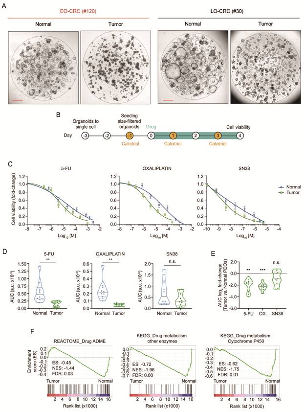
Comparison of dose-response curves of mean values from both types of PDOs revealed that tumor PDOs are significantly more sensitive to 5-FU and oxaliplatin than normal PDOs (Figure 1C, D), thereby demonstrating the utility of human organoids to identify the therapeutic window in vitro. No statistical significance was found for SN38, probably due to the variable response of normal PDOs to this drug (Figure 1C, D). These results were confirmed when analyzing the AUC fold-change between paired normal and tumor cultures, a comparison only possible using paired PDOs (Figure 1E). Interestingly, GSEA of previously published RNA-seq data from paired colon normal and tumor organoids from CRC patients [41] showed that the gene expression profile of normal PDOs was positively associated with drug metabolism-related signatures (Figure 1F). These data constitute a potential explanation for the reduced sensitivity of normal PDOs to chemotherapeutic drugs.
Age-dependent differences in PDOs chemosensitivity
Considering the increasing clinical relevance of EO-CRC, we next evaluated PDO drug responses according to patient age. First, we found that normal organoids from EO-CRC and LO-CRC patients were less sensitive to 5-FU and oxaliplatin than tumor organoids, supporting the existence of a therapeutic window in vitro for these two drugs across age (Figure 2A, B). Second, age-based stratification showed that EO-CRC and LO-CRC PDOs displayed a different response to SN38: in young patients, both normal and tumor PDOs exhibited similar sensitivity to SN38, whereas in aged patients the sensitivity of tumor PDOs seems to be higher than that of normal PDOs, although the difference did not reach statistical significance (Figure 2B). This difference became significant when analyzing the AUC fold-change between paired normal and tumor PDOs, indicating that the therapeutic window for SN38 is maintained in LO-CRC PDOs but it is lost in organoids from EO-CRC patients (Figure 2C).
Age-dependent drug sensitivity of colorectal normal and tumor PDOs. (A) Heatmap illustrating the z-score of drug resistance of age-grouped normal and tumor PDOs treated with 5-FU, oxaliplatin or SN38 (n = 10 paired PDOs). (B) Violin graphs showing the AUC values of age-grouped normal and tumor PDOs treated with 5-FU, oxaliplatin or SN38 using data from Figure S2A, D. Statistical analysis was performed by two-tailed paired t-test comparing normal and tumor PDOs within each age group (n = 5 paired PDOs per group). (C) Violin graph representing the AUC log2 fold-change values (tumor/normal) of tumor organoids from EO-CRC and LO-CRC patients calculated from the data shown in (B). Dashed line indicates the reference value corresponding to normal PDOs. Differences between tumor and paired normal PDOs were evaluated by two-tailed one-sample t-test within each age group. Comparisons between EO-CRC and LO-CRC patients were performed using two-tailed unpaired t-test (n = 5 PDOs per group). (D) GSEA showing the positive association between the transcriptomic profile of normal organoids from EO-CRC and LO-CRC patients and gene signatures related with drug metabolism. (E) Heatmaps illustrating the differential gene expression (tumor vs. normal PDOs) in EO-CRC and LO-CRC patients for the genes included in the drug metabolism signatures identified in (D).
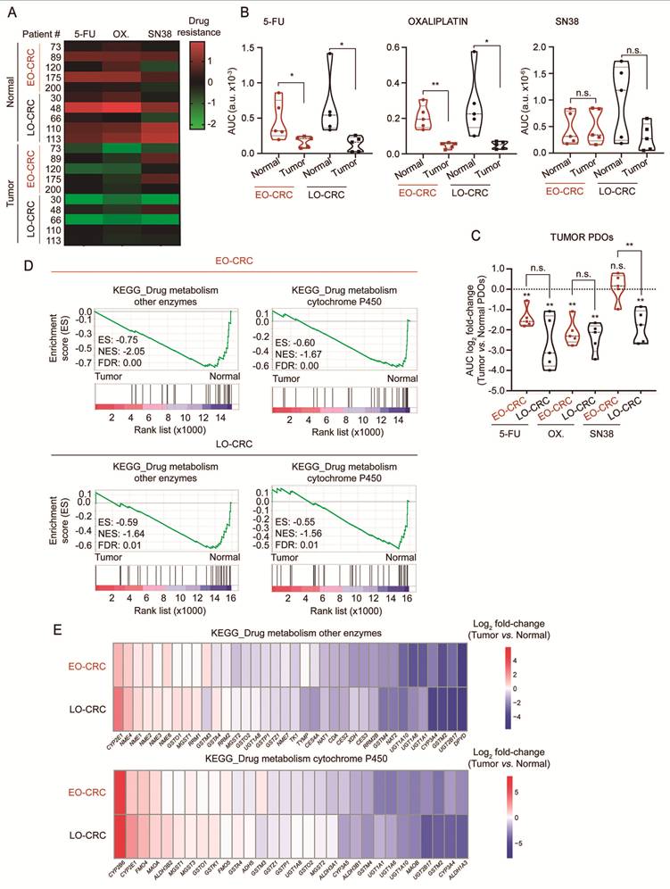
In addition, we performed an RNA-seq analysis of paired normal and tumor PDOs from 5 EO-CRC and 6 LO-CRC patients (Table S2). This study revealed that normal organoids from both young and aged patients exhibit expression profiles enriched in drug metabolism-related signatures (Figure 2D, E), which is in line with the results from our previously published RNA-seq analysis of PDOs lacking age stratification (Figure 1F). No age-related differences were observed in the enrichment of these signatures (Figure 2D, E).
Calcitriol modulates drug resistance of normal and tumor PDOs
Next, we evaluated the role of calcitriol treatment on the chemosensitivity of normal and tumor PDOs. First, we examined the responsiveness to calcitriol of the two types of organoids. As shown in Figure S4A, both normal and tumor PDOs, regardless of patient age, expressed comparable levels of VDR RNA encoding the vitamin D receptor. Accordingly, CYP24A1, the most responsive calcitriol target gene, which encodes the enzyme responsible for its degradation, was upregulated by calcitriol in normal and tumor PDOs from both age groups (Figure S4B). Concordantly with reports of CYP24A1 overexpression in human colorectal tumors [50, 51], the basal expression of CYP24A1 was higher in tumor PDOs than in normal PDOs (Figure S4B).
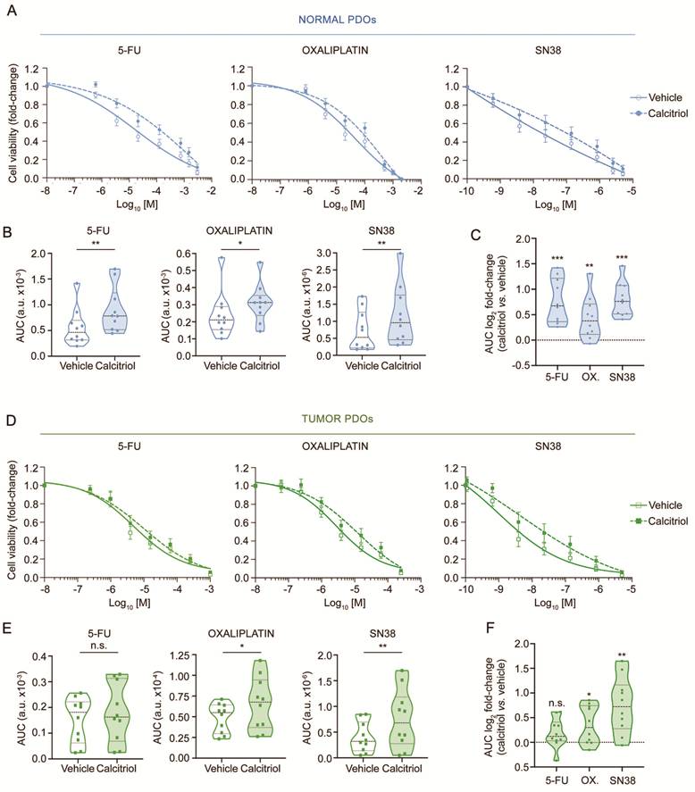
Paired normal and tumor PDOs were treated with 5-FU, oxaliplatin or SN38 in the presence or absence of calcitriol as described in Figure 1B. Strikingly, in normal PDOs calcitriol decreased the sensitivity to the three drugs (Figure 3A-C), suggesting a protective effect on healthy colon mucosa upon chemotherapy. Likewise, in tumor PDOs the sensitivity to oxaliplatin and SN38, but not to 5-FU, was also reduced by calcitriol (Figure 3D-F). In line with this, GSEA of RNA-seq data including calcitriol-treated PDOs revealed that drug metabolism-related signatures were significantly enriched in both normal and tumor PDOs upon calcitriol treatment regardless of patient age (Figure 4A).
Calcitriol modulates drug cytotoxicity in colorectal normal and tumor PDOs. (A) Dose-response curves of normal PDOs treated with 5-FU, oxaliplatin or SN38 in the presence of 100 nM calcitriol or vehicle (ethanol) for 96 h. Mean values ± SEM are plotted (n = 10 PDOs). (B) Violin graphs showing the AUC values of drug assays shown in (A). Statistical analysis was performed by two-tailed paired t-test comparing vehicle- and calcitriol-treated PDOs (n = 10 PDOs). (C) Violin graph representing AUC log2 fold-change values (calcitriol/vehicle) calculated from the data shown in (B). Dashed line represents the reference value corresponding to vehicle-treated normal PDOs. Differences from the reference value were evaluated using two-tailed one-sample t-test (n = 10 PDOs). (D) Dose-response curves of tumor PDOs treated with 5-FU, oxaliplatin or SN38 in the presence of 100 nM calcitriol or vehicle (ethanol) for 96 h. Mean values ± SEM are plotted (n = 10 PDOs). (E) Violin graphs showing AUC values from the drug assays shown in (D). Statistical analysis was performed using two-tailed paired t-tests comparing vehicle- and calcitriol-treated PDOs (n = 10 PDOs). (F) Violin graph displaying AUC log2 fold-change values (calcitriol/vehicle) calculated from the data shown in (E). Dashed line represents the reference value corresponding to vehicle-treated tumor PDOs. Differences from the reference value were evaluated using two-tailed one-sample t-test (n = 10 PDOs).
Differential effect of calcitriol on drug cytotoxicity in EO-CRC and LO-CRC tumor PDOs. (A) Graph showing the association identified by GSEA of RNA-seq data between drug metabolism-related signatures and the gene expression profiles of calcitriol-treated normal and tumor organoids from EO-CRC and LO-CRC patients. Dashed line represents the cut-off for statistical significance. (B) Violin graphs showing AUC values of age-grouped normal and tumor PDOs treated with 5-FU, oxaliplatin or SN38 in the presence of 100 nM calcitriol or vehicle (ethanol) for 96 h. Statistical analysis was performed using two-tailed paired t-test comparing vehicle- and calcitriol-treated PDOs patient within each age group (n = 5 PDOs per group). (C) Violin graphs representing AUC log2 fold-change values (calcitriol/vehicle) calculated from the data shown in (B) from normal and tumor PDOs from EO-CRC and LO-CRC patients. Dashed line indicates the reference value corresponding to vehicle-treated PDOs. Differences from the reference value were evaluated using two-tailed one-sample t-test (n = 5 PDOs per group).
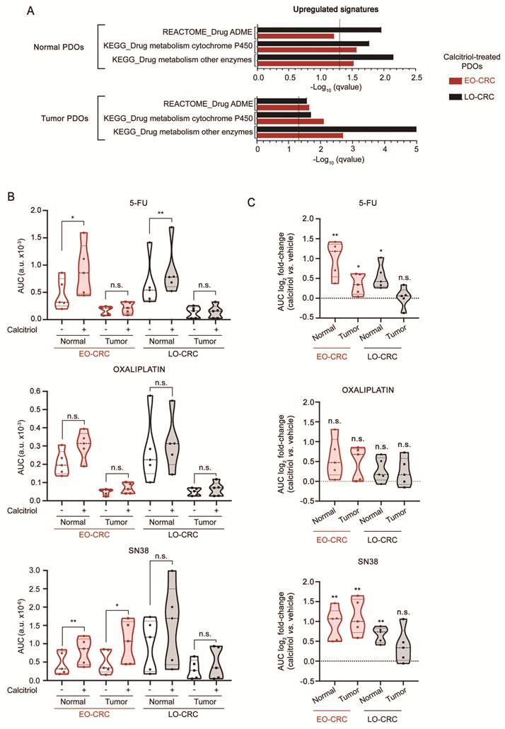
Dose-response analyses of age-stratified normal and tumor PDOs showed that patient age defines the response to chemotherapy under calcitriol treatment. Calcitriol diminished the cytotoxic activity of 5-FU and SN38 in normal organoids from both EO-CRC and LO-CRC patients (Figure S5; Figure 4B, C). Strikingly, calcitriol had age-dependent differential effects on the sensitivity of tumor PDOs to 5-FU and SN38: it diminished their cytotoxicity in young patients but did not alter drug sensitivity in organoids derived from aged patients (Figure S5; Figure 4B, C). These age-related effects were further supported by the analysis of AUC fold-changes between calcitriol- and vehicle-treated PDOs, where the reduction exerted by calcitriol on the sensitivity of LO-CRC normal PDOs to SN38 and that of EO-CRC tumor PDOs to 5-FU reached statistical significance (Figure 4C). Together, these results suggest that calcitriol cotreatment may favor a wider therapeutic window for 5-FU and SN38 specifically in LO-CRC patients. In contrast, calcitriol did not modify oxaliplatin sensitivity in normal or tumor PDOs across age groups (Figure S5; Figure 4B, C), even though this effect was observed when age stratification was not considered (Figure 3B, C, E, F).
Age-dependent differential antiproliferative effect of calcitriol in tumor PDOs
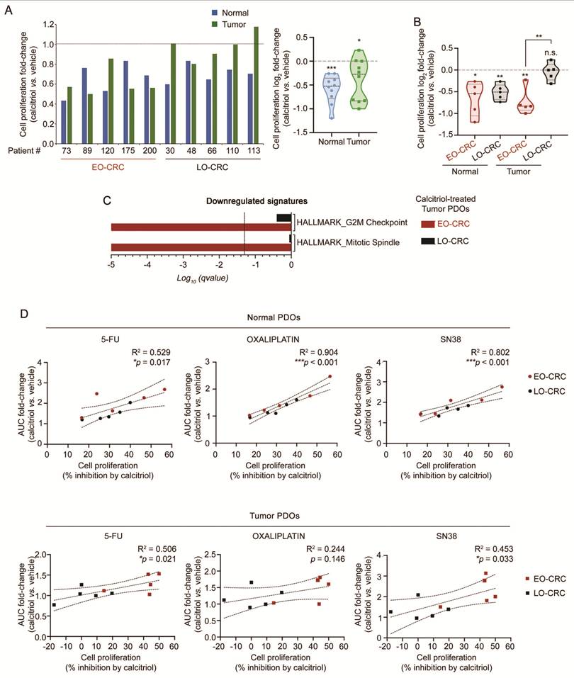
We next explored potential age-dependent differences in cell proliferation inhibition by calcitriol in paired normal and tumor PDOs. Calcitriol consistently inhibited cell proliferation in normal PDOs, while, in contrast, it showed substantial interindividual variability in tumor PDOs (Figure 5A). Stratification by age revealed that calcitriol reduced cell proliferation in EO-CRC and LO-CRC normal PDOs and also in EO-CRC tumor PDOs, but had no effect in LO-CRC tumor PDOs (Figure 5B). In line with these results, GSEA of RNA-seq data revealed a significant negative association between the gene expression profile of calcitriol-treated EO-CRC tumor PDOs and cell proliferation-related signatures, an association that was not observed in LO-CRC tumor PDOs (Figure 5C). These findings indicate that the calcitriol-induced proliferative arrest in tumor PDOs is restricted to young patients. Of note, while no significant correlations between cell proliferation and drug response were observed in vehicle-treated PDOs (Figure S3), significant positive correlations were detected between the antiproliferative effect of calcitriol and the increase in drug resistance exerted by calcitriol in both normal and tumor (except for oxaliplatin) PDOs (Figure 5D).
Relationship between drug cytotoxicity and cell proliferation in colorectal normal and tumor PDOs treated with calcitriol. (A) Cell proliferation assays of paired normal and tumor PDOs treated with 100 nM calcitriol or vehicle (ethanol) for 96 h. Bar (left panel) and violin (right panel) graphs show the effect of calcitriol. Data shown in violin graph are expressed as log2 fold-change values (calcitriol/vehicle). Statistical analysis was performed using two-tailed one-sample t-test comparing values to the reference condition (dashed line) (n = 10 paired PDOs). (B) Violin graph illustrating the log2 fold-change effect (calcitriol/vehicle) on cell proliferation in PDOs from (A) stratified by age group. Differences relative to vehicle-treated organoids were evaluated using two-tailed one-sample t-test within each group (n = 5 per group). Comparisons between EO-CRC and LO-CRC tumor PDOs were performed using two-tailed unpaired t-test with Welch's correction. Dashed line indicates the reference value corresponding to vehicle-treated PDOs. (C) Graph showing the negative association identified by GSEA of RNA-seq data between cell proliferation-related signatures and the gene expression profiles of calcitriol-treated tumor organoids from EO-CRC and LO-CRC patients. Dashed line represents the cut-off for statistical significance. (D) Scattergrams displaying the correlations between the effect of calcitriol on cell proliferation and its impact on the cytotoxicity of 5-FU, oxaliplatin and SN38 in normal (n = 10 PDOs, upper panels) and tumor (n = 10 PDOs, lower panels) organoids from EO-CRC and LO-CRC patients. Statistical correlations were evaluated using Pearson's test.
In summary, our results show that calcitriol exerts distinct, age-dependent effects on the sensitivity of CRC PDOs to 5-FU and SN38 antitumoral drugs. In young EO-CRC patients, calcitriol increases drug resistance in both normal and tumor PDOs, thereby preserving the therapeutic window observed in its absence. Contrarily, in aged LO-CRC patients, calcitriol selectively increases drug resistance in normal PDOs but not in tumor PDOs, resulting in a widened therapeutic window.
Discussion
To our knowledge, this is the first study comparing the response to antitumor drugs of paired normal and tumor colorectal organoids from young and aged CRC patients. Our results show that normal organoids derived from EO-CRC and LO-CRC patients are less sensitive to currently used drugs such as 5-FU and oxaliplatin than their paired tumor counterparts, which defines an in vitro differential sensitivity between tumor and normal cells that reflects the selective tumor cytotoxicity of these agents. We show that this reduced sensitivity is likely attributable to the elevated expression of drug metabolism-related genes in normal PDOs, rather than an age-dependent trait. In the case of SN38, the lower sensitivity of normal vs. tumor organoids only applies to LO-CRC organoids, suggesting higher intestinal toxicity of this drug in young than in aged patients. In addition, we report that tumor organoids from EO-CRC and LO-CRC patients display similar sensitivity to each of the three antitumor drugs commonly used in clinical practice. This is in line with data regarding response to chemotherapy of these two groups of patients [2, 7, 52].
Organoids derived from both normal and tumor tissues of the same patient constitute a unique experimental platform, as they enable the evaluation of drug efficacy and toxicity within a genetically paired system. This dual organoid approach is particularly powerful because it allows the assessment of antitumor drug selectivity, distinguishing their cytotoxic effects on malignant cells from those on healthy epithelia, which cannot be achieved with conventional cancer cell lines. Therefore, the present study not only provides insights into the age-dependent drug response of CRC, but also shows the potential of PDOs to model therapeutic selectivity in a personalized manner. Additionally, testing drug sensitivity in both types of PDOs allows to identify which agents are more effective against tumor cells and also to estimate doses that could minimize adverse effects on healthy (intestinal) tissue. Thus, this in vitro therapeutic window provides a framework to explore personalized treatment strategies and guide dose optimization in the clinical setting. Although translation from in vitro to in vivo remains challenging, these assays provide a useful framework to explore such strategies. It should be noted that normal and tumor PDOs were cultured in distinct media due to their different biological requirements. Although these culture conditions may influence cellular responses, the use of paired organoids from the same patient minimizes this potential bias by allowing direct comparisons within the same individual.
We performed drug assays on fully formed organoids to preserve the 3D structure and cellular interactions. This strategy better recapitulates the in vivo tissue architecture and provides a more physiologically relevant assessment of drug efficacy. In addition, the determination of optimal organoid size and density helps to minimize variability and ensures reproducibility across experiments. This methodological refinement enhances assay robustness and distinguishes the drug assay performed from those developed with traditional 2D cultures or with other 3D systems.
Interestingly, our results reveal that the active vitamin D metabolite calcitriol diminishes the cytotoxicity of 5-FU and SN38 in colorectal normal organoids from both young and aged patients. This result suggests that calcitriol modulates the cellular response to these drugs, potentially reducing their intestinal side effects/toxicity. Calcitriol also reduces the cytotoxic action of 5-FU and SN38 in EO-CRC but not in LO-CRC tumor organoids, which points to a preferential beneficial effect of a combined treatment with calcitriol and either of these two drugs in aged CRC patients. The capacity of calcitriol to diminish drug cytotoxicity in colorectal stem cell-derived organoids differs from observations obtained in established carcinoma cell lines indicating the potentiation by vitamin D compounds of the activity of antitumor drugs [34-38]. Possible explanations for this discrepancy include the usual highly malignant features of cell lines that are established with low success from human tumors and the accumulation of karyotypic aberrations during their long-term culture. Supporting this idea, calcitriol has distinct effects on gene expression in colon carcinoma cells and colon tumor organoids, as it is exemplified by the repression of Wnt/β-catenin target genes in the former but not in the latter system [41, 53].
Calcitriol has been shown to display a wide range of protective actions against CRC that include the inhibition of cancer cell proliferation and invasion, angiogenesis and metastasis, the promotion of cell differentiation and apoptosis, and the potentiation of immune responses [24, 32, 33]. Accordingly, VDR expression is a good prognostic biomarker and correlates with immune infiltration in CRC [54, 55]. Moreover, recent data show that vitamin D positively modulates intestinal microbiome composition and augments immune-dependent resistance to cancers in mice, while in humans the expression of calcitriol-induced genes correlates with improved responses to immune checkpoint inhibitors [56]. In line with all these data, the majority of recent reviews and meta-analyses of epidemiological and supplementation studies support a beneficial effect of an adequate vitamin D status in CRC [24-27]. Here, we report that calcitriol reduces cell proliferation in colorectal PDOs, an effect that is associated with decreased cytotoxicity of antitumor drugs. Of note, this action takes place in normal organoids from all-age patients and in tumor PDOs from EO-CRC patients, but only in a subset of LO-CRC tumor PDOs. These data are consistent with the reported anticancer effect of calcitriol and, moreover, suggest that its combination with anticancer therapies may enhance the functional therapeutic window of antitumor drugs, preferentially in aged patients.
A limitation of our study is the pure epithelial nature of the organoids used, as several features of cancer cells, including their response to drugs, are modulated by the surrounding stroma [57]. Thus, cancer-associated fibroblasts have been shown to induce therapy resistance in cancer cells and CRC organoids [58, 59]. Moreover, the organoid system has other intrinsic and experimental limitations including moderate success rate and difficult standardization, which still need to be optimized [60, 61]. Nevertheless, interpatient variability in drug response across PDOs closely mirrors clinical diversity, representing one of the major strengths of the organoid system for functional drug testing and translational research. Moreover, PDOs retain a significant degree of intratumor heterogeneity, preserving diverse cellular populations and subclonal architectures that are present in the original tumor. In addition, organoid apical-basal orientation might limit certain applications, such as co-culture with microorganisms or nutrient absorption assays, due to the difficulty in accessing the apical pole [62]. However, this feature is less critical in tumor PDOs, which often display compact and non-polarized structures, making drug assays largely unaffected. Overall, PDOs are considered a valuable drug screening platform and a promising system to predict patient response to treatment in a variety of cancer types [17, 63]. Consistent with this, a good correlation between PDOs chemosensitivity and CRC patient response has been reported to some, but not all, chemotherapy drugs [20, 64, 65], and recent studies show that PDOs sensitivity correlates with the response of metastatic CRC patients to 5-FU, oxaliplatin and irinotecan [66].
In conclusion, this study indicates that colorectal normal and tumor PDOs exhibit age-dependent sensitivity to antitumor drugs commonly used for CRC and to the modulatory action of calcitriol on drug activity. Our results also support an age-independent role for calcitriol in attenuating the intestinal side effects of chemotherapy and highlight the potential relevance of considering patient age to evaluate vitamin D status and/or calcitriol cotreatment during chemotherapy.
Supplementary Material
Supplementary figures and tables.

Acknowledgements
We thank the patients and the Biobank of IdiPAZ (PT23/00028), integrated into the Spanish Biobank Network, for their collaboration. This study has been funded by Instituto de Salud Carlos III (ISCIII) through the project ICI20/00057 and co-funded by the European Union. Additional support was provided by grants CIBERONC/CB16/12/00273, CIBERONC/CB16/12/00398, and CIBERES/CB15/00037 funded by the Instituto de Salud Carlos III (ISCIII) - FEDER, UE. This work was also supported by grant PID2019-104867RB-I00 funded by MICIU/AEI/10.13039/501100011033; grant PID2022-136729OB-I00 funded by MICIU/AEI/10.13039/501100011033 and FEDER, UE and grant S2022/BMD-7212 funded by Comunidad de Madrid. SR-M was supported by grant PREP2022-000611 funded by MICIU/AEI/10.13039/501100011033 and FSE+. EA-G was supported by grant FPU24/03135 funded by Ministerio de Ciencia, Innovación y Universidades. AF-B, AB, SR-M, AH-C, EA-G, JMG-S, MJL and AM belong to the Spanish National Research Council (CSIC)'s Cancer Hub.
Author contributions
Conceptualization and design: AF-B, AB, MJL, AM. Methodology and data acquisition: AB, AF-B, SR-M, EA-G. Bioinformatic analysis: LdP, AH-C. Resources: IP, ABu, NR-S, ML-A, DG-O, TO. Funding acquisition: NR-S, JMG-S, MJL, AM, TO. Writing - original draft: AM. Writing - review and editing: AF-B, AB, MJL, JMG-S. All authors approved and agreed to the final manuscript submission.
Data availability
RNA-seq data have been deposited in the European Nucleotide Archive (ENA) at EMBL-EBI under accession number PRJEB102090 (https://www.ebi.ac.uk/ena/browser/view/PRJEB102090). In addition, previously published transcriptomic data analyzed in this study are available at the Gene Expression Omnibus (GEO) database (accession number GSE100785) [41].
Competing Interests
Tomás Olleros is president of Grupo Farmasierra. All other authors have declared no conflicts of interest.
References
1. Takada K, Hotta K, Kishida Y, Ito S, Imai K, Ono H. Comprehensive Analysis of Early-onset Colorectal Cancer: A Review. J Anus Rectum Colon. 2023;7:241-9
2. O'Reilly M, Linehan A, Krstic A, Kolch W, Sheahan K, Winter DC. et al. Oncotherapeutic Strategies in Early Onset Colorectal Cancer. Cancers (Basel). 2023 15
3. Kim S, Keum N. Global trends in early-onset and late-onset cancer incidence. J Public Health (Oxf). 2025
4. Jayakrishnan T, Ng K. Early-Onset Gastrointestinal Cancers: A Review. JAMA. 2025
5. Mi X, Zheng P, Wu X. Differential Analysis of Early-Onset and Late-Onset Colorectal Cancer Based on Multidimensional Evidence Integration: A Review. Cancer Control. 2025;32:10732748251363337
6. Venugopal A, Carethers JM. Epidemiology and biology of early onset colorectal cancer. EXCLI J. 2022;21:162-82
7. Saraiva MR, Rosa I, Claro I. Early-onset colorectal cancer: A review of current knowledge. World J Gastroenterol. 2023;29:1289-303
8. Medici B, Ricco B, Caffari E, Zaniboni S, Salati M, Spallanzani A. et al. Early Onset Metastatic Colorectal Cancer: Current Insights and Clinical Management of a Rising Condition. Cancers (Basel). 2023 15
9. Adnan D, Trinh JQ, Sharma D, Alsayid M, Bishehsari F. Early-onset Colon Cancer Shows a Distinct Intestinal Microbiome and a Host-Microbe Interaction. Cancer Prev Res (Phila). 2024;17:29-38
10. Li J, Pan Y, Guo F, Wang C, Liang L, Li P. et al. Patterns in genomic mutations among patients with early-onset colorectal cancer: an international, multicohort, observational study. Lancet Oncol. 2025;26:1055-66
11. Yan HHN, Chan AS, Lai FP, Leung SY. Organoid cultures for cancer modeling. Cell Stem Cell. 2023;30:917-37
12. Thorel L, Perreard M, Florent R, Divoux J, Coffy S, Vincent A. et al. Patient-derived tumor organoids: a new avenue for preclinical research and precision medicine in oncology. Exp Mol Med. 2024;56:1531-51
13. Heydari Z, Devasahayam Arokia Balaya R, Sarkar G, Boardman L. The Role of Organoids in Advancing Colorectal Cancer Research: Insights and Future Directions. Cancers (Basel). 2025 17
14. Kim SY, van de Wetering M, Clevers H, Sanders K. The future of tumor organoids in precision therapy. Trends Cancer. 2025;11:665-75
15. Rae C, Amato F, Braconi C. Patient-Derived Organoids as a Model for Cancer Drug Discovery. Int J Mol Sci. 2021 22
16. Yao Y, Xu X, Yang L, Zhu J, Wan J, Shen L. et al. Patient-Derived Organoids Predict Chemoradiation Responses of Locally Advanced Rectal Cancer. Cell Stem Cell. 2020;26:17-26 e6
17. Wensink GE, Elias SG, Mullenders J, Koopman M, Boj SF, Kranenburg OW. et al. Patient-derived organoids as a predictive biomarker for treatment response in cancer patients. NPJ Precis Oncol. 2021;5:30
18. Barbachano A, Fernandez-Barral A, Bustamante-Madrid P, Prieto I, Rodriguez-Salas N, Larriba MJ. et al. Organoids and Colorectal Cancer. Cancers (Basel). 2021 13
19. Mertens S, Huismans MA, Verissimo CS, Ponsioen B, Overmeer R, Proost N. et al. Drug-repurposing screen on patient-derived organoids identifies therapy-induced vulnerability in KRAS-mutant colon cancer. Cell Rep. 2023;42:112324
20. Martini G, Belli V, Napolitano S, Ciaramella V, Ciardiello D, Belli A. et al. Establishment of patient-derived tumor organoids to functionally inform treatment decisions in metastatic colorectal cancer. ESMO Open. 2023;8:101198
21. Yan HHN, Siu HC, Ho SL, Yue SSK, Gao Y, Tsui WY. et al. Organoid cultures of early-onset colorectal cancers reveal distinct and rare genetic profiles. Gut. 2020;69:2165-79
22. Grant WB, Boucher BJ, Al Anouti F, Pilz S. Comparing the Evidence from Observational Studies and Randomized Controlled Trials for Nonskeletal Health Effects of Vitamin D. Nutrients. 2022 14
23. Kim H, Yuan C, Nguyen LH, Ng K, Giovannucci EL. Prediagnostic Vitamin D Status and Colorectal Cancer Survival by Vitamin D Binding Protein Isoforms in US Cohorts. J Clin Endocrinol Metab. 2023;108:e223-e9
24. Munoz A, Grant WB. Vitamin D and Cancer: An Historical Overview of the Epidemiology and Mechanisms. Nutrients. 2022 14
25. Lawler TP, Khankari NK, Shu XO, Steinwandel M, Cai Q, Jurutka PW. et al. Prediagnostic 25-Hydroxyvitamin D in Association with Cancer-specific and All-cause Mortality among Low-income, Non-Hispanic Black Americans with Colorectal Cancer. Cancer Epidemiol Biomarkers Prev. 2023;32:1061-8
26. Sha S, Chen LJ, Brenner H, Schottker B. Associations of 25-hydroxyvitamin D status and vitamin D supplementation use with mortality due to 18 frequent cancer types in the UK Biobank cohort. Eur J Cancer. 2023;191:113241
27. Ottaiano A, Facchini S, Santorsola M, Nasti G, Facchini G, Montella L. et al. Circulating Vitamin D Level and Its Impact on Mortality and Recurrence in Stage III Colorectal Cancer Patients: A Systematic Review and Meta-Analysis. Cancers (Basel). 2023 15
28. Kim H, Lipsyc-Sharf M, Zong X, Wang X, Hur J, Song M. et al. Total Vitamin D Intake and Risks of Early-Onset Colorectal Cancer and Precursors. Gastroenterology. 2021;161:1208-17 e9
29. Ferrer-Mayorga G, Larriba MJ, Crespo P, Munoz A. Mechanisms of action of vitamin D in colon cancer. J Steroid Biochem Mol Biol. 2019;185:1-6
30. Carlberg C, Munoz A. An update on vitamin D signaling and cancer. Semin Cancer Biol. 2022;79:217-30
31. Na SY, Kim KB, Lim YJ, Song HJ. Vitamin D and Colorectal Cancer: Current Perspectives and Future Directions. J Cancer Prev. 2022;27:147-56
32. Pereira F, Fernandez-Barral A, Larriba MJ, Barbachano A, Gonzalez-Sancho JM. From molecular basis to clinical insights: a challenging future for the vitamin D endocrine system in colorectal cancer. FEBS J. 2024;291:2485-518
33. Ferrer-Mayorga G, Munoz A, Gonzalez-Sancho JM. Vitamin D and Colorectal Cancer. In: Hewison M, Bouillon R, Giovannucci EL, Goltzman D, Meyer MB, Welsh J, editors. Feldman and Pike's Vitamin D, Fifth Edition. 5 ed: Elsevier. 2024 p. 859-99
34. Bak DH, Kang SH, Choi DR, Gil MN, Yu KS, Jeong JH. et al. Autophagy enhancement contributes to the synergistic effect of vitamin D in temozolomide-based glioblastoma chemotherapy. Exp Ther Med. 2016;11:2153-62
35. Bunch BL, Ma Y, Attwood K, Amable L, Luo W, Morrison C. et al. Vitamin D(3) enhances the response to cisplatin in bladder cancer through VDR and TAp73 signaling crosstalk. Cancer Med. 2019;8:2449-61
36. Negri M, Gentile A, de Angelis C, Monto T, Patalano R, Colao A. et al. Vitamin D-Induced Molecular Mechanisms to Potentiate Cancer Therapy and to Reverse Drug-Resistance in Cancer Cells. Nutrients. 2020 12
37. Segovia-Mendoza M, Garcia-Quiroz J, Diaz L, Garcia-Becerra R. Combinations of Calcitriol with Anticancer Treatments for Breast Cancer: An Update. Int J Mol Sci. 2021 22
38. Wei D, Wang L, Liu Y, Hafley MA, Tan L, Lorenzi PL. et al. Activation of Vitamin D/VDR Signaling Reverses Gemcitabine Resistance of Pancreatic Cancer Cells Through Inhibition of MUC1 Expression. Dig Dis Sci. 2023;68:3043-58
39. Liu G, Hu X, Chakrabarty S. Vitamin D mediates its action in human colon carcinoma cells in a calcium-sensing receptor-dependent manner: downregulates malignant cell behavior and the expression of thymidylate synthase and survivin and promotes cellular sensitivity to 5-FU. Int J Cancer. 2010;126:631-9
40. Colak S, Medema JP. Cancer stem cells-important players in tumor therapy resistance. FEBS J. 2014;281:4779-91
41. Fernandez-Barral A, Costales-Carrera A, Buira SP, Jung P, Ferrer-Mayorga G, Larriba MJ. et al. Vitamin D differentially regulates colon stem cells in patient-derived normal and tumor organoids. FEBS J. 2020;287:53-72
42. Bustamante-Madrid P, Barbachano A, Albandea-Rodriguez D, Rodriguez-Cobos J, Rodriguez-Salas N, Prieto I. et al. Vitamin D opposes multilineage cell differentiation induced by Notch inhibition and BMP4 pathway activation in human colon organoids. Cell Death Dis. 2024;15:301
43. van de Wetering M, Francies HE, Francis JM, Bounova G, Iorio F, Pronk A. et al. Prospective derivation of a living organoid biobank of colorectal cancer patients. Cell. 2015;161:933-45
44. Huch M, Gehart H, van Boxtel R, Hamer K, Blokzijl F, Verstegen MM. et al. Long-term culture of genome-stable bipotent stem cells from adult human liver. Cell. 2015;160:299-312
45. Sachs N, de Ligt J, Kopper O, Gogola E, Bounova G, Weeber F. et al. A Living Biobank of Breast Cancer Organoids Captures Disease Heterogeneity. Cell. 2018;172:373-86 e10
46. Costales-Carrera A, Fernandez-Barral A, Bustamante-Madrid P, Guerra L, Cantero R, Barbachano A. et al. Plocabulin Displays Strong Cytotoxic Activity in a Personalized Colon Cancer Patient-Derived 3D Organoid Assay. Mar Drugs. 2019 17
47. Baek SO, Keuper K, Milletti G, Adelantado-Rubio A, Lisby M, Bartek J. et al. Stepwise DNA damage and repair mechanisms at replication forks in response to topoisomerase I inhibition. DNA Repair (Amst). 2026;157:103914
48. Chen JK, Merrick KA, Kong YW, Izrael-Tomasevic A, Eng G, Handly ED. et al. An RNA damage response network mediates the lethality of 5-FU in colorectal cancer. Cell Rep Med. 2024;5:101778
49. Sutton EC, DeRose VJ. Early nucleolar responses differentiate mechanisms of cell death induced by oxaliplatin and cisplatin. J Biol Chem. 2021;296:100633
50. Anderson MG, Nakane M, Ruan X, Kroeger PE, Wu-Wong JR. Expression of VDR and CYP24A1 mRNA in human tumors. Cancer Chemother Pharmacol. 2006;57:234-40
51. Hobaus J, Hummel DM, Thiem U, Fetahu IS, Aggarwal A, Mullauer L. et al. Increased copy-number and not DNA hypomethylation causes overexpression of the candidate proto-oncogene CYP24A1 in colorectal cancer. Int J Cancer. 2013;133:1380-8
52. Lipsyc-Sharf M, Zhang S, Ou FS, Ma C, McCleary NJ, Niedzwiecki D. et al. Survival in Young-Onset Metastatic Colorectal Cancer: Findings From Cancer and Leukemia Group B (Alliance)/SWOG 80405. J Natl Cancer Inst. 2022;114:427-35
53. Palmer HG, Gonzalez-Sancho JM, Espada J, Berciano MT, Puig I, Baulida J. et al. Vitamin D(3) promotes the differentiation of colon carcinoma cells by the induction of E-cadherin and the inhibition of beta-catenin signaling. J Cell Biol. 2001;154:369-87
54. Xia X, Xu F, Dai D, Xiong A, Sun R, Ling Y. et al. VDR is a potential prognostic biomarker and positively correlated with immune infiltration: a comprehensive pan-cancer analysis with experimental verification. Biosci Rep. 2024 44
55. Ferrer-Mayorga G, Gomez-Lopez G, Barbachano A, Fernandez-Barral A, Pena C, Pisano DG. et al. Vitamin D receptor expression and associated gene signature in tumour stromal fibroblasts predict clinical outcome in colorectal cancer. Gut. 2017;66:1449-62
56. Giampazolias E, Pereira da Costa M, Lam KC, Lim KHJ, Cardoso A, Piot C. et al. Vitamin D regulates microbiome-dependent cancer immunity. Science. 2024;384:428-37
57. Qu Y, Dou B, Tan H, Feng Y, Wang N, Wang D. Tumor microenvironment-driven non-cell-autonomous resistance to antineoplastic treatment. Mol Cancer. 2019;18:69
58. Fiori ME, Di Franco S, Villanova L, Bianca P, Stassi G, De Maria R. Cancer-associated fibroblasts as abettors of tumor progression at the crossroads of EMT and therapy resistance. Mol Cancer. 2019;18:70
59. Ramos Zapatero M, Tong A, Opzoomer JW, O'Sullivan R, Cardoso Rodriguez F, Sufi J. et al. Trellis tree-based analysis reveals stromal regulation of patient-derived organoid drug responses. Cell. 2023;186:5606-19 e24
60. Bose S, Clevers H, Shen X. Promises and Challenges of Organoid-Guided Precision Medicine. Med. 2021;2:1011-26
61. van Renterghem AWJ, van de Haar J, Voest EE. Functional precision oncology using patient-derived assays: bridging genotype and phenotype. Nat Rev Clin Oncol. 2023;20:305-17
62. Co JY, Margalef-Catala M, Monack DM, Amieva MR. Controlling the polarity of human gastrointestinal organoids to investigate epithelial biology and infectious diseases. Nat Protoc. 2021;16:5171-92
63. Zhao Z, Chen X, Dowbaj AM, Sljukic A, Bratlie K, Lin L. et al. Organoids. Nat Rev Methods Primers. 2022 2
64. Ooft SN, Weeber F, Dijkstra KK, McLean CM, Kaing S, van Werkhoven E. et al. Patient-derived organoids can predict response to chemotherapy in metastatic colorectal cancer patients. Sci Transl Med. 2019 11
65. Mo S, Tang P, Luo W, Zhang L, Li Y, Hu X. et al. Patient-Derived Organoids from Colorectal Cancer with Paired Liver Metastasis Reveal Tumor Heterogeneity and Predict Response to Chemotherapy. Adv Sci (Weinh). 2022;9:e2204097
66. Smabers LP, Wensink E, Verissimo CS, Koedoot E, Pitsa KC, Huismans MA. et al. Organoids as a biomarker for personalized treatment in metastatic colorectal cancer: drug screen optimization and correlation with patient response. J Exp Clin Cancer Res. 2024;43:61
Author contact
Corresponding author: Asunción Fernández-Barral or María Jesús Larriba, Instituto de Investigaciones Biomédicas Sols-Morreale, Arturo Duperier, 4, 28029 Madrid, Spain; E-mail: afbarraluam.es / mjlarribauam.es.