Impact Factor ISSN: 1449-2288




Int J Biol Sci 2026; 22(13):7053-7081. doi:10.7150/ijbs.138638 This issue Cite
Review
Post-Translational Modifications in Cancer-Associated CD8⁺ T-Cell Exhaustion: Mechanisms and Therapeutic Opportunities
Yongqi Lv1,*, Qihang Shang1,*, Yang Yu2,*, Yahui Fu1, Lijuan Liu1,3,  , Changgang Sun1,3,4,
1. College of First Clinical Medicine, Shandong University of Traditional Chinese Medicine, Jinan, Shandong, 250000, China.
2. Faculty of Chinese Medicine and State Key Laboratory of Mechanism and Quality of Chinese Medicine, Macau University of Science and Technology, Macau, 999078, China.
3. Department of Oncology, Weifang Traditional Chinese Hospital, Weifang, Shandong, 261000, China.
4. College of Traditional Chinese Medicine, Shandong Second Medical University, Weifang, Shandong, 261000, China.
* Yongqi Lv, Qihang Shang, and Yang Yu have contributed equally to this work and share the first authorship.
Received 2026-6-1; Accepted 2026-7-1; Published 2026-7-30
Abstract
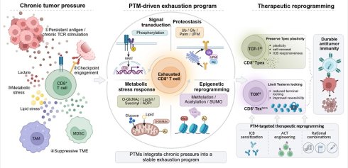
CD8⁺ T-cell exhaustion is a distinct differentiation state driven by persistent antigen stimulation, and its establishment and maintenance are major barriers to effective cancer immunotherapy. Although the transcriptional and epigenetic landscapes of exhausted CD8⁺ T cells have been extensively characterized, it remains unclear how sustained external stimulation is integrated at the level of protein function to produce stable dysfunction and altered cell fate. Post-translational modifications constitute a key regulatory layer of protein function. They form a dynamic network that links persistent antigenic stimulation and tumor microenvironmental stress to cell fate, and therefore provide a critical entry point for understanding how exhaustion is initiated and maintained. In this review, we focus on how post-translational modifications convert persistent antigen stimulation and tumor microenvironmental stress into protein-level dysregulation and ultimately lock CD8⁺ T cells into an exhausted fate. We summarize how multiple classes of post-translational modifications drive exhaustion through effects on signal transduction, protein homeostasis, metabolic stress responses, and epigenetic reprogramming. We then discuss potential intervention strategies centered on critical regulatory nodes that may preserve the plasticity of precursor exhausted CD8⁺ T cells, restrain stabilization of the terminally exhausted state in CD8⁺ T cells, and optimize rational combination therapies. Finally, we outline the translational challenges and future directions of targeting post-translational modifications, and emphasize that identifying actionable modification nodes will be important for patient stratification and combination design in cancer immunotherapy.
Keywords: cancer immunotherapy, CD8⁺ T-cell exhaustion, post-translational modifications, tumor microenvironment, immune checkpoint blockade
1. Introduction
Immunotherapies such as immune checkpoint inhibitors (ICIs) have reshaped the therapeutic landscape of cancer, yet the overall response rate remains limited [1]. Functional impairment of CD8⁺ T cells in the tumor microenvironment (TME) is now recognized as one of the main factors restricting therapeutic efficacy [1, 2]. A central manifestation of this dysfunctional state is CD8⁺ T-cell exhaustion (Tex). The establishment and maintenance of CD8⁺ Tex are jointly driven by persistent antigen stimulation, suppressive cues in the TME, and metabolic stress [2]. Hallmarks of this state include reduced proliferative capacity, diminished production of interleukin-2 (IL-2), interferon-γ (IFN-γ), and tumor necrosis factor-α (TNF-α), sustained upregulation of inhibitory receptors such as programmed cell death protein 1 (PD-1), cytotoxic T-lymphocyte-associated protein 4 (CTLA-4), T-cell immunoglobulin and mucin-domain-containing protein 3 (TIM-3), and lymphocyte activation gene-3 (LAG-3), and broad metabolic and epigenetic reprogramming [2]. These changes do not occur in isolation. Instead, they form an integrated functional network that drives and stabilizes exhaustion, thereby limiting effector function and durability.
Exhausted CD8⁺ T cells display relatively stable molecular and epigenetic features. Even after blockade of the PD-1/programmed death-ligand 1 (PD-L1) axis, functional restoration is often incomplete and difficult to sustain [3]. This observation suggests the presence of a relatively stable "epigenetic scar" in CD8⁺ Tex, which is widely considered a key basis for the stable maintenance of exhaustion and resistance to immunotherapy [3, 4]. In recent years, substantial progress has been made in defining the transcriptional programs, epigenetic landscapes, and lineage heterogeneity of CD8⁺ Tex. However, these studies have largely described state features rather than fully explaining how exhaustion is established and durably maintained. In particular, how persistent antigen stimulation, suppressive signals within the TME, and metabolic pressure are continuously integrated by CD8⁺ T cells and converted into stable dysfunction and altered cell fate remains a central mechanistic question.
Dynamic changes in protein activity, stability, subcellular localization, and molecular interactions represent a critical layer linking external stimulation to long-term fate fixation [5]. Post-translational modifications (PTMs) are one of the major regulatory mechanisms operating at this layer. These include phosphorylation, ubiquitination, acetylation, methylation, lactylation, and other dynamic covalent modifications. With the rapid development of mass spectrometry-based proteomics and chemical proteomics, the systematic identification, quantification, and functional interrogation of PTM sites have advanced considerably [6]. Increasing evidence indicates that PTMs are not merely accompanying events during the development of CD8⁺ Tex. Rather, they constitute a major regulatory layer that governs exhaustion initiation, maintenance, and terminal stabilization. By remodeling signaling pathways, maintaining the homeostasis of checkpoint proteins and other critical factors, regulating transcription factor activity and nuclear dynamics, reprogramming metabolic stress and mitochondrial homeostasis, and shaping epigenetic states, PTMs continuously influence the differentiation trajectory of exhausted CD8⁺ T cells [7]. However, these mechanisms still require systematic integration.
In this review, we therefore examine four major functional layers, including signal transduction, protein homeostasis, metabolic stress responses, and epigenetic states, to summarize how distinct PTM classes regulate the formation, maintenance, and terminal stabilization of CD8⁺ Tex. We further discuss their potential translational value in preserving the precursor exhausted T-cell (Tpex) pool, limiting stabilization of the terminally exhausted state (Texterm), and improving the efficacy of immune checkpoint blockade (ICB), adoptive cell therapy (ACT), radiotherapy, and chemotherapy.
2. Hierarchical differentiation of exhausted CD8⁺ T cells
The concept of "T-cell exhaustion" was first established in models of chronic lymphocytic choriomeningitis virus (LCMV) infection [8]. During acute viral infection, naive T cells become activated after antigen recognition and co-stimulation, differentiate into effector T cells, exert cytotoxic activity, and produce effector cytokines [9]. Under conditions of persistent antigen stimulation, however, CD8⁺ T cells progressively deviate from the canonical effector differentiation trajectory and enter an exhausted state. Cytokine production is lost in a characteristic hierarchical manner: IL-2 is impaired first, followed by TNF-α, whereas IFN-γ is further attenuated under sustained stimulation, reflecting the progressive deepening of CD8⁺ Tex [10].
With the development of tumor immunology and single-cell studies, the hierarchical differentiation framework revealed by chronic infection models has been further extended to the tumor context. Studies using mouse tumor models and human tumor samples have shown that tumor-infiltrating CD8⁺ T cells also contain transcriptional and functional states corresponding to precursor-like, intermediate, and terminal exhaustion, which are associated with clonal expansion and therapeutic responses after immune checkpoint blockade [11-14]. However, cancer-associated CD8⁺ Tex are not simply equivalent to Tex induced by chronic infection. Instead, they constitute a tumor-contextualized exhaustion continuum shaped by persistent tumor antigen/neoantigen exposure, checkpoint ligand signaling, and immunosuppressive and metabolic pressures within the TME [15, 16].
In this tumor context, exhausted CD8⁺ T cells can generally be further divided into Tpex, intermediate exhausted cells (Texint), and highly differentiated Texterm [17]. Tpex are thought to constitute the reservoir population of the exhausted lineage and retain self-renewal and regenerative capacity. They typically express high levels of T-cell factor 1 (TCF-1), relatively low levels of PD-1, and frequently co-express surface markers such as signaling lymphocytic activation molecule family member 6 (SLAMF6), L-selectin (CD62L), and C-X-C motif chemokine receptor 5 (CXCR5) [18]. The proliferative burst observed after PD-1 blockade arises mainly from this precursor-like population [19]. CD8⁺ T cells lacking TCF-1 fail to survive long term and respond poorly to anti-PD-L1 therapy [20]. As TCF-1 expression declines, cells may transition into Texint, a state characterized by high T-bet expression and partial effector-like transcriptional features, which serves as an intermediate stage between Tpex and terminal exhaustion [21]. Texterm may arise from Texint through TOX-mediated antagonism of TBX21 (T-bet), leading to T-bet loss, or may develop directly from Tpex under TCF-1-dependent regulation. This population displays low proliferative activity, severely impaired effector function, and sustained high expression of inhibitory receptors and TOX [21]. At the epigenetic level, Texterm cells exhibit a chromatin landscape distinct from that of effector and memory T cells and acquire a relatively stable "epigenetic scar". This scar is primarily characterized by the persistent retention of an exhaustion-associated epigenetic landscape, involving alterations in chromatin accessibility, DNA methylation, histone modifications, and chromatin remodeling. By maintaining exhaustion-associated transcriptional programs and restricting the reactivation of effector- and memory-associated gene programs, these alterations promote Texterm fate stabilization and limit the functional reversibility of Texterm cells [3, 22-24] (Fig. 1).
Activation, effector differentiation, exhaustion lineage progression, and epigenetic scar formation in CD8⁺ T cells. (A) Naïve CD8⁺ T cells are activated by antigen-presenting cells. (B) Functional effector CD8⁺ T cells mediate antitumor activity through the release of perforin (PFN) and granzyme B (GzmB), FasL-Fas-mediated cytotoxicity, and effector cytokine production. (C) Under persistent antigen stimulation, cells progressively differentiate along the exhaustion lineage. TCF-1hi precursor exhausted T cells (Tpex), which retain self-renewal capacity and form the main reservoir that responds to immune checkpoint blockade (ICB), transition into a T-bethi intermediate exhausted state (Texint) and then into a TOXhi terminally exhausted state (Texterm), accompanied by dynamic changes in inhibitory receptors and differentiation markers. (D) Terminal exhaustion is associated with the formation of an "epigenetic scar", characterized by restricted chromatin accessibility, indicating that the exhausted fate has been stably fixed and is only partially reversible.
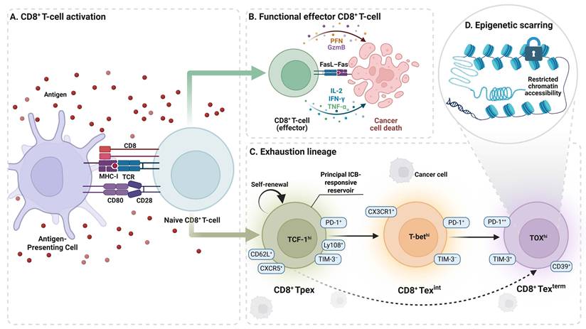
This dynamic differentiation trajectory from Tpex through Texint to Texterm highlights why PTMs are central to fate determination in CD8⁺ Tex. Whether CD8⁺ T cells retain Tpex-like plasticity or progress toward Texterm is not dictated solely by a given gene-expression profile. It also depends on how key functional modules, including signal transduction, protein homeostasis, metabolic stress responses, and epigenetic reprogramming, are regulated by PTMs [25]. For example, phosphorylation mainly shapes T-cell receptor (TCR) signaling and its downstream pathways [26, 27]. Ubiquitination, glycosylation, palmitoylation, and UFMylation participate in maintaining the homeostasis of inhibitory receptors and other critical proteins [28-31]. O-GlcNAcylation, succinylation, lactylation, and ADP-ribosylation can convert metabolic stressors such as nutrient deprivation and lactate accumulation into stable exhaustion programs [32-35]. Methylation, acetylation, and SUMOylation promote the stabilization of exhaustion-associated epigenetic states by reshaping chromatin organization and transcriptional control [36-38]. PTMs should therefore not be viewed as accessory layers during the development of CD8⁺ Tex. Rather, they are decisive regulatory strata that determine fate commitment and terminal stabilization. A systematic framework for these hierarchical mechanisms is essential for understanding how CD8⁺ T-cell exhaustion is initiated, maintained, and stabilized.
3. Layered PTM mechanisms driving CD8⁺ T-cell exhaustion
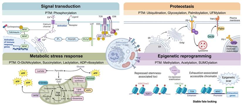
PTMs are covalent chemical modifications that are introduced at specific amino acid residues after protein synthesis through enzyme-dependent or enzyme-independent reactions, including the addition of phosphate, acetyl, or ubiquitin groups [7]. During the development of CD8⁺ Tex driven by persistent antigenic stimulation and TME pressure, PTMs operate in a layered manner. Based on the post-translational modification categories and their predominant functions described above, this section proceeds sequentially. First, we examine how chronic stimulation is sensed through phosphorylation-mediated signal transduction and how activation thresholds are reset. We then describe how inhibitory input is durably maintained at the level of checkpoint molecules and the homeostasis of key proteins through ubiquitination, glycosylation, palmitoylation, and UFMylation. Next, we address how metabolic stress is translated into stable exhaustion programs through metabolism-sensitive post-translational modifications, including O-GlcNAcylation, succinylation, lactylation, and ADP-ribosylation. Finally, we consider how these changes become further fixed at the epigenetic level through methylation, acetylation, and SUMOylation, leading to a relatively stable terminally exhausted state (Fig. 2).
Four functional PTM layers that drive CD8⁺ T-cell exhaustion. Phosphorylation resets activation thresholds through signal transduction. Ubiquitination, glycosylation, palmitoylation, and UFMylation regulate the homeostasis of key proteins. O-GlcNAcylation, succinylation, lactylation, and ADP-ribosylation convert metabolic stress into stable exhaustion programs. Methylation, acetylation, and SUMOylation promote the formation of epigenetic scars. These PTM layers act cooperatively to drive progressive fixation of terminal exhaustion in CD8⁺ T cells.
3.1 Signal transduction: phosphorylation
Phosphorylation is the covalent attachment of a phosphate group from adenosine triphosphate (ATP) to serine (Ser), threonine (Thr), or tyrosine (Tyr) residues of substrate proteins by protein kinases, whereas protein phosphatases catalyze dephosphorylation at the corresponding sites [39]. Together, these opposing reactions create a high-turnover writing-and-erasing cycle that makes phosphorylation the most immediate and sensitive regulatory mode within T-cell signaling networks [40]. Under persistent antigenic stimulation, phosphorylation resets the intensity and duration of proximal TCR signals and their downstream pathways, thereby converting recurrent exogenous input into cumulative functional deviation (Fig. 3).
Phosphorylation-mediated signaling control in CD8⁺ T-cell exhaustion. (A) Proximal TCR signaling is transmitted through the LCK-CD3ζ-ZAP-70-LAT-SLP76-MEK-ERK cascade, whereas threshold-gating molecules such as PSGL-1, INPP4B, PTPN2, and PTPN1 constrain PI3K-AKT-mTOR or JAK-STAT branches and reset the activation window. (B) Persistent TCR stimulation induces Ca²⁺ influx and activates calcineurin, which dephosphorylates nuclear factor of activated T cells (NFAT), promotes its nuclear translocation, and drives TOX transcription, thereby coupling chronic stimulation to exhaustion-associated transcriptional programs. (C) Aberrant phosphorylation further links chronic stimulation to transcriptional and metabolic burden. The MEK-ERK-NELF-A axis promotes RNA polymerase II pause release and nascent transcription, together with increased reactive oxygen species (ROS). Upregulation of PGAM5 and dephosphorylation of DRP1 at Ser637 promote mitochondrial fission, mitochondrial stress, and reduced oxidative phosphorylation (OXPHOS). Signals originating from fibroblast growth factor 2 (FGF2)-SPRY1, TIGIT, and myeloid-derived suppressor cell (MDSC)-derived TSH/cAMP-PKA pathways further suppress phosphorylation output and weaken effector function.
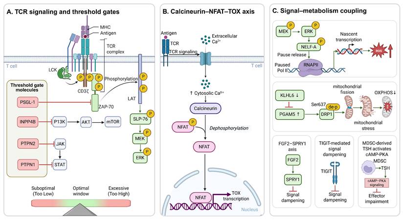
Persistent stimulation can therefore be converted into transcriptional burden and metabolic stress. Sustained mitogen-activated protein kinase kinase (MEK) activation increases phosphorylation of the Ser5 and Ser2 residues within the carboxy-terminal domain of RNA polymerase II (RNAPII-CTD), thereby driving energy-intensive nascent transcription and protein synthesis, together with reactive oxygen species accumulation and metabolic impairment, and ultimately biasing CD8⁺ T cells toward terminal exhaustion [26]. Downregulation or loss of kelch-like family member 6 (KLHL6) also induces dephosphorylation of dynamin-related protein 1 (DRP1) at Ser637, which promotes excessive mitochondrial fission, loss of membrane potential, and decreased oxidative phosphorylation, thereby driving deeper exhaustion in tumor-infiltrating CD8⁺ T cells [41]. Phosphorylation therefore determines not only whether signaling occurs, but also whether persistent stimulation can be converted into a transcriptional and metabolic burden that the cell can no longer tolerate.
At the receptor-proximal level, phosphorylation imbalance is reflected in resetting of the T-cell activation window [42]. Sustained Ca²⁺ influx drives dephosphorylation and nuclear translocation of NFAT, which then induces TOX, TOX2, and PD-1 expression, thereby forming a critical bridge between chronic stimulation and exhaustion-associated transcriptional programs [43]. In parallel, P-selectin glycoprotein ligand 1 (PSGL-1) suppresses proximal phosphorylation cascades involving phosphorylated ζ-chain-associated protein kinase 70 (p-ZAP70), extracellular signal-regulated kinase 1/2 (ERK1/2), and protein kinase B (AKT), thereby restricting signal amplification and stabilizing the inhibitory receptor phenotype [44]. Phosphorylation of Y-box-binding protein 1 (YBX1) at Ser102 by AKT1 activates hypoxia-inducible factor-2α (HIF-2α) and promotes CD8⁺ Tex [45]. Folic acid has also been reported to enhance AKT phosphorylation and activate Notch receptor 1 (NOTCH1) signaling, thereby facilitating CD8⁺ Tex formation [46]. Distinct from this downstream AKT-associated pro-exhaustion mechanism, the intracellular domain of basigin (CD147) acts at the level of proximal TCR signaling by interacting with CD3ε, CD3ζ, and lymphocyte-specific protein tyrosine kinase (LCK), enhancing CD3ζ/LCK/ZAP70 phosphorylation and promoting LCK/ZAP70 recruitment to CD3ζ, thereby causing proximal signaling overload and accumulation of tumor-infiltrating CD8⁺ Tex [47]. These findings suggest that commitment toward CD8⁺ Tex is determined not by the absolute strength of proximal TCR output alone, but by whether phosphorylation output remains within a range that sustains effector function without causing chronic overload.
This threshold shift is not limited to the proximal TCR machinery. It is further reinforced across broader signaling networks by the concerted action of intrinsic negative regulators and extrinsic suppressive signals. Inositol polyphosphate 4-phosphatase type II B (INPP4B) dephosphorylates phosphatidylinositol (3,4)-bisphosphate (PI(3,4)P2) into phosphatidylinositol 3-phosphate (PI(3)P), weakens TCR/CD28-induced PI3K-AKT activation, and positively correlates with exhaustion scores and PDCD1 expression in single-cell analyses [48]. Protein tyrosine phosphatase non-receptor type 2 (PTPN2) and PTPN1, also known as PTP1B, increase the activation threshold of CD8⁺ T cells and consolidate exhausted phenotypes by dephosphorylating proximal TCR kinases and inputs to Janus kinase (JAK)-signal transducer and activator of transcription (STAT) signaling [49]. Stromal fibroblast growth factor 2 (FGF2) promotes dephosphorylation of linker for activation of T cells (LAT) and SH2-domain-containing leukocyte protein of 76 kDa (SLP-76) through a CBL-mediated negative-feedback loop, while simultaneously increasing inhibitory phosphorylation of LCK at Tyr505, thereby driving terminal exhaustion [27]. Signaling related to the ITIM-containing receptor TIGIT further reinforces this suppressed state by weakening TCR-induced ERK phosphorylation and reducing IFN-γ production [50]. Phosphorylation-dependent threshold resetting is therefore not a transient event. It evolves into a durable hyporesponsive state through the joint action of intrinsic brakes and microenvironment-derived inhibitory inputs.
Phosphorylation networks also extend into intercellular suppressive signaling within the TME. Tumor-associated signals activate transforming growth factor-β (TGF-β)-SMAD2/3 and ERK1/2 phosphorylation cascades in Schwann cells, which in turn induce upregulation of PDCD1 and NT5E in CD8⁺ T cells. NT5E encodes ecto-5′-nucleotidase CD73, which cooperates with CD39 to convert extracellular ATP-derived AMP into immunosuppressive adenosine, thereby promoting CD8⁺ Tex [51]. Myeloid-derived suppressor cells (MDSCs) secrete thyroid-stimulating hormone (TSH) and cooperate with tumor-derived exosomes to induce phosphorylated cAMP response element-binding protein (p-CREB), thereby transcriptionally activating genes encoding co-inhibitory receptors such as PDCD1 and HAVCR2 [52]. IFN-γ can also promote phosphorylation of sphingosine kinase 1 (SPHK1) at Ser225 in tumor-associated macrophages (TAMs), amplifying myeloid-cell recruitment and accelerating functional exhaustion of CD8⁺ tumor-infiltrating lymphocytes (TILs) [53]. C-C motif chemokine ligand 23 (CCL23) secreted by M2-like TAMs activates the CCR1-glycogen synthase kinase 3β (GSK-3β) phosphorylation axis and is accompanied by coordinated upregulation of multiple checkpoint molecules [54]. A Siglec-9⁺ TAM-associated SHP-1 pathway has likewise been closely linked to the development of CD8⁺ Tex [55]. These observations indicate that phosphorylation converts stromal and myeloid cells into sustained amplifiers of inhibitory signaling, thereby further shifting the activation window from outside the T cell.
The supply of inhibitory ligands from tumor cells can also be persistently reinforced through phosphorylation, which further stabilizes the already established hyporesponsive state [56]. In tumor cells, PTPN22 blocks K48-linked ubiquitin-mediated degradation of PD-L1 by promoting dephosphorylation of CBL at Tyr700, thereby stabilizing PD-L1 expression [57]. Under high-glucose conditions, ERK2 phosphorylates V-domain Ig suppressor of T-cell activation (VISTA) at Ser248, recruits the deubiquitinase USP22, and stabilizes VISTA through deubiquitination, which promotes CD8⁺ Tex [58]. ERK1/2 also phosphorylates CREB1 at Ser133, upregulates USP8, reduces ubiquitination of TGF-β receptor 2 (TGFBR2), and increases its stability, thereby inducing an exhausted phenotype [59]. Phosphorylation of basic leucine zipper ATF-like transcription factor 2 (BATF2) at Ser227 is a key event for promoting stimulator of interferon genes (STING) oligomerization and amplifying type I interferon (IFN-I) responses. Consistently, high-glutamine states induced by tumor cells suppress BATF2 expression through epigenetic mechanisms, thereby weakening effector expansion and promoting exhaustion [60]. Increased tumor-cell levels of phosphorylated SHP2 at Tyr542 are also associated with upregulation of exhaustion markers such as HAVCR2 and PDCD1 in CD8⁺ Tex [61]. Loss of tumor-cell CD58 results in excessive p-STAT1 activation and concomitant PD-L1 upregulation, thereby strengthening the immunosuppressive barrier [62]. Immune-derived signals can, in turn, further shape the inhibitory output of tumor cells. Extracellular granzyme K (GZMK) triggers AKT-GSK3β-β-catenin and JAK2-STAT1 phosphorylation cascades, upregulates tumor-cell PD-L1, and promotes CD8⁺ Tex formation [63]. Non-canonical phosphorylation of STAT1 at Ser727 can likewise promote CD8⁺ Tex by upregulating CD276 [64]. Similar roles have been reported for phosphorylation-related pathways involving STAT3, GSK-3β, β-catenin, AKT, NF-κB, immunoreceptor tyrosine-based inhibitory motifs (ITIMs), and protein phosphatase 1A (PP1A), all of which participate in PD-L1 upregulation in tumor cells and reinforcement of an inhibitory TME. Phosphorylation therefore promotes CD8⁺ Tex not only through T-cell-intrinsic signaling but also by sustaining the supply of inhibitory ligands from tumor cells.
Phosphorylation-related suppressive signals can even spread beyond the tumor through long-range intercellular transfer. Tumor-derived extracellular vesicles carrying phosphorylated interferon-γ receptor 1 (IFNGR1) can trigger a JAK1-STAT1 phosphorylation cascade, upregulate PD-L1, and induce an exhaustion program in CD8⁺ T cells [65]. At the same time, acidic pH and interleukin-8 (IL-8) within the microenvironment enhance STAT1/STAT3 phosphorylation and are associated with upregulation of the PD-L1/PD-1 axis [66]. These results indicate that phosphorylation-mediated inhibitory remodeling is not confined to the local tumor niche. It can also precondition distant sites and create molecular conditions that favor exhaustion before T cells arrive, thereby extending the impact of persistent antigenic stimulation beyond local spatial and temporal boundaries.
Taken together, the central role of phosphorylation in CD8⁺ Tex lies not in a simple increase or decrease in the activity of any single pathway, but in the continuous reshaping of T-cell responses to chronic stimulation. Conversion of persistent stimulation into transcriptional and metabolic burden, deviation of the proximal TCR activation window, cooperative reinforcement by intrinsic and extrinsic inhibitory signals, and intercellular amplification within both local and distant niches together form the upstream mechanism that initiates the exhaustion program.
3.2 Protein homeostasis
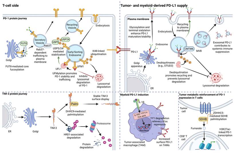
Suppressive signal transduction alone cannot fully explain why the exhausted phenotype is maintained over long periods. Transient inhibitory input becomes chronic pressure because the processing, maturation, membrane localization, stability, and degradation of inhibitory receptors such as PD-1 and TIM-3 and their ligands, including PD-L1, are continuously regulated [67]. In addition, critical proteins functionally coupled to these pathways must also be maintained at high steady-state levels to sustain inhibitory input. At this layer, ubiquitination, glycosylation, palmitoylation, and UFMylation jointly shape the homeostasis of checkpoints and associated proteins through control of degradation, biosynthetic processing, membrane presentation, and PTM crosstalk (Fig. 4), thereby preserving suppressive input over time.
Sustained preservation of inhibitory input in CD8⁺ T-cell exhaustion through ubiquitination, glycosylation, palmitoylation, and UFMylation. Upper left: in T cells, PD-1 undergoes FUT8-mediated core fucosylation, Rab37-dependent membrane transport, and USP5/USP24-mediated recycling and stabilization. UFL1-UFM1-mediated UFMylation further promotes PD-1 stability and trafficking while suppressing lysosomal degradation, whereas K48-linked ubiquitination promotes degradation. Upper right: Tumor- and myeloid cell-derived PD-L1 is stabilized and matured by glycosylation and terminal sialylation. CMTM6 and deubiquitination facilitate recycling after endocytosis and prevent lysosomal degradation, whereas exosomal PD-L1 further strengthens systemic immune suppression. Lower left: TIM-3 remains stably expressed on the membrane after DHHC9-mediated palmitoylation, whereas HRD1-associated ubiquitination promotes its proteasomal degradation. Lower center: in TAMs, the IL-1β-IL1R2-YY1-c-Fos axis promotes surface PD-L1 upregulation. Lower right: tumor-derived metabolic input can further enhance PD-1 expression in T cells through ZDHHC5-mediated palmitoylation of SDHB, fumarate accumulation, and H3K27ac-associated activation of PDCD1 transcription.
3.2.1 Ubiquitination
Ubiquitination is mediated by the E1-E2-E3 enzyme cascade, which covalently conjugates ubiquitin to lysine residues on substrate proteins. Depending on chain type and context, it can direct proteasomal degradation, receptor turnover, or reorganization of signaling complexes, whereas deubiquitinases (DUBs) reversibly remove ubiquitin chains. Ubiquitination is therefore a key determinant of protein half-life and cell-surface receptor homeostasis [68]. In CD8⁺ Tex, its major significance lies in stabilizing checkpoint molecules and other critical proteins, thereby preserving inhibitory input and progressively fixing a stable functionally suppressed state.
At the T-cell-intrinsic level, inflammatory signals within the TME can alter PD-1 protein homeostasis. CAF-derived IL-8 downregulates the E3 ligase FBXO38 in CD8⁺ T cells and suppresses ubiquitin-mediated degradation of PD-1 [25, 69]. Interleukin-6 (IL-6) derived from TAMs and tumor cells induces the deubiquitinase USP24 through STAT3/NF-κB signaling, enhances removal of K48-linked chains from PD-1, and markedly prolongs the PD-1 half-life, thereby sustaining a PD-1hi exhausted TIL state [70]. In addition, ERK-mediated site-specific phosphorylation promotes the interaction between PD-1 and the deubiquitinase USP5, leading to removal of degradation-associated ubiquitin chains and further stabilization of the PD-1 protein [28]. Defects in core ubiquitin-system components similarly impair PD-1 degradation and allow PD-1 accumulation in TILs [71]. Sustained PD-1 expression is thus not only a transcriptional phenomenon, but also a steady-state consequence of restricted degradation and enhanced protein stability.
Persistent suppressive input is also reflected at the ligand-supply side. OTU domain-containing protein 3 (OTUD3) removes K48-linked ubiquitin chains from PD-L1 and blocks its proteasomal degradation [72]. Extracellular GZMK activates protease-activated receptor 2 (PAR2) in tumor cells, inhibits PD-L1 ubiquitin-dependent degradation, and maintains high PD-L1 abundance [63]. Similar deubiquitinating mechanisms can affect non-classical checkpoints. USP2a removes K48/K63-linked chains from B7 homolog 4 (B7-H4), increases its stability, and is associated with exhaustion-related infiltrates [73]. Moreover, sustained expression of inhibitory ligands is not limited to single-cell-intrinsic processes. It can be amplified through cross-cellular cooperation. TAM-derived IL-1β activates IL-1R2, whose intracellular domain drives ubiquitin-proteasome-mediated degradation of YY1, thereby synchronously increasing PD-L1 in TAMs and tumor cells and deepening TIL exhaustion [74].
Once inhibitory ligand supply is persistently maintained, changes in the homeostasis of T-cell-intrinsic signaling molecules and exhaustion-associated proteins further determine whether the exhaustion program can be sustained. Upregulation of the E3 ligase Pellino 1 (Peli1) promotes K48-linked ubiquitination and degradation of protein kinase C-θ (PKC-θ), thereby weakening TCR signaling and maintaining the PD-1hi Tex phenotype [75]. By contrast, loss of the E3 ligases NEURL3, RNF149, and WSB1 causes abnormal K48-linked ubiquitination, accumulation of unfolded proteins, and enhanced unfolded protein response (UPR), leading to loss of the Tpex pool and accelerated terminal exhaustion [76]. The deubiquitinase USP7 stabilizes recombination signal binding protein for immunoglobulin kappa J region (RBPJ) through deubiquitination and drives an interferon regulatory factor 4 (IRF4)/tumor necrosis factor receptor superfamily member 1B (TNFRSF1B) axis that maintains exhaustion programs [77]. In parallel, there are also ubiquitin-dependent restraints that limit terminal exhaustion. KLHL6 promotes K48-linked ubiquitination and degradation of TOX, thereby constraining the transition from Tpex to Texterm [41].
Notably, ubiquitin-dependent control extends beyond classical signaling or transcriptional regulators to metabolic and mitochondrial nodes that determine whether suppression can be maintained. Tumor-derived exosomes deliver β-transducin repeat-containing protein (β-TrCP) into CD8⁺ T cells, which induces ubiquitination-mediated degradation of yes-associated protein 1 (YAP1), suppresses expression of amino acid transporters SLC7A5 and SLC38A1, and is accompanied by increased inhibitory receptor expression and impaired effector function [78]. Valosin-containing protein (VCP) stabilizes glycerol-3-phosphate dehydrogenase 1-like protein (GPD1L) by preventing its ubiquitin-dependent degradation, leading to glycerol-3-phosphate accumulation and enhanced inhibitory phosphorylation of LCK at Tyr505. In this way, abnormal metabolic protein homeostasis is converted into sustained hyporesponsive signaling [79]. NFATc1 upregulates the mitochondrial deubiquitinase USP30, which counteracts PINK1/Parkin-mediated ubiquitination of mitochondrial proteins, inhibits mitophagy, promotes accumulation of damaged mitochondria, and thereby drives both formation and maintenance of CD8⁺ Tex [80]. Similarly, dipeptidyl peptidase 7 (DPP7) in TAMs blocks TRIM25-mediated ubiquitination of carnitine palmitoyltransferase 1A (CPT1A), stabilizes CPT1A, enhances fatty acid β-oxidation (FAO)/OXPHOS, and maintains an immunosuppressive macrophage phenotype that favors accumulation of CD8⁺ Tex [81]. K48-linked ubiquitination and proteasomal degradation of tropomyosin receptor kinase A (TRKA) suppress glycolysis in CD8⁺ T cells and promote exhaustion [82]. Ubiquitination therefore sustains inhibitory input not only by stabilizing checkpoints themselves, but also by fixing a broad set of proteins that regulate TCR output, mitochondrial quality control, and immunosuppressive myeloid states.
Tumor-driven abnormal signaling can likewise induce ubiquitination imbalance in critical proteins and thereby exacerbate CD8⁺ Tex. High expression of the long intergenic non-coding RNA LINC01116 in tumor cells blocks RAD18-mediated ubiquitination and degradation of EWS RNA-binding protein 1 (EWSR1). This promotes the preferential uptake of long-chain fatty acids, especially linoleic acid, by tumor cells, causing lipid nutrient competition with CD8⁺ T cells in the TME and ultimately exacerbating CD8⁺ Tex [83]. Circulating tumor cells induce CD8⁺ Tex by triggering CBL-mediated ubiquitination and degradation of epidermal growth factor receptor (EGFR) [84]. In tumor cells, follistatin-like 3 (FSTL3) translocates into the nucleus, binds c-Myc, prevents its ubiquitin-mediated degradation, upregulates PD-L1, and weakens responsiveness to anti-PD-1 therapy [85]. The tumor metabolite kynurenine suppresses aryl hydrocarbon receptor (AhR) ubiquitin-dependent degradation in CD8⁺ T cells and promotes its nuclear translocation, leading to increased PD-1, LAG-3, and TIM-3 and reduced IFN-γ/TNF production [86]. In tumor cells, fucosyltransferase 2 (FUT2) promotes FBXO2-mediated ubiquitination and degradation of nuclear receptor subfamily 2 group F member 2 (NR2F2). Radiotherapy suppresses FUT2 through METTL14-dependent N6-methyladenosine (m6A), ultimately inducing CD8⁺ Tex [87]. High expression of the E3 ligase bifunctional apoptosis regulator (BFAR) in tumor cells drives K48-linked ubiquitination and degradation of pre-mRNA processing factor 19 (PRPF19), promoting surface PD-L1 expression on tumor-associated neutrophils [88]. Likewise, high tumor-cell expression of ependymin-related protein 1 (EPDR1) interferes with TRIM21-mediated ubiquitination and autophagic degradation of IKKβ, enhances NF-κB-dependent PD-L1 transcription, and promotes CD8⁺ Tex [89]. Ring finger protein 26 (RNF26) also promotes K48-linked ubiquitin-mediated degradation of glucose-regulated protein 78 (GRP78), induces persistent endoplasmic reticulum stress, upregulates PD-L1, and deepens CD8⁺ Tex [90]. These observations indicate that tumor-cell ubiquitin abnormalities do not simply affect scattered metabolic or stress pathways. They stabilize key proteins that control checkpoint expression, antigen presentation, and recruitment of immunosuppressive cells, thereby maintaining suppressive input at high load.
Taken together, the central role of ubiquitination in CD8⁺ Tex is to convert transient inhibitory input into a sustained molecular state by controlling the degradation and stability of PD-1, PD-L1, and other critical proteins.
3.2.2 Glycosylation
Whereas ubiquitination mainly determines degradation and retention of checkpoints and related proteins, glycosylation primarily controls their maturation and cell-surface homeostasis along the endoplasmic reticulum-Golgi route. N-glycosylation, O-glycosylation, and related glycan modifications participate in protein folding, quality control, intracellular trafficking, membrane localization, and ligand recognition, and thus directly determine the maturation efficiency and surface availability of membrane proteins [91]. In CD8⁺ Tex, glycosylation is critical because it maintains the processing, trafficking, and membrane residence of checkpoint molecules and other key proteins, thereby supporting persistent inhibitory input and stabilizing the exhausted phenotype.
At the T-cell-intrinsic level, abnormal glycosylation can sustain inhibitory receptor burden by affecting both effector-molecule secretion and checkpoint membrane homeostasis. In tumor-infiltrating CD8⁺ T cells, downregulation of the oligosaccharyltransferase (OST) complex leads to insufficient occupancy of N-glycosylation sites and creates a mismatch in which IFN-γ transcripts increase but protein secretion declines. Because IFN-γ exerts negative-feedback inhibition on PDCD1, this secretory defect weakens PD-1 restraint and promotes exhaustion [92]. Glycosylation can also directly determine the stable membrane residence of PD-1. FUT8-mediated core fucosylation maintains PD-1 surface expression, whereas FUT8 inhibition reduces membrane PD-1 and enhances T-cell activation [93]. Complementing this glycan effect, PD-1 glycosylation facilitates its loading into Rab37-positive vesicles and subsequent plasma membrane presentation. Consistently, Rab37 deficiency reduces PD-1 and TIM-3 expression and is accompanied by restoration of T-cell effector function [29]. In addition, glycosylation may interact with other PTMs to influence the steady-state levels of exhaustion-associated proteins. Short-term fasting blocks N-glycosylation of the fatty acid transporter CD36, increases AMP-activated protein kinase (AMPK) phosphorylation, reduces UFM1 modification of the deubiquitinase USP7, enhances ubiquitin-mediated USP7 degradation, and ultimately promotes degradation of RBPJ and suppression of the IRF4/TNFRSF1B exhaustion axis [77]. Thus, glycosylation prolongs suppressive signaling both by maintaining maturation and membrane presentation of receptors such as PD-1 and by influencing key homeostatic proteins through PTM crosstalk.
Persistent suppressive input is also reflected on the supply side through stable maintenance of inhibitory ligands and related membrane proteins. In tumor cells, either the β-glucuronidase (GUSB) H351Q mutation or glycosyltransferase 8 domain-containing protein 2 (GLT8D2) promotes aberrant N-glycosylation of PD-L1, thereby enhancing PD-L1 stability and increasing infiltration by exhausted CD8⁺ T cells [94]. Loss of tuberous sclerosis complex subunit 1 (TSC1) enhances α2,6-sialylation of PD-L1, increases its affinity for PD-1, and ultimately promotes CD8⁺ Tex formation [95]. Beyond PD-L1, aberrant glycosylation can also sustain suppressive input by stabilizing other immunoregulatory membrane proteins at the cell surface. FUT8-mediated core fucosylation of semaphorin 7A (SEMA7A) in tumor cells promotes differentiation of CD8⁺ T cells along the exhaustion trajectory and increases the expression of multiple checkpoint molecules [96]. These data indicate that glycosylation abnormalities in tumor cells do not simply increase the abundance of a single ligand. Rather, they maintain long-term suppressive load by enhancing the maturation, surface stability, and receptor-binding capacity of inhibitory molecules.
Notably, the glycome of intratumoral CD8⁺ T cells changes early during exhaustion. Increased β1,6-GlcNAc-branched N-glycans are associated with a PD-1⁺TIM-3⁺ phenotype [97]. This finding suggests that glycosylation not only maintains already established checkpoint homeostasis, but may also preconfigure a cell state that is intrinsically more prone to acquiring a persistent inhibitory receptor phenotype.
Taken together, the key role of glycosylation in CD8⁺ Tex is to extend suppressive input over time and stabilize it in space by controlling the maturation, intracellular trafficking, membrane presentation, and receptor-ligand binding capacity of checkpoints and associated proteins.
3.2.3 Palmitoylation
Palmitoylation is a reversible lipid modification in which palmitate and related long-chain fatty acids are covalently linked to cysteine residues through thioester bonds. This modification amplifies signaling output by altering membrane affinity, subcellular localization, and protein stability, thereby reinforcing persistent inhibitory effects mediated by checkpoints and associated proteins [98]. Palmitoylation is mainly written by the zinc finger DHHC-type palmitoyltransferase family (ZDHHCs) and erased by depalmitoylating enzymes, forming a high-turnover cycle [99].
The contribution of this PTM to CD8⁺ Tex first becomes evident at the level of inhibitory receptors themselves. In TIM-3-expressing CD8⁺ T cells, the palmitoyltransferase DHHC9 has been shown to catalyze the palmitoylation of TIM-3 at Cys296, thereby suppressing its ubiquitination-mediated degradation and maintaining TIM-3 stability and surface expression, ultimately contributing to CD8⁺ Tex [30]. Beyond directly stabilizing checkpoint receptors, palmitoylation can indirectly sustain suppressive programs by stabilizing key metabolic signaling nodes. ZDHHC5-mediated palmitoylation of succinate dehydrogenase complex subunit B (SDHB) prevents its lysosomal degradation, promotes fumarate production, enhances H3K27ac-dependent PD-1 transcription, and ultimately facilitates CD8⁺ Tex formation [100]. Likewise, after palmitate enters CD8⁺ T cells through CD36, it increases STAT3 palmitoylation and membrane localization, transcriptionally activates TOX, and promotes differentiation toward Texterm [101]. These findings indicate that palmitoylation not only extends the membrane half-life of inhibitory receptors, but also stabilizes upstream proteins that continuously support checkpoint expression and exhaustion-associated transcriptional programs.
Taken together, the central role of palmitoylation in CD8⁺ Tex is to prolong the effective lifetime of inhibitory molecules and exhaustion-associated nodes by enhancing membrane residence, suppressing degradation, and stabilizing functional protein sites.
3.2.4 UFMylation
UFMylation is a ubiquitin-like modification mediated by UFM1. In the present context, it appears to reinforce protein homeostasis mainly through crosstalk with other PTMs. Although the available evidence remains limited, its position in CD8⁺ Tex is becoming clearer. UFMylation does not seem to create an entirely independent exhaustion-driving pathway. Rather, it acts as a cross-regulatory component within the homeostatic network that controls checkpoints and related proteins [102]. By directly stabilizing PD-1 and acting in concert with glycosylation and ubiquitination, UFMylation helps preserve suppressive molecules and prolong the period during which inhibitory input is stably maintained [31, 67].
Taken together, ubiquitination, glycosylation, palmitoylation, and UFMylation act through distinct molecular mechanisms, yet they converge on a common outcome in CD8⁺ Tex: maintenance of the homeostasis of checkpoint molecules and associated critical proteins. At this layer, the threshold shift initiated by altered signaling is translated into durable checkpoint overexpression and abnormal steady-state control of key proteins, thereby forming a major basis for the maintenance of CD8⁺ Tex.
3.3 Metabolic stress responses
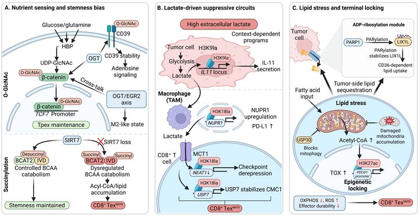
The mechanisms operating at the levels of signal transduction and protein homeostasis are not sufficient to explain how metabolic stress is further integrated into a durable exhaustion program. Hypoxia, nutrient restriction, lactate accumulation, lipid overload, and NAD⁺-related stress in the TME do not spontaneously become stable exhausted states. They must first be sensed and inscribed through specific PTMs before they can be deposited as functional defects and stable fate changes. Based on current evidence, O-GlcNAcylation, succinylation, lactylation, and ADP-ribosylation translate metabolic stress into persistent exhaustion programs by reshaping T-cell fate, rewiring metabolic pathways, reinforcing suppressive niches, and disrupting mitochondrial homeostasis, thereby jointly promoting the formation and progression of CD8⁺ Tex (Fig. 5).
O-GlcNAcylation, succinylation, lactylation, and ADP-ribosylation mediate metabolic stress responses in CD8⁺ T-cell exhaustion. (A) O-GlcNAcylation and succinylation inscribe nutrient sensing into stemness maintenance and immunosuppressive bias. The hexosamine biosynthetic pathway (HBP)-O-GlcNAc transferase (OGT) axis supports β-catenin/TCF7-driven Tpex maintenance and stabilizes CD39 to enhance adenosine signaling. β-Catenin also crosstalks with the OGT-EGR2 axis to promote an M2-like state. Loss of SIRT7 is associated with abnormal succinylation of BCAT2 and IVD, disrupted branched-chain amino acid catabolism, and terminal exhaustion. (B) A high-lactate microenvironment establishes a suppressive circuit among tumor cells, TAMs, and CD8⁺ T cells through lactylation. In tumor cells, H3K9la activates the IL11 locus and promotes IL-11 secretion. In TAMs, H3K18la-associated NUPR1 upregulation enhances PD-L1 expression. Lactate uptake through monocarboxylate transporter 1 (MCT1) induces H3K18la-associated NEAT1 downregulation and USP7 upregulation in T cells; USP7 stabilizes CMC1, and these changes promote terminal exhaustion. (C) ADP-ribosylation and lipid stress jointly aggravate mitochondrial imbalance and facilitate terminal locking. Poly(ADP-ribose) polymerase 1 (PARP1)-dependent PARylation stabilizes LIX1L and enhances CD36-dependent lipid uptake. USP30 suppresses mitophagy, leading to accumulation of damaged mitochondria, increased acetyl-CoA, TOX upregulation, and enhanced H3K27ac at the PDCD1 promoter, ultimately causing reduced OXPHOS, elevated ROS, and formation of CD8⁺ Texterm.
3.3.1 O-GlcNAcylation
O-GlcNAcylation mainly occurs on serine or threonine residues of nuclear, cytoplasmic, and mitochondrial proteins. It is written by O-GlcNAc transferase (OGT), which uses uridine diphosphate N-acetylglucosamine (UDP-GlcNAc) as a donor, and erased by O-GlcNAcase (OGA) [103]. In the context of CD8⁺ T-cell exhaustion, O-GlcNAcylation translates nutrient sensing into T-cell metabolic programming, fate maintenance, and remodeling of immunosuppressive niches, with its functional outcome determined by the modified substrate, cell type, and tumor metabolic context.
At the T-cell-intrinsic level, O-GlcNAcylation links nutrient status to metabolic rewiring and maintenance of a precursor-like program, thereby influencing the tendency of CD8⁺ T cells to differentiate toward exhaustion under chronic stimulation. D-mannose has been shown to reduce glycolysis and enhance mitochondrial function while increasing OGT-mediated O-GlcNAcylation and stabilization of β-catenin. This preserves Tcf7 expression and epigenetic stemness, thereby promoting a stem-like program and restricting exhaustion differentiation [32]. By contrast, substrate-specific O-GlcNAcylation may also support exhaustion-associated phenotypes. Increased OGT activity promotes O-GlcNAcylation of CD39 at Ser239, inscribing elevated hexosamine biosynthetic pathway activity into high CD39 expression, which is accompanied by reduced cytotoxic molecules and increased exhaustion markers [104].
O-GlcNAcylation also acts outside T cells by converting glucose-metabolic signals into tumor-promoting differentiation programs in myeloid cells. OGT-mediated O-GlcNAcylation of early growth response 2 (EGR2) at Ser299 promotes the formation of protumorigenic TAM subsets and thereby enhances CD8⁺ Tex features [105]. In tumor cells, O-GlcNAcylation can further reshape secretory programs that support immunosuppressive niches. OGT-mediated O-GlcNAcylation of Wilms tumor 1-associated protein (WTAP), together with USP7-mediated deubiquitination, stabilizes WTAP, drives myeloid macrophages toward an M2-like program, and promotes CD8⁺ Tex [106]. Clinical correlation analyses also suggest that high expression of EGF domain-specific O-linked N-acetylglucosamine transferase (EOGT) is associated with CD8⁺ Tex in tumors, although a direct causal mechanism remains to be established [107].
Taken together, O-GlcNAcylation does not uniformly promote CD8⁺ Tex, but shapes exhaustion-related programs in a substrate-, cell-type-, and tumor metabolic context-dependent manner.
3.3.2 Lysine succinylation
Succinylation is an acyl modification that introduces a succinyl group onto the epsilon-amino group of lysine residues. This changes residue charge and thereby influences protein conformation, enzymatic activity, and interaction networks [108]. Current direct evidence connecting succinylation to CD8⁺ Tex centers on sirtuin 7 (SIRT7), which restrains branched-chain amino acid catabolism and subsequent lipid accumulation through desuccinylation. Loss of SIRT7 in T cells promotes metabolic imbalance and drives CD8⁺ T cells toward exhaustion [34].
3.3.3 Lactylation
Lactylation is an acyl modification in which a lactyl group is introduced onto the epsilon-amino group of lysine residues. It can form specific epigenetic marks such as H3K9la and H3K18la and translate increased glycolysis and lactate accumulation into altered transcriptional output [109]. In CD8⁺ Tex, lactylation can contribute importantly by establishing a lactate-driven suppressive chain among tumor cells, myeloid cells, and T cells, thereby expanding local metabolic pressure into a durable immunosuppressive network.
In tumor cells, lactylation converts metabolic pressure into altered secretory programs and immunosuppressive output [110]. Tumor-cell forkhead box K1 (FOXK1)-driven glycolipid metabolic reprogramming increases lactate production and modulates TOX-induced histone lactylation in CD8⁺ T cells, thereby promoting CD8⁺ Tex formation and immune evasion [33]. Intratumoral lactate accumulation enriches H3K9la at the IL-11 transcriptional start region, promotes IL-11 secretion, increases multiple inhibitory receptors on CD8⁺ T cells, and suppresses effector output [111]. Non-histone lactylation also contributes to this process. In tumor cells, hepatitis B virus (HBV)-induced lactate production promotes lactylation of spectrin alpha, non-erythrocytic 1 (SPTAN1) at K1952/K1957 in an alanyl-tRNA synthetase 1 (AARS1)-dependent manner, thereby facilitating accumulation of CD8⁺ Tex [112]. In myeloid cells, tumor-derived lactate enters TAMs and induces H3K18la, which increases PD-L1/PD-L2 expression and ultimately promotes accumulation of PD-1⁺ CD8⁺ Tex [113]. Loss of claudin 7 (CLDN7) can also remodel neutrophil metabolism, increase glycolysis and histone lactylation together with PD-L1 upregulation, and thereby induce CD8⁺ Tex [114].
Within CD8⁺ T cells, lactylation under high-lactate conditions can directly reshape exhaustion-associated transcriptional programs and disturb mitochondrial homeostasis. Lactate entry increases pan-lactylation (Pan-Kla) and H3K18la at the NEAT1 promoter, suppresses NEAT1 transcription, and drives exhaustion programs [115]. Lactate can also accelerate terminal differentiation through crosstalk between lactylation and protein homeostasis. It induces Pan-Kla enrichment at the Usp7 promoter and upregulates USP7, which then deubiquitinates and stabilizes cytochrome c oxidase assembly factor CMC1, rendering CD8⁺ T cells more prone to enter terminal exhaustion [116].
Thus, in a tumor microenvironment enriched in lactate, lactylation converts local metabolic pressure into a mutually reinforcing immunosuppressive network spanning tumor cells, myeloid cells, and T cells, while further promoting terminal exhaustion and mitochondrial dysfunction in CD8⁺ T cells.
3.3.4 ADP-ribosylation
ADP-ribosylation is a modification in which ADP-ribosyltransferases transfer ADP-ribose groups from NAD⁺ onto substrate proteins. It can occur as mono-ADP-ribosylation (MARylation) or poly-ADP-ribosylation (PARylation) [117]. In tumor cells, poly(ADP-ribose) polymerase 1 (PARP1) mediates PARylation of limb expression 1-like protein (LIX1L), increases its stability, and promotes CD36-mediated lipid uptake. This remodels a protumorigenic microenvironment and is consistent with impaired maintenance of CD8⁺ T-cell effector function and deviation toward exhaustion [35]. This suggests that ADP-ribosylation may link tumor-cell metabolic remodeling to CD8⁺ Tex-associated changes in the immune microenvironment. However, direct evidence for T cell-intrinsic regulation of CD8⁺ Tex programs by ADP-ribosylation remains limited.
Taken together, O-GlcNAcylation, succinylation, lactylation, and ADP-ribosylation involve different metabolic substrates and cellular processes, yet they converge in CD8⁺ Tex to sense, transmit, and inscribe metabolic stress into persistent exhaustion programs, thereby driving sustained exhaustion and progression toward terminal states.
3.4 Epigenetic reprogramming
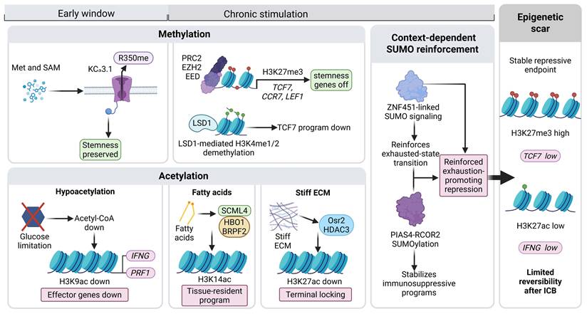
Mechanisms at the levels of signal transduction, protein homeostasis, and metabolic stress still do not fully explain why CD8⁺ Tex ultimately enter a relatively stable and difficult-to-reverse lineage state. The crucial issue is whether the upstream abnormalities can be further inscribed into chromatin organization, enhancer activity, and transcriptional regulatory complexes so that dynamic responses become fixed as stable epigenetic programs. At this layer, methylation, acetylation, and SUMOylation progressively close stemness-associated programs, maintain exhaustion-related transcriptional networks, and ultimately drive CD8⁺ T cells into a terminally exhausted state (Fig. 6).
Methylation, acetylation, and SUMOylation drive epigenetic reprogramming in CD8⁺ T-cell exhaustion. Upper left, early window: methionine/S-adenosylmethionine (Met/SAM) supply sustains KCa3.1 R350 methylation and preserves stemness programs. Upper middle, chronic stimulation stage: the polycomb repressive complex 2 (PRC2)-EZH2-EED axis deposits H3K27me3 and suppresses stemness-associated genes, whereas lysine-specific demethylase 1 (LSD1) removes H3K4me1/2 and further attenuates stemness programs. Lower section: aberrant acetylation inscribes metabolic and microenvironmental stress into chromatin states. Glucose restriction lowers acetyl-CoA and H3K9ac, thereby suppressing effector genes. The fatty acid-associated SCML4-HBO1-BRPF2 axis sustains H3K14ac and supports tissue-resident programs in CD8⁺ T cells. A stiff extracellular matrix (ECM) decreases H3K27ac through the Osr2-HDAC3 axis and promotes terminal locking. Middle right: SUMOylation further reinforces inhibitory epigenetic programs under chronic stimulation. Far right: these changes are ultimately deposited as epigenetic scars that show only limited reversibility after ICB.
3.4.1 Methylation
Methylation can occur on histone and non-histone lysine or arginine residues, generating mono-, di-, or trimethylation states. These marks are written mainly by protein arginine methyltransferases (PRMTs) and SET-domain-containing methyltransferases and are erased by lysine-specific demethylase 1 (LSD1) and the lysine demethylase (KDM) family [118]. By creating cumulative epigenetic marks, methylation influences both the retention of CD8⁺ T-cell plasticity and the fixation of the exhausted lineage.
This role is first evident in the early activation phase, when T cells are highly sensitive to methyl-donor availability. Within approximately 30 minutes of initial activation, CD8⁺ T cells become acutely dependent on methionine (Met)/S-adenosylmethionine (SAM) supply. When Met/SAM is sufficient, dimethylation of KCa3.1 at Arg350 is maintained, excessive Ca²⁺ influx and NFAT1 nuclear translocation are restrained, and initiation of the exhaustion program is suppressed. By contrast, even transient methionine insufficiency can bias cells toward terminal exhaustion [119]. Within the TME, this early deviation can be further amplified by nutrient competition. Tumor cells competitively consume methionine, leading to reduced H3K79me2 in T cells and impaired STAT5 function, whereas methionine supplementation can at least partially restore this axis and improve the exhausted state [120].
During chronic stimulation, methylation further pushes CD8⁺ T cells from reversible suppression toward lineage fixation by silencing stemness-associated genes and maintaining exhaustion-related transcriptional programs. In specific models, increased activity of polycomb repressive complex 2 (PRC2) deposits H3K27me3 at stemness-associated loci such as Tcf7 and drives the Tpex-to-Texterm transition [121]. PRC2 can also suppress C-X-C motif chemokine ligand 10 (CXCL10) through H3K27me3, limit recruitment of CXCR3⁺ CD8⁺ T cells, reduce peripheral effector T-cell replenishment, and thereby reinforce the immunosuppressive microenvironment [122]. As the counterpart to methyltransferase activity, demethylation also determines whether this state can be reversed. LSD1 removes H3K4me1/2, antagonizes TCF-1-dependent maintenance programs, silences stemness networks, and promotes terminal exhaustion [123]. In PD-1⁺ CD8⁺ T cells, nuclear phosphorylated LSD1 co-localizes with eomesodermin (EOMES) and regulates its modification state, thereby maintaining EOMES-associated exhaustion-like transcriptional output [124]. In addition, the macrophage IRG1 (ACOD1)-itaconate axis promotes H3K4me3 at the EOMES promoter, increases PD-1/TIM-3, and drives exhaustion [125]. Oxidative stress can also induce SET7/9-mediated methylation of sirtuin 1 (SIRT1) and block its secretion, promoting accumulation of PD-L1⁺ M2-like TAMs and thereby deepening CD8⁺ Tex [126].
Thus, methylation drives CD8⁺ T cells from functional suppression toward epigenetic lineage fixation through three coordinated processes: early restriction of methyl-donor availability, chronic silencing of stemness-associated genes, and maintenance of exhaustion-related transcriptional programs.
3.4.2 Acetylation
Acetylation mainly occurs on the epsilon-amino group of lysine residues. It is written by lysine acetyltransferases (KATs), erased by histone deacetylases (HDACs), and read by bromodomain-containing proteins, thereby regulating chromatin accessibility, assembly of transcriptional complexes, and gene-expression programs [127]. During the formation of CD8⁺ Tex, acetylation integrates metabolic substrate availability, tissue stress, and tumor-derived inhibitory signals into relatively stable epigenetic states.
One manifestation of this role is the sustained maintenance of exhaustion-associated enhancer activity. Enhancers or super-enhancers adjacent to exhaustion-associated genes such as Pdcd1, Havcr2, and Tox are frequently enriched for H3K27ac, often together with H3K4me3, which is a recurrent epigenetic feature of CD8⁺ Tex [37]. In human studies, elderly patients with diffuse large B-cell lymphoma (DLBCL) more often harbor abnormalities in regulators such as TET2 and CREBBP. These changes are accompanied by reduced enhancer activity, decreased histone acetylation, reduced major histocompatibility complex class I (MHC-I), fewer naive T cells, and increased CD8⁺ Tex [128]. Such observations suggest that acetylation does not merely affect isolated loci, but participates in defining the enhancer landscape characteristic of CD8⁺ Tex.
Mechanistically, acetylation directly inscribes nutrient restriction and altered metabolic pathways into chromatin remodeling. Prolonged glucose limitation reduces acetyl-CoA production, lowers H3K9/14ac and H3K27ac, compresses chromatin accessibility at effector-gene loci, and leads to exhaustion-like functional decline in CD8⁺ T cells [129]. Under chronic antigenic stimulation, nuclear acetyl-CoA becomes more dependent on ATP-citrate lyase (ACLY), and redistribution of H3K27ac, H3K9ac, and H4ac mediated by KAT2A promotes the transition from Tpex to Texterm [130]. In contrast, fatty-acid metabolism can limit terminal exhaustion by maintaining expression of SCM polycomb group protein-like 4 (SCML4), increasing H3K14ac, and enhancing chromatin accessibility [131].
In addition to substrate availability, mechanical cues within the TME and tumor-derived suppressive signals can further consolidate exhaustion through acetylation. A stiff extracellular matrix activates an odd-skipped related transcription factor 2 (Osr2) axis that recruits HDAC3 and decreases H3K27ac at cytotoxic gene loci such as Prf1 and Ifng, thereby suppressing cytotoxic programs and promoting Texterm differentiation [132]. In tumor cells, casein kinase 2 β (CK2B) progressively increases along the Tpex-to-Texterm trajectory and upregulates HDAC8, which suppresses the T-bet program by reducing H3K27ac at the TBX21 promoter and thereby promotes a PD-1⁺ TIM-3⁺ terminally exhausted state [133]. Tumor-cell acetylation abnormalities may also indirectly shape suppressive niches by altering secretory programs. Aldolase B (ALDOB) translocates into the nucleus and interacts with KAT2A, thereby decreasing H3K9ac at the TGFB1 promoter and suppressing TGFB1 transcription. Conversely, ALDOB deficiency increases TGF-β expression and promotes an exhaustion-like phenotype in tumor-infiltrating CD8⁺ T cells [134]. Beyond histones, acetylation of non-histone substrates can reshape tumor metabolism and immunogenicity. Under chemotherapy stress, Tat-interactive protein 60 kDa (TIP60) mediates acetylation of glycerol-3-phosphate acyltransferase 3 (GPAT3) at K316, enhances lipid synthesis and lipid-droplet accumulation, and is accompanied by an increased PD-1⁺ TIM-3⁺ exhausted-like population [135]. Importantly, although HDAC inhibitors can enhance effector-molecule release by activated CD8⁺ T cells in vitro, they may also produce co-existing effector-like and exhausted-like populations as well as increased myeloid and regulatory T-cell components in vivo, highlighting strong state dependence and timing dependence [136].
Overall, acetylation promotes a more stable terminally exhausted state in CD8⁺ T cells by reshaping enhancer activity and chromatin accessibility, thereby integrating substrate availability, mechanical stress, and tumor-derived inhibitory signals into exhaustion-associated transcriptional programs.
3.4.3 SUMOylation
SUMOylation is a reversible modification in which small ubiquitin-like modifier (SUMO) proteins are conjugated to lysine residues on substrates. It affects gene-expression programs by modulating protein-protein interactions, subcellular localization, and the stability of nuclear transcriptional complexes [137]. More broadly, SUMOylation is often associated with transcriptional repression. It can facilitate recruitment of HDAC-containing repressor complexes by modifying transcription factors and co-repressors and can also participate in regulating enhancer acetylation marks and chromatin states [138]. At present, however, direct evidence that SUMOylation mediates epigenetic fixation in CD8⁺ Tex remains limited. It is better regarded as a reinforcement mechanism for inhibitory epigenetic programs than as a simple regulator of protein homeostasis.
Available evidence indicates that hypoxia can upregulate REST corepressor 2 (RCOR2) through hypoxia-inducible factor-1α (HIF-1α) and promote PIAS4-mediated SUMO2 modification of RCOR2 at Lys60. This suppresses RCOR2 ubiquitin-dependent degradation and stabilizes it in the nucleus. Stable RCOR2 then enhances transcription of leukemia inhibitory factor (LIF), drives M2-like polarization of TAMs, and promotes accumulation of CD8⁺ Tex [139]. These findings indicate that SUMOylation can contribute to maintenance and reinforcement of inhibitory epigenetic programs by stabilizing nuclear transcriptional corepressor networks. Single-cell analyses have also implicated the SUMO E3 ligase ZNF451 in transitions of the CD8⁺ Tex state, although its direct link to epigenetic fixation remains to be defined [140].
Taken together, methylation, acetylation, and SUMOylation act on different substrates and through different biochemical mechanisms, yet they converge on a shared outcome: they convert the earlier layers of altered signaling, protein homeostasis, and metabolic stress responses into stable epigenetic programs.
In summary, CD8⁺ T-cell exhaustion is a progressive process shaped cooperatively by multiple PTM classes across different layers. This view suggests that intervention at PTM nodes should move beyond isolated target blockade and toward combination strategies guided by mechanistic layering, temporal matching, and complementary modes of action.
4. Therapeutic opportunities and rational combination strategies to alleviate CD8⁺ Tex
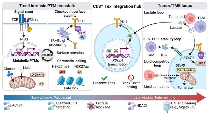
Increasing evidence indicates that CD8⁺ T-cell exhaustion is not driven by a single modification event. Rather, it reflects a dynamic process jointly shaped by T-cell-intrinsic PTM crosstalk networks and tumor- or TME-derived extrinsic feedback loops. By altering signal transduction, protein homeostasis, metabolic stress responses, and epigenetic reprogramming, diverse PTMs influence the differentiation trajectory from Tpex to Texterm, and therefore offer opportunities to enhance ICB, ACT, radiotherapy, and chemotherapy-based combinations (Fig. 7). Representative PTM-oriented intervention strategies aimed at alleviating CD8⁺ T-cell exhaustion are summarized in Table 1.
Representative PTM-based intervention strategies currently reported to alleviate CD8⁺ T-cell exhaustion
| PTMs | Agents | Targets | Mechanism of action | Combination | Ref |
|---|---|---|---|---|---|
| Phosphorylation | SRPK1 inhibitor SPHINX31 | SRSF1 | Phosphorylation-dependent activation of the splicing factor SRSF1 biases PD-1 mRNA toward the pro-exhaustion membrane isoform (flPD1). | - | [141] |
| Phosphorylation | Ginsenoside Rg5 | STAT2 | Suppresses STAT2 phosphorylation and downregulates PD-L1 expression. | Anti-PD-1 antibody | [143] |
| Phosphorylation | Curcumin (PTPN22 inhibitor) | CBL | Blocks CBL Tyr700 dephosphorylation and lowers PD-L1 stability in tumor cells. | Anti-PD-L1 antibody; anti-CTLA-4 antibody | [57] |
| Phosphorylation | Factor Xa inhibitor rivaroxaban | STAT2 | Downregulates PD-L1 by reducing STAT2 phosphorylation. | Anti-PD-1 antibody | [145] |
| Phosphorylation | PTPN inhibitor AC484 (ABBV-CLS-484) | PTPN2/PTPN1 | Blocks PTPN2/PTPN1-mediated dephosphorylation, enhances JAK/STAT signaling, lowers the TCR activation threshold, and broadens effector function. | Anti-PD-1 antibody | [49] |
| Phosphorylation | PTP1B allosteric inhibitor MSI-1436 | STAT5 | Inhibits PTP1B activity and enhances STAT5 Tyr694 phosphorylation. | Anti-PD-1 antibody | [147] |
| Phosphorylation | PKC-θ inhibitor nPKC-θi2 | PKC-θ | Interferes with nuclear PKC-θ-related serine/threonine phosphorylation programs and disrupts the ZEB1/PKC-θ inhibitory complex. | - | [148] |
| Phosphorylation | Euphorbia helioscopia L. | STAT3 | Reduces STAT3 phosphorylation and increases TCF-1 expression. | - | [151] |
| Phosphorylation | JAK3 inhibitor PF-06651600 | STAT5 | Suppresses IL-2R downstream STAT5 phosphorylation and weakens chronic cytokine signaling. | - | [149] |
| Phosphorylation | AURKB inhibitor AZD1152 | H3K9me3S10ph at the NCEH1 promoter | Reduces the H3K9me3S10ph mark at the NCEH1 promoter, restores H3K9me3/HP1 repression, and lowers cholesterol accumulation. | Chemotherapy (gemcitabine or cisplatin) and anti-PD-1 antibody | [152] |
| Phosphorylation | MEK inhibitor | RNAPII CTD | Reduces Ser5/Ser2 phosphorylation of the RNAPII-CTD, suppresses energy-intensive nascent transcription, and alleviates mitochondrial NADH/ROS accumulation. | - | [26] |
| Phosphorylation | Zuojin capsules | mTOR, eIF4E, p70S6K | Suppresses phosphorylation-dependent activation of mTOR, eIF4E, and p70S6K and downregulates cyclin-dependent kinase 1 (CDK1), thereby remodeling the immunosuppressive TME. | - | [158] |
| Phosphorylation | GSK3 inhibitor SB415286 | GSK3β | Blocks M2 TAM-derived CCL23/CCR1-GSK3β phosphorylation signaling in CD8⁺ T cells, reducing CTLA-4, TIGIT, TIM-3, LAG-3, and PD-1 expression. | - | [54] |
| Phosphorylation | MERTK inhibitor MRX-2843 | MERTK | Inhibits MERTK receptor tyrosine phosphorylation, expands CD8α⁺ dendritic cells, and enhances antigen presentation. | - | [155] |
| Phosphorylation | Licochalcone B | ERK5, NF-κB | Breaks an NCAM1-associated immunosuppressive lineage by inhibiting ERK5 and NF-κB p65 phosphorylation and inducing ferroptosis. | - | [157] |
| Ubiquitination | USP5 inhibitor EOAI3402143 | USP5/PD-1 | Inhibits USP5 deubiquitinase activity, promotes K48-linked ubiquitination and degradation of PD-1, and reduces PD-1 expression on CD8⁺ T cells. | Anti-CTLA-4 antibody | [28] |
| Ubiquitination | USP24 inhibitor USP24-i-101 | USP24/PD-1 | Inhibits USP24-mediated deubiquitination and lowers PD-1 protein stability. | Anti-CTLA-4 antibody | [70] |
| Ubiquitination | OTUD3 inhibitor rupatadine | OTUD3/PD-L1 | Inhibits OTUD3-mediated deubiquitination, promotes K48-linked ubiquitination and degradation of PD-L1, and lowers tumor-cell PD-L1 expression. | Anti-PD-L1 antibody | [72] |
| Ubiquitination | MARCH5 inhibitor pitavastatin calcium | γc | Inhibits MARCH5-mediated K27-linked polyubiquitination and lysosomal degradation of γc, thereby upregulating γc and enhancing IL-2/STAT5 signaling. | Anti-PD-1 antibody | [176] |
| Ubiquitination | USP8 inhibitor | TβRII | Inhibits deubiquitination of TβRII and lowers its plasma membrane stability, markedly reducing the burden of circulating tumor-derived extracellular vesicles. | Chemotherapy (paclitaxel) and anti-PD-L1 antibody | [177] |
| Ubiquitination | Ellagic acid | NRF2 | Reduces ubiquitin-proteasome degradation of NRF2, activates the NRF2-TFAM axis, and enhances OXPHOS/ATP supply. | Anti-PD-1 antibody | [178] |
| Ubiquitination | USP30 inhibitor ST-539 | USP30 | Suppresses USP30 transcription and protein expression, thereby relieving PINK1/Parkin pathway-mediated defects in mitophagy. | - | [80] |
| Palmitoylation | FASN inhibitor C75 | STAT3 | Inhibits fatty acid synthase (FASN) and reduces STAT3 palmitoylation and activation. | Anti-PD-1 antibody | [101] |
| O-GlcNAcylation | OGT inhibitor OSMI-1 | WTAP | Inhibits OGT and OGT-mediated O-GlcNAcylation of WTAP, weakens WTAP stability, and suppresses M2-like TAM differentiation. | - | [106] |
| O-GlcNAcylation | D-mannose | β-catenin | Increases UDP-GlcNAc supply, enhances OGT-mediated O-GlcNAcylation of β-catenin, promotes its nuclear translocation, and drives stemness-associated transcriptional programs such as Tcf7. | - | [32] |
| Lactylation | NUPR1 inhibitor | NUPR1 | Reactivates ERK/JNK-MAPK signaling in TAMs, reverses M2-like polarization, and reduces PD-L1/SIRPA-mediated immunosuppressive signaling. | Anti-PD-1 antibody | [113] |
| Methylation | KCa3.1 inhibitor TRAM-34 | KCa3.1 | Maintains dimethylation of KCa3.1 Arg350, restricts excessive Ca²⁺ influx and NFAT1 nuclear translocation, and suppresses initiation of exhaustion programs such as Pdcd1/Lag3. | Anti-PD-1 antibody | [119] |
| Methylation | PRC2 inhibitor EED226; EZH2 inhibitor GSK126; tazemetostat | H3K27me3 | Inhibits EZH2/PRC2, lowers H3K27me3, relieves silencing of stemness-associated genes such as Tcf7, and expands the TCF-1⁺ Tpex pool. | Anti-PD-1 antibody | [121, 122, 184] |
| Methylation | Ibuprofen | H3K4me3 at the EOMES promoter | Inhibits the macrophage IRG1/itaconate axis and blocks the itaconate-succinate-H3K4me3-EOMES exhaustion program. | Anti-PD-1 antibody | [125] |
| Methylation | LSD1 inhibitors GSK2879552 and ORY-1001 | LSD1, H3K4me1/2 | Inhibits LSD1-mediated demethylation, increases H3K4me1/2, and expands the intratumoral TCF-1⁺ Tpex pool. | Anti-PD-1 antibody | [122] |
| Methylation | Fumarate | KDM5/H3K4me3 | Inhibits KDM5-mediated H3K4me3 demethylation, maintains H3K4me3 and TCF-1 expression at the Tcf7 promoter. | Anti-PD-L1 antibody | [186] |
| Methylation | KDM4C inhibitor SD70 | KDM4C; H3K36me3 at the CXCL10 promoter | Increases H3K36me3 enrichment at the CXCL10 promoter by inhibiting KDM4C, upregulates CXCL10, enhances CD8⁺ T-cell migration and activation, and lowers PD-1/CD39 expression. | Radiotherapy and anti-PD-L1 antibody | [187] |
| Acetylation | HDAC6 inhibitors ACY-1215 and ACY-241 | HDAC6 | Downregulates the GATA3-driven T helper 2 (Th2) transcriptional program, weakens immunosuppressive programs, and increases T-bet. | - | [191] |
| Acetylation | HDAC inhibitor SAHA (vorinostat) | HDAC, JAK1 | Globally increases acetylation and remodels the exhaustion trajectory; may also weaken the FGL1-LAG3 axis through JAK1 Lys1109 acetylation. | - | [192] |
| Acetylation | CK2 inhibitor CX4945; HDAC8 inhibitor PCI-34051 | H3K27ac at the TBX21 promoter | Restores H3K27ac at the TBX21 promoter and upregulates T-bet. | Anti-PD-1 antibody | [133] |
| Acetylation | HDAC3 inhibitor RGFP966 | HDAC3/H3K27ac at the Prf1 and Ifng promoters | Blocks Osr2-mediated recruitment of HDAC3, restores H3K27ac at loci including Prf1 and Ifng, and releases epigenetic repression of cytotoxic genes. | - | [132] |
| Acetylation | Class I HDAC inhibitor entinostat (MS-275) | HDAC1/3 | Inhibits HDAC1/3-mediated histone deacetylation, restores immune-cell function in tumors, and suppresses immunosuppressive pathways driven by increasing tumor burden. | - | [190] |
| Acetylation | KAT2A inhibitor MB3 | KAT2A/H3K9ac at the TGFB1 promoter | Inhibits the histone acetyltransferase activity of KAT2A, lowers H3K9ac at the TGFB1 promoter, and restricts TGF-β output. | - | [134] |
| Acetylation | ACLY inhibitor BMS-303141 | ACLY | Blocks ACLY-KAT2A-mediated citrate-dependent nuclear acetyl-CoA supply. | Anti-PD-L1 antibody | [130] |
An integrated hub formed by T-cell-intrinsic PTM crosstalk and tumor/TME feedback circuits suggests stage-specific intervention strategies for CD8⁺ Tex. The left panel depicts the intrinsic PTM network in T cells: phosphorylation resets signaling thresholds; glycosylation, UFMylation, and ubiquitination regulate PD-1 surface stability; metabolism-related PTMs reshape mitochondrial states; and chromatin-related PTMs promote fate stabilization. The central hub highlights the joint nuclear control of PDCD1 transcription by lactylation and acetylation and identifies preserving Tpex while blocking Texterm locking as a key therapeutic objective. The right panel shows tumor- and TME-derived feedback loops, including a lactate circuit, an IL-6-p-STAT3-USP24-mediated PD-1 stabilization loop, and lipid competition-driven metabolic stress. The lower panel proposes two intervention windows along disease progression: an early “pulse-reset” window that focuses on correcting signaling thresholds and checkpoint homeostasis, and a late “fate-rewiring” window that focuses on releasing epigenetic constraints and integrating ACT-based engineering strategies.
4.1 Phosphorylation
Because phosphorylation encodes proximal TCR/CD3ζ immunoreceptor tyrosine-based activation motif (ITAM) signaling as well as downstream co-stimulatory and cytokine receptor signals such as JAK-STAT and AKT-mTOR, it can influence both initiation and progression of exhaustion by resetting activation thresholds and the duration of signal transmission. Among PTM classes, phosphorylation related pathways are currently among the most tractable therapeutic entry points, although their effects remain highly context dependent.
Phosphorylation networks can reduce extrinsic inhibitory input by suppressing the generation or upregulation of inhibitory receptors and ligands along the PD-1/PD-L1 axis. In T cells, the SRPK1 inhibitor SPHINX31 blocks phosphorylation of serine/arginine-rich splicing factor 1 (SRSF1) and functionally achieves an exhaustion-relieving effect similar to PD-1 blockade [141]. Under acidic tumor conditions, NaHCO3 buffering combined with the eukaryotic translation initiation factor 4A (eIF4A) inhibitor silvestrol reduces STAT1 phosphorylation and lowers PD-L1 expression in tumor cells [66]. Tumor cells also harbor druggable upstream transcriptional nodes. Asiaticoside suppresses SMAD2/3 phosphorylation and ultimately reduces PD-1, TIM-3, and LAG-3 expression on CD8⁺ T cells [142]. Ginsenoside Rg5 inhibits the NFKB2/STAT2 axis and downregulates PD-L1 [143]. Curcumin suppresses PTPN22, restores CBL-mediated K48-linked ubiquitination and degradation of PD-L1, and decreases PD-L1 stability [57]. Conventional treatments may also dampen checkpoint supply by interfering with phosphorylation-related pathways. ISP-I combined with temozolomide (TMZ) downregulates PD-L1 through remodeling of GSK-3β/β-catenin-associated phosphorylation signaling [144], and rivaroxaban, a factor Xa (FXa) inhibitor, also reduces STAT2 phosphorylation together with PD-L1 expression [145].
In addition to reducing inhibitory input, phosphorylation-targeted interventions can delay Texterm formation by resetting T-cell-intrinsic phosphorylation hierarchies spanning the proximal TCR network, cytokine receptor pathways, and nuclear programs. ATP depletion under chronic stimulation preferentially damages distal ITAM phosphorylation and thereby promotes exhaustion, suggesting that receptor engineering to reset ITAM phosphorylation hierarchies may improve ICB or ACT efficacy [146]. The dual PTPN2/PTPN1 inhibitor AC484 lowers exhaustion by blocking dephosphorylation along the JAK-STAT axis and has entered a phase I clinical trial (NCT04777994) [49]. The allosteric PTP1B inhibitor MSI-1436 reduces the proportion of PD-1⁺ TIM-3hi CD8⁺ Tex in tumors by enhancing STAT5 Tyr694 phosphorylation [147]. The selective inhibitor nPKC-θi2 blocks nuclear translocation of protein kinase C-θ (PKC-θ), interferes with exhaustion-associated nuclear serine/threonine phosphorylation programs, and relieves Tex [148]. Inflammatory STAT-maintenance circuits can also be pharmacologically corrected. Low-dose continuous PF-06651600, a JAK3 inhibitor, reduces terminally exhausted phenotypes by suppressing persistent IL-2R-JAK3-STAT5 phosphorylation [149]. Tumor-derived Epstein-Barr virus-induced gene 3 (EBI3) promotes STAT4 phosphorylation and upregulates IL10/CCL5 to induce CD8⁺ Tex, whereas the antagonistic peptide VYLHWHD competitively binds EBI3 and reverses the induced phenotype [150]. An aqueous extract of Euphorbia helioscopia L. attenuates CD8⁺ Tex in inflammation-driven tumors by lowering STAT3 phosphorylation [151]. Some interventions act even further downstream at nuclear and translational phosphorylation outputs. The aurora kinase B (AURKB) inhibitor AZD1152 reduces H3K9me3-S10ph-associated marks and decreases CD8⁺ Tex [152]. Perioperative low-dose sirolimus suppresses phosphorylation of ribosomal protein S6 (rpS6), thereby mitigating surgery-induced CD8⁺ Tex [153]. MEK or MAPK13 inhibition regulates phosphorylation of the RNAPII-CTD and TCF-1 and helps maintain TCF-1⁺ Tpex cells [26, 154].
Beyond T-cell-intrinsic pathways, phosphorylation-directed interventions can alleviate CD8⁺ Tex indirectly by remodeling tumor-cell and myeloid-cell programs. The GSK3α/β inhibitor SB415286 attenuates exhaustion-like phenotypes by interfering with the GSK3β Ser9 phosphorylation axis [54]. The MERTK inhibitor MRX-2843 suppresses receptor tyrosine phosphorylation and alleviates Tex through a dendritic cell-T-cell axis [155]. TCP-1-targeted liposomal delivery of parthenolide (PTL) induces phosphorylation-dependent necroptosis in tumor cells and secondarily improves CD8⁺ Tex [156]. Licochalcone B remodels the Tex landscape by suppressing phosphorylation of ERK5 and NF-κB p65 [157]. At the level of herbal formulations, Zuojin capsules improve Tex by inhibiting phosphorylation-dependent activation of mTOR, eIF4E, and p70S6K [158], whereas Xiaoshui Formula suppresses TAM M2 polarization through inhibition of NF-κB p65 phosphorylation and thereby alleviates CD8⁺ Tex [159].
Correction of phosphorylation circuits related to hypoxia and metabolic stress represents another strategy for alleviating CD8⁺ Tex through improvement of the TME and mitochondrial homeostasis. Hypoxic TME drives CD8⁺ T cells toward PD-1hi terminal exhaustion. Escherichia coli Nissle 1917 carrying gold-palladium (Au-Pd) nanocatalysts reduces hypoxia-associated exhaustion by suppressing pCREB signaling and synergizes with stereotactic body radiotherapy (SBRT) and anti-PD-L1 treatment [160]. Ultrasound-targeted microbubble cavitation (UTMC) induces phosphorylation of endothelial nitric oxide synthase (eNOS) at Ser1177 and Ser633, alleviates hypoxia, reduces CD8⁺ Tex, and can further enhance anti-PD-L1 efficacy [161]. Mitochondrial metabolic adaptation is another point of leverage. Diisopropylamine dichloroacetate (DADA) inhibits phosphorylation of the pyruvate dehydrogenase complex (PDC), enhances OXPHOS and mitochondrial fitness, and suppresses terminal exhaustion [162]. Under fractionated radiotherapy, the combination of metformin and 2-deoxyglucose (2-DG) liposomes suppresses incomplete mitochondrial outer membrane permeabilization (iMOMP)-integrated stress response (ISR) adaptation and reduces eIF2α phosphorylation, thereby attenuating radiotherapy-induced adaptive PD-L1 upregulation [163].
During ACT manufacturing and engineering, the ex vivo time window provides a more controllable context in which phosphorylation circuits can be preset to improve the Tpex-like features and persistence of reinfused cells. CAR-T cells have been shown to express vascular endothelial growth factor receptor 2 (VEGFR2), suggesting that vascular endothelial growth factor (VEGF)-VEGFR2 signaling may act directly on CAR-T cells rather than being confined to its conventional anti-angiogenic context. Short-term pretreatment with axitinib, a small-molecule VEGFR inhibitor, suppresses VEGFR2 phosphorylation in CAR-T cells and reduces degradation-associated phosphorylation of β-catenin at Ser33/Ser37/Thr41, thereby stabilizing β-catenin, activating Wnt/β-catenin signaling, and alleviating CAR-T cell terminal differentiation and exhaustion [164]. Engineered expression of the interleukin-21 receptor (IL-21R) maintains key JAK-STAT phosphorylation signals in the absence of exogenous IL-21, enhances proliferation and cytotoxicity, and reduces inhibitory receptor expression [165]. Similarly, interleukin-17A (IL-17A) has been associated with restoration of AKT phosphorylation and a reduction in PD-1⁺ TIM-3⁺ CD8⁺ Tex [166]. Relief of dephosphorylation-mediated repression can also be engineered. CRISPR-mediated deletion of SHP-1 enhances cytotoxic output and shows stronger antitumor efficacy when combined with simvastatin [167]. Engineering viral tyrosine kinase-interacting protein (TIP) triggers STAT5 phosphorylation, sustains survival and effector function in the absence of IL-2, and suppresses exhaustion differentiation [168]. At the mitochondrial level, forkhead box P3 (FOXP3) engineering reduces phosphorylation of DRP1 at Ser616, suppresses mitochondrial fission, rewires metabolism, and ultimately mitigates CD8⁺ Tex [169].
Importantly, phosphorylation-targeted therapy is not inherently equivalent to ICB sensitization. The net effect depends on the functional position of the target as well as timing and dosing intensity. Excessive anabolic signaling can sustain high AKT-mTOR-S6 phosphorylation and accelerate overstimulation and exhaustion-like differentiation of CD8⁺ T cells [170]. Ex vivo analyses of patient-derived cells show that dasatinib simultaneously suppresses TCR and STAT5 phosphorylation and has differential effects in TIM-3⁺ T-cell subsets, suggesting marked subset dependence of Src-family kinase-related interventions [171]. By contrast, intermittent interleukin-2-inducible T-cell kinase (ITK) inhibition can reactivate exhausted cytotoxic T lymphocytes in multiple ICB-resistant solid-tumor models and enhance ICB efficacy through remodeling of proximal TCR phosphorylation [172]. Yet inhibition of protein kinase D (PKD) suppresses AKT phosphorylation in T cells and antagonizes anti-PD-1 therapy [173]. Tumor-derived extracellular vesicles can also promote exhaustion through non-canonical pathways. For example, exosomal lncRNA HDAC2-AS2 suppresses cytoplasmic cyclin-dependent kinase 9 (CDK9) in CD8⁺ T cells, reduces p38 phosphorylation, increases PD-1 expression, and aggravates exhaustion [174].
Thus, the key to phosphorylation-directed intervention is not to globally enhance or suppress phosphorylation, but to select targets, delivery modes, and combinations that reset activation thresholds, prevent chronic stimulation from being converted into transcriptional or metabolic burden, block TME-derived amplification of inhibitory phosphorylation, and reduce the supply of inhibitory ligands from tumor cells.
4.2 Ubiquitination
Compared with phosphorylation, ubiquitination more directly controls the half-life and cell-surface abundance of inhibitory receptors or ligands, cytokine receptors, and proteins involved in mitochondrial quality control, thereby influencing whether suppressive load can be durably maintained. Because the ubiquitin-proteasome system (UPS) is broadly required for cell survival and stress responses, strategies to alleviate CD8⁺ Tex are better suited to targeting specific DUBs or E3 ligases than to globally inhibiting the UPS.
Targeting selected DUBs and E3 ligases can reduce PD-1 stability in T cells while also diminishing PD-L1 supply from tumors or the microenvironment, thereby reducing sustained suppressive input. USP5 has been identified as a deubiquitinase for PD-1, and its inhibitor EOAI3402143 promotes PD-1 degradation [28]. USP24 also deubiquitinates and stabilizes PD-1, whereas USP24-i-101 reduces PD-1 stability and decreases PD-1⁺ TIM-3⁺ CD8⁺ Tex [70]. OTUD3 stabilizes PD-L1 through deubiquitination, and the antiallergic drug rupatadine has been reported to inhibit OTUD3 and promote PD-L1 degradation [72]. The fusion peptide UM-6 blocks N-glycosylation of PD-L1 and facilitates its ubiquitin-mediated degradation, thereby attenuating PD-1-associated CD8⁺ Tex [175]. Microenvironment-driven UPS loops can also be interrupted. A neutralizing antibody against IL-1R2 blocks the ICD-IL1R2-YY1 UPS axis, lowers tumor-cell PD-L1, and enhances anti-PD-1 efficacy [74].
Ubiquitin-dependent regulation can also restore upstream signaling support required for T-cell activation by resetting the homeostasis of cytokine receptors and alternative inhibitory axes. The MARCH5 inhibitor pitavastatin calcium suppresses K27-linked ubiquitination and lysosomal degradation of the common γ-chain (γc), thereby strengthening cytokine signaling and improving exhaustion-associated phenotypes [176]. On the bypass-suppression side, USP8 inhibition lowers the steady-state level of TGF-β receptor II (TβRII), alleviates extracellular vesicle-mediated TGF-β signaling, and improves CD8⁺ T-cell function [177].
At the Texterm stage, deficient mitochondrial function together with ROS accumulation and reduced ATP supply forms a major metabolic bottleneck, and the UPS-mitochondrial quality-control axis provides an entry point for metabolic rescue. T-cell-specific high mobility group box 2 (HMGB2) promotes UPS-dependent degradation of nuclear factor erythroid 2-related factor 2 (NRF2), weakens mitochondrial OXPHOS, and can be counteracted by ellagic acid, which synergizes with anti-PD-1 therapy to reduce terminal exhaustion [178]. The USP30 inhibitor ST-539 enhances ubiquitination of mitochondrial proteins and mitophagy, improves mitochondrial adaptation, and decreases Texterm [80].
In ACT settings, ubiquitination also affects stability of stem-like populations through chromatin-associated marks. CRISPR disruption of chromatin regulators such as additional sex combs-like 1 (ASXL1) maintains H2AK119ub1 and stabilizes the TCF-1⁺Ly108⁺ Tpex pool, which can synergize with anti-PD-L1 therapy [179]. However, because chromatin regulators are pleiotropic, such strategies require more systematic evaluation of off-target effects and long-term phenotypic consequences.
4.3 Glycosylation
Glycosylation-directed interventions may alleviate CD8⁺ Tex and enhance treatment response at three levels: restoration of T-cell function, ex vivo glycoengineering, and optimization of delivery. N-glycosylation defects in T cells are associated with impaired effector function, and compensating for the oligosaccharyltransferase (OST) complex can restore IFN-γ production and alleviate CD8⁺ Tex [92]. In ex vivo engineering and delivery settings, glycoengineering provides a more direct and controllable means of product optimization. CRISPR knockout of Mgat5 removes β1,6-GlcNAc-branched N-glycans, reduces exhaustion phenotypes, and enhances cytotoxic activity [97]. In addition, site-specific conjugation based on glycosylation sites has enabled αPD-1-(iRGD)2 to improve antibody penetration into "cold tumors" and to work in combination with ACT [180].
4.4 Palmitoylation
Targeting palmitoylation and its upstream lipid-supply pathways can reduce the burden of terminal exhaustion and strengthen antitumor activity. For example, the competitive peptide TIM-3-Palm-WT blocks TIM-3 palmitoylation, reduces PD-1⁺ TIM-3⁺ TOX⁺ TCF-1- CD8⁺ Texterm, and enhances the antitumor killing capacity of TILs, CAR-T cells, and natural killer (NK) cells [30]. The fatty acid synthase (FASN) inhibitor C75 reduces STAT3 palmitoylation and activation, thereby decreasing PD-1hi TIM-3⁺/CD39⁺ CD8⁺ Texterm [101].
4.5 O-GlcNAcylation
Within immunosuppressive niches, the OGT inhibitor OSMI-1 may attenuate CD8⁺ Tex by weakening WTAP-dependent immunosuppressive signaling; in translational studies, its combination with LOXL2 antagonism further improves anti-PD-1 responses [106]. In contrast, D-mannose supplementation represents a more T-cell-intrinsic strategy by enhancing β-catenin O-GlcNAcylation, preserving Tcf7-associated stem-like programs, and limiting CD8⁺ Tex progression [32]. These findings suggest that the therapeutic effect of O-GlcNAcylation on CD8⁺ Tex depends on the cellular context and modified substrate rather than simple unidirectional inhibition or enhancement.
4.6 Lactylation
Inhibition of lactate-driven histone lactylation, particularly the reduction of key lactylation marks such as H3K18la, may suppress the transcription of exhaustion-associated genes and provide a therapeutic entry point for restoring CD8⁺ T-cell effector function. NUPR1 inhibition reduces H3K18la, increases IFN-γ⁺/GzmB⁺ CD8⁺ T cells, and decreases PD-1⁺ CD8⁺ T cells [113]. Recent studies have further expanded the therapeutic relevance of the p300 inhibitor C646, suggesting that, beyond its established role in inhibiting p300/CBP histone acetyltransferase activity, it can also be leveraged for lactylation-associated immune reprogramming. CD8α⁺ nanovesicle-mediated delivery of C646 enables targeted entry into exhausted CD8⁺ T cells, reduces H3K18la enrichment at the PDCD1 promoter, suppresses PDCD1 transcription and PD-1 expression, and restores IFN-γ and GZMB expression [181].
4.7 ADP-ribosylation
Current studies more strongly support ADP-ribosylation as a microenvironment-remodeling or combination-sensitizing module than as a direct reversal strategy for terminal exhaustion. PARP14 inhibition combined with PD-1 blockade regulates macrophage polarization and induces a quiescent state in cytotoxic CD8⁺ T cells, thereby mitigating terminal exhaustion while preserving functionality [182]. Whether this class can reverse terminal exhaustion as monotherapy remains unclear.
4.8 Methylation
Methylation-oriented strategies often emphasize short-course intervention and precise stratification so as to avoid long-term disruption of the global epigenetic landscape. Brief methionine supplementation or short-term treatment with the KCa3.1 inhibitor TRAM-34 maintains dimethylation of KCa3.1 Arg350 and suppresses initiation of the CD8⁺ Tex program [119]. In dietary-metabolic interventions, α-ketoglutarate supplementation shows marked sex dependence. It decreases H3K27me3 and H3K9me3 in females, but may increase H3K27me3 and remodel exhaustion features in males, suggesting that strict stratification and timing control are required [183].
The PRC2-H3K27me3 axis directly participates in the transcriptional repression and reactivation of exhaustion-associated genes. The PRC2 inhibitor EED226 reduces H3K27me3, relieves the epigenetic repression of memory/naïve-associated genes, and downregulates selected exhaustion-associated programs [121]. Within the EZH2-H3K27me3-CXCL10 axis, the EZH2 inhibitor GSK126 may delay exhaustion progression by relieving the epigenetic repression of CXCL10 [122]. Moreover, short-term tazemetostat treatment during ex vivo expansion preferentially reduces H3K27me3 at the Tcf7 promoter, expands PD-1⁺ TIM-3- TCF-1⁺ Tpex, and decreases Texterm [184]. In contrast, engineering strategies further suggest that EZH2 activity can also be used to precondition T cells against tumor-imposed metabolic stress. In tumor-specific CD8⁺ T cells, expression of the gain-of-function EZH2Y641F mutant enhances PRC2-mediated H3K27me3, improves energy metabolism and cytotoxic function, and attenuates the progression of metabolic stress-associated exhaustion [185]. These findings suggest that the therapeutic effects of the EZH2-H3K27me3 axis depend on the intervention window, target gene loci, and exhaustion stage.
Besides H3K27me3, H3K4- and H3K36-related marks also participate in maintenance of stemness, recruitment, and exhaustion control in CD8⁺ T cells. Ibuprofen lowers H3K4me3 enrichment at the EOMES promoter and slows progression toward Texterm [125]. LSD1 inhibitors such as GSK2879552 and ORY-1001 expand the intratumoral TCF-1⁺ Tpex pool by inhibiting removal of H3K4me1/2 [36, 123]. Fumarate inhibits KDM5, maintains H3K4me3 at the Tcf7 promoter, expands Tpex, and suppresses Texterm [186]. The KDM4C inhibitor SD70 restores H3K36me3, enhances CD8⁺ T-cell recruitment, alleviates exhaustion, promotes inflammatory conversion of the TME, and shows stronger tumor control when combined with radiotherapy and anti-PD-L1 therapy [187].
4.9 Acetylation
Because acetylation directly determines the accessibility of enhancers and promoters and whether core transcriptional networks can be reactivated, it can be used both to loosen exhaustion-associated chromatin states and to suppress bypass exhaustion axes or tumor-derived inhibitory output through non-histone acetylation. The small molecule KI-TOX-A3 selectively disrupts the interaction between TOX and lysine acetyltransferase 7 (KAT7), relieves TOX-mediated suppression of histone acetylation, restores H3K14ac, and thereby relaxes exhaustion-associated chromatin constraints [188]. Calcitriol increases H3K27ac at the Cd28 promoter through the vitamin D receptor (VDR) and is associated with downregulation of exhaustion-related genes and enhancement of effector function, suggesting that restoration of co-stimulatory accessibility may improve the exhausted phenotype [189].
Compared with targeting a single acetylation node, HDAC inhibition can more broadly reprogram dysfunction linked to histone and non-histone deacetylation. In ACT settings, the class I HDAC inhibitor entinostat (MS-275) reduces exhaustion-associated output and improves antitumor efficacy through epigenetic reprogramming and correction of the TME [190]. At a more specific level, the HDAC3 inhibitor RGFP966 interrupts Osr2-mediated recruitment of HDAC3, relieves repression of cytotoxic genes, and suppresses the exhaustion program [132]. Selective HDAC6 inhibitors ACY-1215 and ACY-241 also reduce exhaustion-associated phenotypes [191]. Upstream correction of the HDAC axis is likewise possible: CK2 inhibition with CX4945 or HDAC8 inhibition with PCI-34051 restores H3K27ac at the TBX21 promoter and lowers terminal exhaustion [133]. The broad-spectrum HDAC inhibitor SAHA (vorinostat) offers an even wider remodeling range and, in some models, both reverses exhaustion-related microenvironmental programs and preserves memory-like features of infiltrating CD8⁺ T cells while suppressing their transition toward exhaustion [192]. Beyond this, targeting upstream transcriptional gates may provide better control. The Helios (IKZF2-encoded) inhibitor BNTX enhances accessibility of stemness-associated chromatin, maintains TCF-1⁺ Tpex, and limits progression from Tpex to Texterm [193].
Correcting acetylation can also directly suppress tumor-derived inhibitory output and provide pharmacodynamic readouts in clinical settings. SAHA increases acetylation of JAK1 at Lys1109 and lowers STAT3-driven transcription of fibrinogen-like protein 1 (FGL1), thereby alleviating exhaustion [194]. In tumor cells, the KAT2A inhibitor MB3 lowers H3K9ac at the TGFB1 promoter, restricts TGF-β output, and improves the CD8⁺ Tex phenotype [134]. Clinically, a phase I/II study of the broad-spectrum HDAC inhibitor belinostat combined with PAC (cisplatin, doxorubicin, and cyclophosphamide) detected increased acetylation in peripheral CD3⁺ T cells together with immunophenotypic changes, suggesting that acetylation-targeted treatment can serve as a monitorable module in combination regimens [195].
Microbial metabolites can also affect T-cell stemness by inhibiting HDAC activity and remodeling histone acetylation, although their direction of action is strongly dependent on metabolite species, concentration, and model context. Butyrate derived from Fusobacterium nucleatum can suppress PD-1 and alleviate CD8⁺ TIL exhaustion through the HDAC3/8-TBX21 axis [196]. In contrast, in a solid-tumor ROR1-CAR-T organ-on-chip model, propionate and butyrate induced PD-1 and TIM-3 and weakened infiltration and function, whereas valerate better preserved cytotoxicity together with reduced inhibitory receptors [197]. Oral D-mannose can enrich Faecalibaculum rodentium and increase propionate and butyrate, thereby enhancing histone acetylation and expanding TCF-1⁺ Tpex.[198] Supplementation with Lactobacillus johnsonii or its metabolite indole-3-propionic acid (IPA) increases H3K27ac at the Tcf7 super-enhancer and promotes Tpex maintenance [199].
In addition to direct effects on T cells, some microbial-metabolite axes and acetyl-CoA supply pathways may indirectly affect exhaustion by reshaping tumor output or nutrient-driven histone acetylation programs. The efficacy gain observed with capecitabine plus anti-PD-1 has been associated with enrichment of enterotoxigenic Bacteroides fragilis (ETBF) and increased microbiota-derived S1P, which inhibits HDAC1, enhances H3K56ac, and is accompanied by a reduction in exhausted subpopulations [200]. Under glucose restriction, acetate supplementation can replenish acetyl-CoA through acetyl-CoA synthetase 2 (ACSS2), restore histone acetylation, and improve IFN-γ production by CD8⁺ T cells [129]. By contrast, during exhaustion differentiation, acetyl-CoA supply shifts from acetate dependence to a citrate-dependent route, and the ACLY inhibitor BMS-303141 suppresses Texterm programs and increases Tpex by blocking ACLY-KAT2A-mediated nuclear acetyl-CoA production [130].
4.10 SUMOylation
Because SUMOylation mainly contributes to maintenance and reinforcement of inhibitory epigenetic programs in CD8⁺ Tex, current interventions are more appropriately viewed as weakening SUMO-dependent immunosuppressive circuits than as directly reversing terminal stabilization of exhaustion. Preclinical studies show that the SUMO E1 inhibitor TAK-981 (ML-792) lowers SUMO2/3 conjugation in tumor cells and reduces CD8⁺ Tex [201]. In TCR-T settings, TAK-981 combined with the DNA methyltransferase inhibitor 5-Aza-2'-dC enhances CD8⁺ TCR-T expansion and persistence while suppressing exhaustion-like differentiation [202]. In addition, β-cryptoxanthin inhibits the SUMO E3 ligase ZNF451 and shows synergy with cisplatin in an osteosarcoma resistance model. Because ZNF451 overexpression is associated with increased PD-1 on CD8⁺ T cells, interference with SUMO homeostasis may help weaken maintenance of suppressive phenotypes [140].
Overall, PTM-based therapeutic opportunities should not be understood as single-target solutions. Their rational use depends on disease stage, differentiation state, target position within the PTM network, and compatibility with combination strategies. Interventions that preserve Tpex, prevent progression into Texterm, and simultaneously reduce tumor- and TME-derived suppressive reinforcement are likely to be the most promising translational route.
5. Conclusions and future perspectives
As discussed throughout this review, formation of CD8⁺ T-cell exhaustion is not driven by a single inhibitory receptor or an isolated signaling abnormality. Instead, it represents a dynamic lineage-evolution process in which persistent antigen stimulation and TME-derived stress are progressively amplified and eventually fixed within PTM networks. PTM biology not only helps explain how CD8⁺ Tex emerge, but also why exhaustion persists and deepens over time. Future work should move beyond cataloguing which modifications are present and instead determine which PTM nodes causally drive exhaustion and at what stage. KLHL6 provides an illustrative example. It promotes TOX polyubiquitination and preserves mitochondrial homeostasis, thereby restricting the transition of precursor exhausted T cells toward terminal exhaustion [41].
At present, however, most evidence still comes from single time points and population-averaged measurements, which makes it difficult to resolve site occupancy, dynamic PTM crosstalk, and spatial heterogeneity across different exhaustion stages. A longitudinal proteomic study tracking antigen-specific CD8⁺ T cells in acute and chronic infection identified more than 180 exhaustion-related proteins and over 900 differentially phosphorylated sites [203]. This kind of longitudinal sampling should become a standard framework for future work. Single-cell and spatial multi-omics have further revealed the trajectories and regulators of early versus terminally exhausted T cells [204]. Functional genomic screens, including CRISPR interference libraries, have also begun to identify candidate regulatory nodes of exhaustion [205]. In addition, proteoform analysis can detect functional protein species defined by specific combinations of modifications and may therefore outperform conventional protein quantification [206]. Together, these approaches should enable PTM maps with temporal and spatial resolution and thereby clarify the causal hierarchy among key modification events.
Recent studies also emphasize that post-translational control cannot be separated from protein homeostasis and translational burden in CD8⁺ Tex. A Nature study described a proteotoxic stress response in exhausted T cells (Tex-PSR) that is distinct from canonical stress programs and is characterized by elevated global translation, increased molecular chaperones, protein aggregation, and enhanced autophagy-dependent protein degradation. Perturbing proteostasis itself can push effector T cells toward exhaustion, whereas targeting Tex-PSR-related chaperones improves antitumor immunity [207]. Another study showed that RNA-binding protein LARP4 mediates "hypertranslation" that selectively increases translation of nuclear-encoded oxidative phosphorylation genes, disrupts mitochondrial subunit stoichiometry, and causes dysfunction. Deletion of Larp4 reduces translational burden, restores mitochondrial function, and alleviates exhaustion [208]. These findings indicate that transcriptional information alone cannot fully reconstruct the true state of CD8⁺ Tex. Future studies should therefore incorporate site-resolved PTM maps, protein homeostasis, and their coupling to translational regulation into the core analytical framework.
Clinical translation of PTM-targeted therapy still faces major challenges. Many modifying enzymes are broadly involved in physiological immune homeostasis and tissue biology, so systemic intervention may cause off-target toxicity, compensatory signaling, and mismatch in treatment timing, as several studies have already cautioned [209, 210]. In addition, the biological consequences of a given PTM class are not unidirectional. They depend strongly on the cell type in which the modification occurs, the protein substrate involved, and the differentiation state of the cell. For example, in CD8⁺ T cells, O-GlcNAcylation can maintain Tcf7 expression and a stem-like program by enhancing O-GlcNAcylation of β-catenin and thus restrain exhaustion differentiation [32], whereas in TAMs it promotes protumor polarization through O-GlcNAcylation of EGR2 and dampens CD8⁺ T-cell antitumor responses [105]. Likewise, acetylation can have opposite effects depending on context. Histone acetylation driven by the ACLY-KAT2A complex promotes expression of Tex-associated genes and exhaustion differentiation [130], whereas the SCML4-associated H3K14ac program sustains multifunctional effector properties of tumor-infiltrating CD8⁺ T cells and restrains exhaustion [131]. These examples underscore that future PTM-targeted interventions must prioritize cell specificity, stage dependence, and therapeutic windows. Compared with broadly applied monotherapies, more realistic routes may involve precise delivery systems, local intervention, engineered T-cell platforms, and composite biomarkers built from modification-layer information to enable selective remodeling of key PTM networks.
In summary, the value of PTM research in CD8⁺ T-cell exhaustion does not lie in listing ever more modification phenomena. Its real significance lies in establishing a mechanistic framework centered on signal transduction, protein homeostasis, metabolic stress responses, and epigenetic reprogramming. The next key task is to identify PTM nodes that combine causal importance, temporal relevance, spatial specificity, and therapeutic accessibility, and to use them to guide more refined patient stratification and rational combination therapies. Only through this shift can PTM biology move from mechanistic description toward clinical implementation and provide a stronger foundation for overcoming CD8⁺ T-cell exhaustion and improving both the depth and durability of cancer immunotherapy.
Abbreviations
ACLY: ATP-citrate lyase; ACT: adoptive cell therapy; AKT: protein kinase B; ATP: adenosine triphosphate; AURKB: aurora kinase B; BATF2: basic leucine zipper ATF-like transcription factor 2; CAR-T: chimeric antigen receptor T cell; CBL: Casitas B-lineage lymphoma proto-oncogene; CCL23: C-C motif chemokine ligand 23; CK2B: casein kinase 2 β; CPT1A: carnitine palmitoyltransferase 1A; CRISPR: clustered regularly interspaced short palindromic repeats; CTLA-4: cytotoxic T-lymphocyte-associated protein 4; CXCL10: C-X-C motif chemokine ligand 10; DRP1: dynamin-related protein 1; DUB(s): deubiquitinase(s); EBI3: Epstein-Barr virus-induced gene 3; EGR2: early growth response 2; eIF4E: eukaryotic translation initiation factor 4E; EOMES: eomesodermin; ERK: extracellular signal-regulated kinase; EZH2: enhancer of zeste homolog 2; FASN: fatty acid synthase; FGF2: fibroblast growth factor 2; FGL1: fibrinogen-like protein 1; FUT2: fucosyltransferase 2; FUT8: fucosyltransferase 8; GSK-3β: glycogen synthase kinase 3β; GZMK: granzyme K; HAVCR2: hepatitis A virus cellular receptor 2; HDAC(s): histone deacetylase(s); ICB: immune checkpoint blockade; IFN-γ: interferon-γ; INPP4B: inositol polyphosphate 4-phosphatase type II B; IRF4: interferon regulatory factor 4; ITAM: immunoreceptor tyrosine-based activation motif; ITIM(s): immunoreceptor tyrosine-based inhibitory motif(s); JAK: Janus kinase; KAT2A: lysine acetyltransferase 2A; KDM: lysine demethylase; KLHL6: kelch-like family member 6; LAG-3: lymphocyte activation gene-3; LAT: linker for activation of T cells; LCK: lymphocyte-specific protein tyrosine kinase; LIX1L: limb expression 1-like protein; LSD1: lysine-specific demethylase 1; MARCH5: membrane-associated RING-CH-type finger 5; MDSC(s): myeloid-derived suppressor cell(s); MEK: mitogen-activated protein kinase kinase; MERTK: MER proto-oncogene tyrosine kinase; mTOR: mechanistic target of rapamycin; NAD⁺: nicotinamide adenine dinucleotide; NCEH1: neutral cholesterol ester hydrolase 1; NEAT1: nuclear paraspeckle assembly transcript 1; NFAT: nuclear factor of activated T cells; NFAT1: nuclear factor of activated T cells 1; NRF2: nuclear factor erythroid 2-related factor 2; NUPR1: nuclear protein 1; OGT: O-GlcNAc transferase; OST: oligosaccharyltransferase; OTUD3: OTU deubiquitinase 3; Osr2: odd-skipped related transcription factor 2; OXPHOS: oxidative phosphorylation; Pan-Kla: pan-lysine lactylation; PARP1: poly(ADP-ribose) polymerase 1; PARylation: poly-ADP-ribosylation; PD-1: programmed cell death protein 1; PD-L1: programmed death-ligand 1; PD-L2: programmed death-ligand 2; PDCD1: programmed cell death protein 1 gene; PI3K: phosphoinositide 3-kinase; PINK1: PTEN-induced kinase 1; PKC-θ: protein kinase C-θ; PRC2: polycomb repressive complex 2; Prf1: perforin 1; PSGL-1: P-selectin glycoprotein ligand 1; PTM(s): post-translational modification(s); PTP1B: protein tyrosine phosphatase 1B; PTPN: protein tyrosine phosphatase non-receptor type; PTPN22: protein tyrosine phosphatase non-receptor type 22; Rab37: Ras-related protein Rab-37; RBPJ: recombination signal binding protein for immunoglobulin kappa J region; RCOR2: REST corepressor 2; RNAPII-CTD: RNA polymerase II carboxy-terminal domain; ROS: reactive oxygen species; SAM: S-adenosylmethionine; SCML4: SCM polycomb group protein-like 4; SDHB: succinate dehydrogenase complex subunit B; SHP-1: Src homology region 2 domain-containing phosphatase-1; SIRT7: sirtuin 7; SMAD: SMAD family members; SRPK1: serine/arginine-rich protein-specific kinase 1; SRSF1: serine/arginine-rich splicing factor 1; STAT: signal transducer and activator of transcription; TAM(s): tumor-associated macrophage(s); TBX21: T-box transcription factor 21; TCF-1: T-cell factor 1; TCF7/Tcf7: T-cell factor 7; TCR: T-cell receptor; TCR-T: T-cell receptor-engineered T cell; Tex: exhausted T cells; Texint: intermediate exhausted T cells; Texterm: terminally exhausted T cells; TGF-β: transforming growth factor-β; TGFB1: transforming growth factor-β1 gene; TIGIT: T-cell immunoreceptor with Ig and ITIM domains; TIL(s): tumor-infiltrating lymphocyte(s); TIM-3: T-cell immunoglobulin and mucin-domain-containing protein 3; TME: tumor microenvironment; TNF: tumor necrosis factor; TNF-α: tumor necrosis factor-α; TNFRSF1B: tumor necrosis factor receptor superfamily member 1B; TOX: thymocyte selection-associated high mobility group box; TβRII: transforming growth factor-β receptor II; TSH: thyroid-stimulating hormone; UFM1: ubiquitin-fold modifier 1; UPS: ubiquitin-proteasome system; VISTA: V-domain Ig suppressor of T-cell activation; WTAP: Wilms tumor 1-associated protein; YY1: Yin Yang 1; ZAP70: ζ-chain-associated protein kinase 70; ZDHHC5: zinc finger DHHC-type palmitoyltransferase 5; ZNF451: zinc finger protein 451.
Acknowledgements
Thanks to BioRender (https://biorender.com/) for helping with the figures in this review.
Funding
This work was supported by the Collaborative Scientific and Technological Project of the Science and Technology Department of the National Administration of Traditional Chinese Medicine (Grant No. GZY-KJS-SD-2023-023), Taishan Scholars Distinguished Expert Program of Shandong Province (Grant No. tstp20221166).
Authors' contributions
Yongqi Lv: Writing-original draft
Qihang Shang and Yang Yu: Writing-review& editing
Yahui Fu: Visualization and Investigation
Lijuan Liu: Supervision
Changgang Sun: Funding acquisition, Project administration
Competing Interests
The authors have declared that no competing interest exists.
References
1. Aliazis K, Christofides A, Shah R, Yeo YY, Jiang S, Charest A. et al. The tumor microenvironment's role in the response to immune checkpoint blockade. Nat Cancer. 2025;6:924-37
2. Chen Z, Zhu Z, Hu T, Yao C, Wu T. Regulation of T cell exhaustion and stemness: molecular mechanisms and implications for cancer immunotherapy. Cell Mol Immunol. 2026;23:1-14
3. Yates KB, Tonnerre P, Martin GE, Gerdemann U, Al Abosy R, Comstock DE. et al. Epigenetic scars of CD8(+) T cell exhaustion persist after cure of chronic infection in humans. Nat Immunol. 2021;22:1020-9
4. Chow A, Perica K, Klebanoff CA, Wolchok JD. Clinical implications of T cell exhaustion for cancer immunotherapy. Nat Rev Clin Oncol. 2022;19:775-90
5. Deribe YL, Pawson T, Dikic I. Post-translational modifications in signal integration. Nat Struct Mol Biol. 2010;17:666-72
6. Guo T, Steen JA, Mann M. Mass-spectrometry-based proteomics: from single cells to clinical applications. Nature. 2025;638:901-11
7. Suskiewicz MJ. The logic of protein post-translational modifications (PTMs): Chemistry, mechanisms and evolution of protein regulation through covalent attachments. Bioessays. 2024;46:e2300178
8. Moskophidis D, Lechner F, Pircher H, Zinkernagel RM. Virus persistence in acutely infected immunocompetent mice by exhaustion of antiviral cytotoxic effector T cells. Nature. 1993;362:758-61
9. Koh CH, Lee S, Kwak M, Kim BS, Chung Y. CD8 T-cell subsets: heterogeneity, functions, and therapeutic potential. Exp Mol Med. 2023;55:2287-99
10. Zajac AJ, Blattman JN, Murali-Krishna K, Sourdive DJ, Suresh M, Altman JD. et al. Viral immune evasion due to persistence of activated T cells without effector function. J Exp Med. 1998;188:2205-13
11. Miller BC, Sen DR, Al Abosy R, Bi K, Virkud YV, LaFleur MW. et al. Subsets of exhausted CD8(+) T cells differentially mediate tumor control and respond to checkpoint blockade. Nat Immunol. 2019;20:326-36
12. Siddiqui I, Schaeuble K, Chennupati V, Fuertes Marraco SA, Calderon-Copete S, Pais Ferreira D. et al. Intratumoral Tcf1(+)PD-1(+)CD8(+) T Cells with Stem-like Properties Promote Tumor Control in Response to Vaccination and Checkpoint Blockade Immunotherapy. Immunity. 2019;50:195-211.e10
13. Sade-Feldman M, Yizhak K, Bjorgaard SL, Ray JP, de Boer CG, Jenkins RW. et al. Defining T Cell States Associated with Response to Checkpoint Immunotherapy in Melanoma. Cell. 2018;175:998-1013.e20
14. Liu B, Hu X, Feng K, Gao R, Xue Z, Zhang S. et al. Temporal single-cell tracing reveals clonal revival and expansion of precursor exhausted T cells during anti-PD-1 therapy in lung cancer. Nat Cancer. 2022;3:108-21
15. Thommen DS, Schumacher TN. T Cell Dysfunction in Cancer. Cancer Cell. 2018;33:547-62
16. van der Leun AM, Thommen DS, Schumacher TN. CD8(+) T cell states in human cancer: insights from single-cell analysis. Nat Rev Cancer. 2020;20:218-32
17. Yu Y, Yao X, Wang Q, Yang M, Li R, Qin J. et al. T Cell Exhaustion in Cancer Immunotherapy: Heterogeneity, Mechanisms, and Therapeutic Opportunities. Adv Sci (Weinh). 2026: e20634.
18. Paley MA, Kroy DC, Odorizzi PM, Johnnidis JB, Dolfi DV, Barnett BE. et al. Progenitor and terminal subsets of CD8+ T cells cooperate to contain chronic viral infection. Science. 2012;338:1220-5
19. Im SJ, Hashimoto M, Gerner MY, Lee J, Kissick HT, Burger MC. et al. Defining CD8+ T cells that provide the proliferative burst after PD-1 therapy. Nature. 2016;537:417-21
20. Utzschneider DT, Charmoy M, Chennupati V, Pousse L, Ferreira DP, Calderon-Copete S. et al. T Cell Factor 1-Expressing Memory-like CD8(+) T Cells Sustain the Immune Response to Chronic Viral Infections. Immunity. 2016;45:415-27
21. Beltra JC, Manne S, Abdel-Hakeem MS, Kurachi M, Giles JR, Chen Z. et al. Developmental Relationships of Four Exhausted CD8(+) T Cell Subsets Reveals Underlying Transcriptional and Epigenetic Landscape Control Mechanisms. Immunity. 2020;52:825-41.e8
22. Pauken KE, Sammons MA, Odorizzi PM, Manne S, Godec J, Khan O. et al. Epigenetic stability of exhausted T cells limits durability of reinvigoration by PD-1 blockade. Science. 2016;354:1160-5
23. Abdel-Hakeem MS, Manne S, Beltra JC, Stelekati E, Chen Z, Nzingha K. et al. Author Correction: Epigenetic scarring of exhausted T cells hinders memory differentiation upon eliminating chronic antigenic stimulation. Nat Immunol. 2021;22:1465
24. Sen DR, Kaminski J, Barnitz RA, Kurachi M, Gerdemann U, Yates KB. et al. The epigenetic landscape of T cell exhaustion. Science. 2016;354:1165-9
25. Meng X, Liu X, Guo X, Jiang S, Chen T, Hu Z. et al. FBXO38 mediates PD-1 ubiquitination and regulates anti-tumour immunity of T cells. Nature. 2018;564:130-5
26. Mitra T, Rahman J, Hwee M, Jesus Faustino Ramos RJ, Liu H, Hartman T. et al. MEK-dependent bioenergetic demand drives terminal CD8(+) T cell exhaustion. bioRxiv. 2025
27. Chen QY, Li YN, Wang XY, Zhang X, Hu Y, Li L. et al. Tumor Fibroblast-Derived FGF2 Regulates Expression of SPRY1 in Esophageal Tumor-Infiltrating T Cells and Plays a Role in T-cell Exhaustion. Cancer Res. 2020;80:5583-96
28. Xiao X, Shi J, He C, Bu X, Sun Y, Gao M. et al. ERK and USP5 govern PD-1 homeostasis via deubiquitination to modulate tumor immunotherapy. Nat Commun. 2023;14:2859
29. Kuo WT, Kuo IY, Hsieh HC, Wu ST, Su WC, Wang YC. Rab37 mediates trafficking and membrane presentation of PD-1 to sustain T cell exhaustion in lung cancer. J Biomed Sci. 2024;31:20
30. Zhang Z, Ren C, Xiao R, Ma S, Liu H, Dou Y. et al. Palmitoylation of TIM-3 promotes immune exhaustion and restrains antitumor immunity. Sci Immunol. 2024;9:eadp7302
31. He C, Xing X, Chen HY, Gao M, Shi J, Xiang B. et al. UFL1 ablation in T cells suppresses PD-1 UFMylation to enhance anti-tumor immunity. Mol Cell. 2024;84:1120-38.e8
32. Qiu Y, Su Y, Xie E, Cheng H, Du J, Xu Y. et al. Mannose metabolism reshapes T cell differentiation to enhance anti-tumor immunity. Cancer Cell. 2025;43:103-21.e8
33. Li M. Mechanisms of FOXK1-regulated glycolipid metabolism in mediating TOX-induced histone lactylation to promote CD8⁺ T cell exhaustion in high-grade serous ovarian cancer. Sci Rep. 2026;16:5390
34. Hu Z, Chen Y, Lei J, Wang K, Pan Z, Zhang L. et al. SIRT7 regulates T-cell antitumor immunity through modulation BCAA and fatty acid metabolism. Cell Death Differ. 2025;32:1777-90
35. Leng Y, Zhang Y, Cheng Y, Ye S, Zheng Y, He M. et al. LIX1L aggravates MASH-HCC progression by reprogramming of hepatic metabolism and microenvironment via CD36. Pharmacol Res. 2025;211:107567
36. Liu Y, Debo B, Li M, Shi Z, Sheng W, Shi Y. LSD1 inhibition sustains T cell invigoration with a durable response to PD-1 blockade. Nat Commun. 2021;12:6831
37. Huang H, Baxter AE, Zhang Z, Good CR, Alexander KA, Chen Z. et al. Deciphering the role of histone modifications in memory and exhausted CD8 T cells. Sci Rep. 2025;15:17359
38. Jia W, Wang J, Yang W, Ding Z, Liang L, Xu C. et al. Hypoxia-induced RCOR2 promotes macrophage M2 polarization and CD8(+) T-cell exhaustion by enhancing LIF transcription in hepatocellular carcinoma. J Immunother Cancer. 2025 13
39. Shah K, Al-Haidari A, Sun J, Kazi JU. T cell receptor (TCR) signaling in health and disease. Signal Transduct Target Ther. 2021;6:412
40. Castro-Sanchez P, Teagle AR, Prade S, Zamoyska R. Modulation of TCR Signaling by Tyrosine Phosphatases: From Autoimmunity to Immunotherapy. Front Cell Dev Biol. 2020;8:608747
41. Cheng H, Su Y, Pan X, Xu Y, Xie E, Du J. et al. The ubiquitin ligase KLHL6 drives resistance to CD8(+) T cell dysfunction. Nature. 2026;651:451-61
42. Rudloff MW, Zumbo P, Favret NR, Roetman JJ, Detrés Román CR, Erwin MM. et al. Hallmarks of CD8(+) T cell dysfunction are established within hours of tumor antigen encounter before cell division. Nat Immunol. 2023;24:1527-39
43. Fu Y, Wang J, Liu C, Liao K, Gao X, Tang R. et al. Glycogen synthase kinase 3 controls T-cell exhaustion by regulating NFAT activation. Cell Mol Immunol. 2023;20:1127-39
44. Hope JL, Otero DC, Bae EA, Stairiker CJ, Palete AB, Faso HA. et al. PSGL-1 attenuates early TCR signaling to suppress CD8(+) T cell progenitor differentiation and elicit terminal CD8(+) T cell exhaustion. Cell Rep. 2023;42:112436
45. Gui C, Liu J, Chen J, Zhou X, Feng H, Fu L. et al. Targeting the NSUN5-circPPAP2A axis reverses immunosuppression and sensitizes renal cell carcinoma to anti-PD-1 therapy. Cancer Lett. 2026: 218466.
46. Qing S, Ling Y, Lai Y, Hu Y, Zhong Y, Liu Y. et al. Folate promotes colorectal cancer progression by impairing CD8(+) T cell function and recruitment via SLC19A1/p-AKT/NOTCH1 axis and CCL5 downregulation. J Transl Med. 2026
47. Shu T, Wang D, Wang H, Zou J, Qin H, Liang X. et al. CD147 regulates CD8(+) T cell cytotoxic activity by facilitating phosphorylation and subsequent recruitment of proximal TCR signaling kinases. iScience. 2026;29:114739
48. Peng T, Fang Q, Zhao Z, Chang Y, Zhao X, Li C. Multi-omics integration analysis identifies INPP4B as a T-cell-specific activation suppressor. Clin Transl Med. 2025;15:e70430
49. Baumgartner CK, Ebrahimi-Nik H, Iracheta-Vellve A, Hamel KM, Olander KE, Davis TGR. et al. The PTPN2/PTPN1 inhibitor ABBV-CLS-484 unleashes potent anti-tumour immunity. Nature. 2023;622:850-62
50. Josefsson SE, Huse K, Kolstad A, Beiske K, Pende D, Steen CB. et al. T Cells Expressing Checkpoint Receptor TIGIT Are Enriched in Follicular Lymphoma Tumors and Characterized by Reversible Suppression of T-cell Receptor Signaling. Clin Cancer Res. 2018;24:870-81
51. Shurin GV, Vats K, Kruglov O, Bunimovich YL, Shurin MR. Tumor-Induced T Cell Polarization by Schwann Cells. Cells. 2022 11
52. Zeng S, Hu H, Li Z, Hu Q, Shen R, Li M. et al. Local TSH/TSHR signaling promotes CD8(+) T cell exhaustion and immune evasion in colorectal carcinoma. Cancer Commun (Lond). 2024;44:1287-310
53. Zhan Y, Xu J, Zhang Z, Hu Y, Li Y, Qian J. et al. Targeting SPHK1 in macrophages remodels the tumor microenvironment and enhances anti-PD-1 immunotherapy efficacy in colorectal cancer liver metastasis. Cancer Commun (Lond). 2025;45:1203-28
54. Kamat K, Krishnan V, Dorigo O. Macrophage-derived CCL23 upregulates expression of T-cell exhaustion markers in ovarian cancer. Br J Cancer. 2022;127:1026-33
55. Wang Y, He M, Zhang C, Cao K, Zhang G, Yang M. et al. Siglec-9(+) tumor-associated macrophages delineate an immunosuppressive subset with therapeutic vulnerability in patients with high-grade serous ovarian cancer. J Immunother Cancer. 2023 11
56. Feng C, Zhang L, Chang X, Qin D, Zhang T. Regulation of post-translational modification of PD-L1 and advances in tumor immunotherapy. Front Immunol. 2023;14:1230135
57. Jin T, Ma J, Wang L, Liu X, Wu M, Wang B. et al. PTPN22 Dephosphorylates CBL to Inhibit PD-L1 Ubiquitination and Drive Immunosuppression in Renal Cell Carcinoma. Int J Biol Sci. 2026;22:663-83
58. Liu X, Liu Q, Guo D, Zhao Z, Wang J, Xiang Y. et al. Hyperglycemia drives lmmune evasion in colorectal cancer through VISTA stabilization. Cell Commun Signal. 2026;24:88
59. Bhoumick A, Ghosh A, Singh A, Mandal S, Agarwal T, Halder R. et al. Sphingosine-1-phosphate promotes CD8 T cell exhaustion in breast cancer via exosomal transfer of TGFBR2. Cell Death Dis. 2026 17
60. Gong W, Taner HF, Wu Y, He Y, Zhou X, Li Z. et al. BATF2 is a glutamine-responsive tumour suppressor required for type-I interferon-dependent anti-tumour immunity. Nat Commun. 2025;17:1271
61. Chen H, Cresswell GM, Libring S, Ayers MG, Miao J, Zhang ZY. et al. Tumor Cell-Autonomous SHP2 Contributes to Immune Suppression in Metastatic Breast Cancer. Cancer Res Commun. 2022;2:1104-18
62. Xu X, Zhang Y, Lu Y, Zhang X, Zhao C, Wang J. et al. CD58 Alterations Govern Antitumor Immune Responses by Inducing PDL1 and IDO in Diffuse Large B-Cell Lymphoma. Cancer Res. 2024;84:2123-40
63. Feng HM, Zhao Y, Zhai KR, Li B, Song TN, Meng YQ. et al. Extracellular granzyme K enhances PD-L1 transcription and stability via F2RL1 activation to facilitate tumor immune evasion in lung adenocarcinoma. J Immunother Cancer. 2026 14
64. Wang Y, Chen S, Liu S, Liang Z, Zheng H, Li C. et al. FLT3-ITD scaffolds PKCι-STAT1 to drive noncanonical S727 phosphorylation and CD276-driven CD8+ T-cell exhaustion in AML. Blood. 2026
65. Han N, Zhou D, Ruan M, Yan M, Zhang C. Cancer cell-derived extracellular vesicles drive pre-metastatic niche formation of lymph node via IFNGR1/JAK1/STAT1-activated-PD-L1 expression on FRCs in head and neck cancer. Oral Oncol. 2023;145:106524
66. Knopf P, Stowbur D, Hoffmann SHL, Hermann N, Maurer A, Bucher V. et al. Acidosis-mediated increase in IFN-γ-induced PD-L1 expression on cancer cells as an immune escape mechanism in solid tumors. Mol Cancer. 2023;22:207
67. Wu Z, Cao Z, Yao H, Yan X, Xu W, Zhang M. et al. Coupled deglycosylation-ubiquitination cascade in regulating PD-1 degradation by MDM2. Cell Rep. 2023;42:112693
68. Liu J, Cheng Y, Zheng M, Yuan B, Wang Z, Li X. et al. Targeting the ubiquitination/deubiquitination process to regulate immune checkpoint pathways. Signal Transduct Target Ther. 2021;6:28
69. Li X, Zhai J, Shen Y, Zhang T, Wang Y, He Y. et al. Tumor-derived IL-8 facilitates lymph node metastasis of gastric cancer via PD-1 up-regulation in CD8(+) T cells. Cancer Immunol Immunother. 2022;71:3057-70
70. Hsieh HC, Young MJ, Chen KY, Su WC, Lin CC, Yen YT. et al. Deubiquitinase USP24 activated by IL-6/STAT3 enhances PD-1 protein stability and suppresses T cell antitumor response. Sci Adv. 2025;11:eadt4258
71. Ye G, Sun X, Li J, Pu M, Zhang J. Parkin modulates the hepatocellular carcinoma microenvironment by regulating PD-1/PD-L1 signalling. J Adv Res. 2025;76:731-44
72. Sui Y, Shen Z, Li X, Lu Y, Feng S, Ma R. et al. Rupatadine-inhibited OTUD3 promotes DLBCL progression and immune evasion through deubiquitinating MYL12A and PD-L1. Cell Death Dis. 2024;15:561
73. Lu Y, Sun Y, Zhang J, Kong M, Zhao Z, Sun B. et al. The deubiquitinase USP2a promotes tumor immunosuppression by stabilizing immune checkpoint B7-H4 in lung adenocarcinoma harboring EGFR-activating mutants. Cancer Lett. 2024;596:217020
74. Xia J, Zhang L, Peng X, Tu J, Li S, He X. et al. IL1R2 Blockade Alleviates Immunosuppression and Potentiates Anti-PD-1 Efficacy in Triple-Negative Breast Cancer. Cancer Res. 2024;84:2282-96
75. Park J, Lee SY, Jeon Y, Kim KM, Lee JK, Ko J. et al. The Pellino1-PKCθ Signaling Axis Is an Essential Target for Improving Antitumor CD8+ T-lymphocyte Function. Cancer Immunol Res. 2022;10:327-42
76. Scharping NE, Ge X, Matias MI, Jiang F, Cafferata A, Heeg M. et al. Proteostasis sustains T cell differentiation potential and tumor-infiltrating lymphocyte function. bioRxiv. 2026
77. Pan B, Chen S, Wu H, Zhang X, Zhang Z, Ye D. et al. Short-term starvation inhibits CD36 N-glycosylation and downregulates USP7 UFMylation to alleviate RBPJ-maintained T cell exhaustion in liver cancer. Theranostics. 2025;15:5931-52
78. Li G, Wen Z, Xiong S. Microenvironmental β-TrCP negates amino acid transport to trigger CD8(+) T cell exhaustion in human non-small cell lung cancer. Cell Rep. 2025;44:115128
79. Cheng C, Zha Q, Sun L, Cui T, Guo X, Xing C. et al. VCP downstream metabolite glycerol-3-phosphate (G3P) inhibits CD8(+)T cells function in the HCC microenvironment. Signal Transduct Target Ther. 2025;10:26
80. Zhang R, Gao F, Li J, Jin J, Chen K, Chaudhuri S. et al. USP30 inhibition augments mitophagy to prevent T cell exhaustion. Sci Adv. 2025;11:eadv6902
81. Chang J, Niu Y, Zhou S, Zhu W, Zhang Z, Xiu H. et al. DPP7 promotes fatty acid β-oxidation in tumor-associated macrophages and determines immunosuppressive microenvironment in colorectal cancer. Int J Biol Sci. 2025;21:6305-25
82. Jiang T, Zhou P, Shen Y, Lin J, Li Y, Shen X. et al. Targeting ANXA1/TRKA axis enhances immunotherapy sensitivity in neural invasion-positive gastric cancer. Mol Biomed. 2026 7
83. Ma K, Chu J, Liu Y, Sun L, Zhou S, Li X. et al. Hepatocellular Carcinoma LINC01116 Outcompetes T Cells for Linoleic Acid and Accelerates Tumor Progression. Adv Sci (Weinh). 2024;11:e2400676
84. Liu X, Tan J, Wu C, Huang G, Cheng Y, Hu J. et al. GPNMB Drives Brain Metastasis by Sculpting a Pathological Endothelial-Immune Interactome. Cancer Discov. 2026
85. Li H, Zheng N, Guo A, Tang W, Li M, Cao Y. et al. FSTL3 promotes tumor immune evasion and attenuates response to anti-PD1 therapy by stabilizing c-Myc in colorectal cancer. Cell Death Dis. 2024;15:107
86. Lou F, Yan L, Luo S, Dong Y, Xu J, Kang N. et al. Dysbiotic oral microbiota-derived kynurenine, induced by chronic restraint stress, promotes head and neck squamous cell carcinoma by enhancing CD8(+) T cell exhaustion. Gut. 2025;74:935-47
87. Chen J, Chen Y, Lin Z, Liang Z, Yu H, Wang C. et al. FUT2 enhances anti-tumor immunity in pancreatic cancer radiotherapy by driving FBXO2-mediated degradation of NR2F2. Cell Death Dis. 2025;17:126
88. Ma X, Liu Y, Chen Y, Wang J, Zhang F, Liang W. et al. BFAR Promotes Neutrophil Infiltration and Immunosuppressive Reprogramming through the PRP19-YBX1 Axis to Induce Immune Evasion in Gastric Cancer. Cancer Immunol Res. 2025;13:2037-53
89. Qian X, Cai J, Zhang Y, Shen S, Wang M, Liu S. et al. EPDR1 promotes PD-L1 expression and tumor immune evasion by inhibiting TRIM21-dependent ubiquitylation of IkappaB kinase-β. Embo j. 2024;43:4248-73
90. Luo J, Zhuo H, Zheng M, Fang Y, Wang X, Jin Y. et al. RNF26 regulating tumor immunogenicity of hepatocellular carcinoma by degrading GRP78 and instigating ER stress. Drug Resist Updat. 2026;86:101375
91. Xu X, Yin K, Wu R. Systematic Investigation of the Trafficking of Glycoproteins on the Cell Surface. Mol Cell Proteomics. 2024;23:100761
92. Kim S, Min H, Nah J, Jeong J, Park K, Kim W. et al. Defective N-glycosylation in tumor-infiltrating CD8(+) T cells impairs IFN-γ-mediated effector function. Immunol Cell Biol. 2023;101:610-24
93. Okada M, Chikuma S, Kondo T, Hibino S, Machiyama H, Yokosuka T. et al. Blockage of Core Fucosylation Reduces Cell-Surface Expression of PD-1 and Promotes Anti-tumor Immune Responses of T Cells. Cell Rep. 2017;20:1017-28
94. Xu Y, Huang S, Chen J, Tang Z, Fu Y, Chen Z. et al. GLT8D2-mediated PD-L1 N-glycosylation promotes tumor immune evasion in ovarian cancer peritoneal metastasis. Cancer Lett. 2026: 218478.
95. Xu X, Liu J, Qin W, Ding C, Cai Z, Shu D. et al. ST6GAL1-Mediated Sialylation Stabilizes PD-L1 and Drives Immunosuppressive Tumor Microenvironment in Colorectal Cancer. Adv Sci (Weinh). 2025;12:e06225
96. Liu Z, Meng X, Zhang Y, Sun J, Tang X, Zhang Z. et al. FUT8-mediated aberrant N-glycosylation of SEMA7A promotes head and neck squamous cell carcinoma progression. Int J Oral Sci. 2024;16:26
97. Azevedo CM, Xie B, Gunn WG, Peralta RM, Dantas CS, Fernandes-Mendes H. et al. Reprogramming CD8+ T-cell Branched N-Glycosylation Limits Exhaustion, Enhancing Cytotoxicity and Tumor Killing. Cancer Immunol Res. 2025;13:1655-73
98. Yan Y, Yu J, Wang W, Xu Y, Tison K, Xiao R. et al. Palmitoylation prevents B7-H4 lysosomal degradation sustaining tumor immune evasion. Nat Commun. 2025;16:4254
99. Greaves J, Chamberlain LH. DHHC palmitoyl transferases: substrate interactions and (patho)physiology. Trends Biochem Sci. 2011;36:245-53
100. Huang F, Li C, Chen Y, Lu J, Fang J, Zhang Z. et al. Succinate dehydrogenase B palmitoylation promotes T cell exhaustion through the H3K27ac-PD1 axis in pancreatic cancer. Cancer Lett. 2026;642:218277
101. Liang J, Liao J, Chang R, Jia W, Li G, Chen Z. et al. Riplet promotes lipid metabolism changes associated with CD8 T cell exhaustion and anti-PD-1 resistance in hepatocellular carcinoma. Sci Immunol. 2025;10:eado3485
102. Wang X, Lv X, Ma J, Xu G. UFMylation: An integral post-translational modification for the regulation of proteostasis and cellular functions. Pharmacol Ther. 2024;260:108680
103. Yang X, Qian K. Protein O-GlcNAcylation: emerging mechanisms and functions. Nat Rev Mol Cell Biol. 2017;18:452-65
104. Zhen H, Yang H, Yao Y. GFPT1 facilitates immune escape in breast cancer by promoting the OGT-mediate O-GlcNAcylation of CD39. Autoimmunity. 2025;58:2593829
105. Zhang Y, Li H, Hao Y, Chen J, Chen X, Yin H. EGR2 O-GlcNAcylation orchestrates the development of protumoral macrophages to limit CD8(+) T cell antitumor responses. Cell Chem Biol. 2025;32:809-25.e7
106. Qiu J, Zhao R, Ma C, Wang Q, Li B, Qi Y. et al. O-GlcNAcylation stabilized WTAP promotes GBM malignant progression in an N6-methyladenosine-dependent manner. Neuro Oncol. 2025;27:900-15
107. Shu Y, He L, Gao M, Xiao F, Yang J, Wang S. et al. EOGT Correlated With Immune Infiltration: A Candidate Prognostic Biomarker for Hepatocellular Carcinoma. Front Immunol. 2021;12:780509
108. Hirschey MD, Zhao Y. Metabolic Regulation by Lysine Malonylation, Succinylation, and Glutarylation. Mol Cell Proteomics. 2015;14:2308-15
109. Zhang D, Tang Z, Huang H, Zhou G, Cui C, Weng Y. et al. Metabolic regulation of gene expression by histone lactylation. Nature. 2019;574:575-80
110. Huang ZW, Zhang XN, Zhang L, Liu LL, Zhang JW, Sun YX. et al. STAT5 promotes PD-L1 expression by facilitating histone lactylation to drive immunosuppression in acute myeloid leukemia. Signal Transduct Target Ther. 2023;8:391
111. Wang R, Li C, Cheng Z, Li M, Shi J, Zhang Z. et al. H3K9 lactylation in malignant cells facilitates CD8(+) T cell dysfunction and poor immunotherapy response. Cell Rep. 2024;43:114957
112. Wang Z, Ye D, Wu L, Liu J, Pan B, Zhang Z. et al. Lactylated SPTAN1 Accelerates Hepatocellular Carcinoma Progression by Promoting NOTCH1/HES1 Activation and Immunosuppression. Adv Sci (Weinh). 2026;13:e07068
113. Cai J, Zhang P, Cai Y, Zhu G, Chen S, Song L. et al. Lactylation-Driven NUPR1 Promotes Immunosuppression of Tumor-Infiltrating Macrophages in Hepatocellular Carcinoma. Adv Sci (Weinh). 2025;12:e2413095
114. Liang X, Yuan D, Zhao S, Zhou J, Wang K, Liu X. et al. Claudin-7 deficiency induces metabolic reprogramming of neutrophils in the colorectal cancer microenvironment. Cell Death Dis. 2025;16:728
115. Li K, Niu L, Zhang X, Li T, Zhou X, Wang L. et al. NEAT1 is a therapeutic target for reversing T-cell exhaustion in bladder cancer. J Immunother Cancer. 2025 13
116. Chen Y, Gao J, Ma M, Wang K, Liu F, Yang F. et al. The potential role of CMC1 as an immunometabolic checkpoint in T cell immunity. Oncoimmunology. 2024;13:2344905
117. Lüscher B, Bütepage M, Eckei L, Krieg S, Verheugd P, Shilton BH. ADP-Ribosylation, a Multifaceted Posttranslational Modification Involved in the Control of Cell Physiology in Health and Disease. Chem Rev. 2018;118:1092-136
118. Lanouette S, Mongeon V, Figeys D, Couture JF. The functional diversity of protein lysine methylation. Mol Syst Biol. 2014;10:724
119. Sharma P, Guo A, Poudel S, Boada-Romero E, Verbist KC, Palacios G. et al. Early methionine availability attenuates T cell exhaustion. Nat Immunol. 2025;26:1384-96
120. Yang C, Ou Y, Zhou Q, Liang Y, Li W, Chen Y. et al. Methionine orchestrates the metabolism vulnerability in cisplatin resistant bladder cancer microenvironment. Cell Death Dis. 2023;14:525
121. Samareh P, Agudelo-Garcia P, Zhang Z, Gilbert M, Mendoza M, Huang H. et al. Chromatin repression by PRC2 results in reduced gene expression driving key features of CD8 T cell exhaustion. J Immunol. 2025;214:2987-3002
122. Zheng Z, Wang W, Feng M, Chen X, Ren F, Hou Y. The mechanism of EZH2/H3K27me3 downregulating CXCL10 to affect CD8(+) T cell exhaustion to participate in the transformation from myelodysplastic syndrome to acute myeloid leukaemia. Br J Haematol. 2025;206:1335-49
123. Qiu F, Jiang P, Zhang G, An J, Ruan K, Lyu X. et al. Priming with LSD1 inhibitors promotes the persistence and antitumor effect of adoptively transferred T cells. Nat Commun. 2024;15:4327
124. Tu WJ, McCuaig RD, Tan AHY, Hardy K, Seddiki N, Ali S. et al. Targeting Nuclear LSD1 to Reprogram Cancer Cells and Reinvigorate Exhausted T Cells via a Novel LSD1-EOMES Switch. Front Immunol. 2020;11:1228
125. Gu X, Wei H, Suo C, Shen S, Zhu C, Chen L. et al. Itaconate promotes hepatocellular carcinoma progression by epigenetic induction of CD8(+) T-cell exhaustion. Nat Commun. 2023;14:8154
126. Wang Z, Guo W, Zhang X, Wei Y, Zhang W, Du N. et al. Tumor microenvironment-associated oxidative stress impairs SIRT1 secretion to suppress anti-tumor immune response. Cell Rep. 2025;44:115679
127. Filippakopoulos P, Knapp S. Targeting bromodomains: epigenetic readers of lysine acetylation. Nat Rev Drug Discov. 2014;13:337-56
128. Zhu Y, Fu D, Shi Q, Shi Z, Dong L, Yi H. et al. Oncogenic Mutations and Tumor Microenvironment Alterations of Older Patients With Diffuse Large B-Cell Lymphoma. Front Immunol. 2022;13:842439
129. Qiu J, Villa M, Sanin DE, Buck MD, O'Sullivan D, Ching R. et al. Acetate Promotes T Cell Effector Function during Glucose Restriction. Cell Rep. 2019;27:2063-74.e5
130. Ma S, Dahabieh MS, Mann TH, Zhao S, McDonald B, Song WS. et al. Nutrient-driven histone code determines exhausted CD8(+) T cell fates. Science. 2025;387:eadj3020
131. Feng M, Liu X, Hao X, Ren Y, Dong G, Tian J. et al. Fatty Acids Support the Fitness and Functionality of Tumor-Resident CD8+ T Cells by Maintaining SCML4 Expression. Cancer Res. 2023;83:3368-84
132. Zhang J, Li J, Hou Y, Lin Y, Zhao H, Shi Y. et al. Osr2 functions as a biomechanical checkpoint to aggravate CD8(+) T cell exhaustion in tumor. Cell. 2024;187:3409-26.e24
133. Liu S, Ma S, Liu G, Hou L, Guan Y, Liu L. et al. CK2B Induces CD8(+) T-Cell Exhaustion through HDAC8-Mediated Epigenetic Reprogramming to Limit the Efficacy of Anti-PD-1 Therapy in Non-Small-Cell Lung Cancer. Adv Sci (Weinh). 2025;12:e2411053
134. Yin C, Zhang C, Wang Y, Liu G, Wang N, Liang N. et al. ALDOB/KAT2A interactions epigenetically modulate TGF-β expression and T cell functions in hepatocellular carcinogenesis. Hepatology. 2025;81:77-93
135. Wang Y, Xu C, Yang X, Liu X, Guo Z, Lin X. et al. Glycerol-3-phosphate acyltransferase 3-mediated lipid droplets accumulation confers chemoresistance of colorectal cancer. MedComm (2020). 2024;5:e486
136. Ibrahim ML, Zheng H, Barlow ML, Latif Y, Chen Z, Yu X. et al. Histone Deacetylase Inhibitors Directly Modulate T Cell Gene Expression and Signaling and Promote Development of Effector-Exhausted T Cells in Murine Tumors. J Immunol. 2024;212:737-47
137. Gareau JR, Lima CD. The SUMO pathway: emerging mechanisms that shape specificity, conjugation and recognition. Nat Rev Mol Cell Biol. 2010;11:861-71
138. Ryu HY, Hochstrasser M. Histone sumoylation and chromatin dynamics. Nucleic Acids Res. 2021;49:6043-52
139. Kersten K, Hu KH, Combes AJ, Samad B, Harwin T, Ray A. et al. Spatiotemporal co-dependency between macrophages and exhausted CD8(+) T cells in cancer. Cancer Cell. 2022;40:624-38.e9
140. Tang N, Deng W, Wu Y, Deng Z, Wu X, Xiong J. et al. Single-Cell Spatial-Temporal Analysis of ZNF451 in Mediating Drug Resistance and CD8(+) T Cell Dysfunction. Research (Wash D C). 2024;7:0530
141. Wahid M, Pratoomthai B, Egbuniwe IU, Evans HR, Babaei-Jadidi R, Amartey JO. et al. Targeting alternative splicing as a new cancer immunotherapy-phosphorylation of serine arginine-rich splicing factor (SRSF1) by SR protein kinase 1 (SRPK1) regulates alternative splicing of PD1 to generate a soluble antagonistic isoform that prevents T cell exhaustion. Cancer Immunol Immunother. 2023;72:4001-14
142. Jiang W, Cao J, Li Q, Xie Y, Li X, Pan X. et al. Asiaticoside enhances the antitumor efficacy of MSLN-targeted CAR-T cells in ovarian cancer. J Transl Med. 2026
143. Zhang J, Ma F, Li Z, Li Y, Sun X, Song M. et al. NFKB2 mediates colorectal cancer cell immune escape and metastasis in a STAT2/PD-L1-dependent manner. MedComm (2020). 2024;5:e521
144. Luo X, Zhong X, Zeng T, Li X, Yang T, Yue Q. et al. Isovalerylspiramycin I Reprograms the Immunosuppressive and Temozolomide-Resistant Microenvironment by Inhibiting the Frizzled-5/Wnt/β-Catenin Pathway in Glioblastoma. Research (Wash D C). 2025;8:0828
145. Li X, Gao L, Wang B, Hu J, Yu Y, Gu B. et al. FXa-mediated PAR-2 promotes the efficacy of immunotherapy for hepatocellular carcinoma through immune escape and anoikis resistance by inducing PD-L1 transcription. J Immunother Cancer. 2024 12
146. Chen H, Lou J, Chen W. Lipid-Gated "Phosphorylation Code" for TCR Graded Signaling and T-cell exhaustion. Protein Cell. 2025
147. Wiede F, Lu KH, Du X, Zeissig MN, Xu R, Goh PK. et al. PTP1B Is an Intracellular Checkpoint that Limits T-cell and CAR T-cell Antitumor Immunity. Cancer Discov. 2022;12:752-73
148. Dunn J, McCuaig RD, Tan AHY, Tu WJ, Wu F, Wagstaff KM. et al. Selective Targeting of Protein Kinase C (PKC)-θ Nuclear Translocation Reduces Mesenchymal Gene Signatures and Reinvigorates Dysfunctional CD8(+) T Cells in Immunotherapy-Resistant and Metastatic Cancers. Cancers (Basel). 2022 14
149. Dammeijer F, van Gulijk M, Klaase L, van Nimwegen M, Bouzid R, Hoogenboom R. et al. Low-Dose JAK3 Inhibition Improves Antitumor T-Cell Immunity and Immunotherapy Efficacy. Mol Cancer Ther. 2022;21:1393-405
150. Yan YJ, Liu X, Wang D, Shen Z, Zhang W, Zhang Z. et al. Tumor-Derived EBI3 Promotes CD8+ T-cell Exhaustion via STAT4-IL10/CCL5 in Gastric Cancer. Cancer Immunol Res. 2025;13:1873-86
151. Duan W, Zhou Z, Huang Y, Cui Y, Jin X, Liu R. et al. Euphorbia helioscopia L. inhibits lung tumorigenesis through alleviating exhausted T cell induced by chronic inflammation. J Ethnopharmacol. 2025;338:119097
152. Liu F, Chen W, Zhang Z, Zeng W, Hu H, Ning S. et al. Targeting Aurora kinase B regulates cholesterol metabolism and enhances chemoimmunotherapy in cholangiocarcinoma. Gut. 2026;75:353-66
153. Svatek RS, Ji N, de Leon E, Mukherjee NZ, Kabra A, Hurez V. et al. Rapamycin Prevents Surgery-Induced Immune Dysfunction in Patients with Bladder Cancer. Cancer Immunol Res. 2019;7:466-75
154. Sun L, Ma Z, Zhao X, Tan X, Tu Y, Wang J. et al. LRP11 promotes stem-like T cells via MAPK13-mediated TCF1 phosphorylation, enhancing anti-PD1 immunotherapy. J Immunother Cancer. 2024 12
155. Huelse JM, Bhasin SS, Jacobsen KM, Yim J, Thomas BE, Branella GM. et al. MERTK inhibition selectively activates a DC - T-cell axis to provide anti-leukemia immunity. Leukemia. 2024;38:2685-98
156. Yao W, Wang X, Yang Y, You J, Jin J, Zeng P. et al. Liposome nanoparticles for targeted delivery of parthenolide induce colorectal cancer necroptosis to ameliorate tumor-infiltrating T cell exhaustion in mice. Nan Fang Yi Ke Da Xue Xue Bao. 2023;43:1674-81
157. Wang S, Liang F, Wei H, Huang X, Lin H, Chen F. et al. Licochalcone B targets neural cell adhesion molecule 1 to inhibit osteosarcoma progression and ferroptosis resistance via extracellular signal-regulated kinase 5 and nuclear factor kappa B pathways. Phytomedicine. 2026;150:157584
158. Hao L, Li S, Deng J, Hu X. Zuojin capsule improves T cell exhaustion and tumor immune microenvironment of hepatocellular carcinoma through the mTOR-eIF4E/p70S6K-CDK1 pathway. Front Immunol. 2025;16:1617604
159. Ge S, Qi R, Zhang Y, Zhao Y, Wei J, Huang Q. et al. XIAOSHUI formula inhibits malignant pleural effusion by targeting the STC1/p65/CXCL5 axis to reprogram tumor-associated macrophages. Phytomedicine. 2026;155:158072
160. Li X, Zhao Q, Feng X, Cui P, Yu J, Liang B. et al. Surface d-Band Modulation via Biodirected Mineralization Enables Nanoenzymes to Inhibit Radiation-Induced T-Cell Exhaustion and Potentiate Immunoradiotherapy. ACS Nano. 2025;19:36353-72
161. Zhao Z, Ba L, Li S, Wang J, Luo Y, Wang S. et al. Ultrasound-targeted microbubble cavitation enhances anti-PD-L1 therapy in TNBC via eNOS-mediated reoxygenation. JCI Insight. 2026
162. Bi M, Li F, Jiang H, Gu L, Sun M, Lu K. et al. DADA Enhances CD8(+) T Cell Stemness to Improve Anti-Tumor Immunity and Immunotherapy Efficacy. Adv Sci (Weinh). 2026: e19765.
163. Li J, Wang Y, Shen W, Zhang Z, Su Z, Guo X. et al. Mitochondria-Modulating Liposomes Reverse Radio-Resistance for Colorectal Cancer. Adv Sci (Weinh). 2024;11:e2400845
164. Wu S, Wei Y, Qiu Y, Ai K, Chen M, Wang H. et al. Inhibition of VEGF signaling prevents exhaustion and enhances anti-leukemia efficacy of CAR-T cells via Wnt/β-catenin pathway. J Transl Med. 2025;23:494
165. Zhu W, Zhang Z, Chen J, Chen X, Huang L, Zhang X. et al. A novel engineered IL-21 receptor arms T-cell receptor-engineered T cells (TCR-T cells) against hepatocellular carcinoma. Signal Transduct Target Ther. 2024;9:101
166. Liu S, Meng Y, Liu L, Lv Y, Yu W, Liu T. et al. CD4(+) T cells are required to improve the efficacy of CIK therapy in non-small cell lung cancer. Cell Death Dis. 2022;13:441
167. Liu H, Ge W, Yu X, Luo J, Zhang J, Yang M. et al. CRISPR/Cas9-mediated SHP-1-knockout T cells combined with simvastatin enhances anti-tumor activity in humanized-PDX HCC model. iScience. 2025;28:112266
168. Zheng Y, Gu Z, Shudde CE, Piper TL, Wang X, Aleck GA. et al. An engineered viral protein activates STAT5 to prevent T cell suppression. Sci Immunol. 2025;10:eadn9633
169. Niu C, Wei H, Pan X, Wang Y, Song H, Li C. et al. Foxp3 confers long-term efficacy of chimeric antigen receptor-T cells via metabolic reprogramming. Cell Metab. 2025;37:1426-41.e7
170. Eldeen MA, Alshaya DS, Abdulsahib WK, Almulla N, Ali MA, Aljumaa MA. et al. Antioxidant activity of 2-mercaptoethanol protects against CD8(+) T cell overstimulation or accelerated exhaustion: evidence from an in vitro exhausted CD8(+) T model and in vivo adoptive cell transfer. Int Immunopharmacol. 2026;172:116183
171. Harrington P, Dillon R, Radia D, Rousselot P, McLornan DP, Ong M. et al. Differential inhibition of T-cell receptor and STAT5 signaling pathways determines the immunomodulatory effects of dasatinib in chronic phase chronic myeloid leukemia. Haematologica. 2023;108:1555-66
172. Zhao M, Li L, Kiernan CH, Castro Eiro MD, Dammeijer F, van Meurs M. et al. Overcoming immune checkpoint blockade resistance in solid tumors with intermittent ITK inhibition. Sci Rep. 2023;13:15678
173. Miyamoto K, Hayabuchi H, Tokifuji Y, Ando M, Onishi N, Okamura T. et al. A protein kinase D inhibitor suppresses AKT on T cells and antagonizes cancer immunotherapy by anti-PD-1. Int Immunol. 2022;34:609-19
174. Gao Y, Zhang Z, Huang X, You M, Du C, Li N. et al. HBV-associated hepatocellular carcinomas inhibit antitumor CD8(+) T cell via the long noncoding RNA HDAC2-AS2. Nat Commun. 2025;16:2055
175. Wang D, He J, Wu S, Liu S, Wang H, Xu T. UM-6 remodels the tumor immune microenvironment by blocking PD-L1 N-glycosylation and promoting ERAD-mediated degradation in cervical cancer. Bioorg Chem. 2026;175:109788
176. Liu R, Zeng LW, Li HF, Shi JG, Zhong B, Shu HB. et al. PD-1 signaling negatively regulates the common cytokine receptor γ chain via MARCH5-mediated ubiquitination and degradation to suppress anti-tumor immunity. Cell Res. 2023;33:923-39
177. Xie F, Zhou X, Li H, Su P, Liu S, Li R. et al. USP8 promotes cancer progression and extracellular vesicle-mediated CD8+ T cell exhaustion by deubiquitinating the TGF-β receptor TβRII. Embo j. 2022;41:e108791
178. Qu WF, Zhu GQ, Yang R, Chu TH, Guan ZQ, Huang R. et al. Targeting HMGB2 acts as dual immunomodulator by bolstering CD8(+) T cell function and inhibiting tumor growth in hepatocellular carcinoma. Sci Adv. 2025;11:eads8597
179. Kang TG, Lan X, Mi T, Chen H, Alli S, Lim SE. et al. Epigenetic regulators of clonal hematopoiesis control CD8 T cell stemness during immunotherapy. Science. 2024;386:eadl4492
180. Pan YF, Ren SJ, Liu WQ, Cai YR, Luo YT, Cao YN. et al. Application effects of programmed cell death protein 1 antibody-internalizing Arg-Gly-Asp peptide conjugate combined with adoptive T cell therapy in cold tumors. Zhonghua Yi Xue Za Zhi. 2025;105:4295-304
181. Xiang Z, Zhang X, Liu X, Lv X, Hu Y, Zhang C. et al. CD8a antibody-functionalized biomimetic red blood cell membrane ectosomes delivering C646 reverse CD8⁺ T Cell exhaustion via H3K18la histone delactylation in gastric cardia adenocarcinoma. J Nanobiotechnology. 2026;24:156
182. Leshem R, Sefton KN, Wong CW, Lin IH, Isaac DT, Niepel M. et al. Combined PARP14 inhibition and PD-1 blockade promotes cytotoxic T cell quiescence and modulates macrophage polarization in relapsed melanoma. J Immunother Cancer. 2025 13
183. Alcaraz M, Pandey A, Santiago M, Dumitras C, Pradis L, Gomez E. et al. Dietary α-Ketoglutarate delays lung adenocarcinoma in females and modulates TBX5-associated transcriptional programs and the immune microenvironment. bioRxiv. 2025
184. Hou Y, Zak J, Shi Y, Pratumchai I, Dinner B, Wang W. et al. Transient EZH2 Suppression by Tazemetostat during In Vitro Expansion Maintains T-Cell Stemness and Improves Adoptive T-Cell Therapy. Cancer Immunol Res. 2025;13:47-65
185. Koss B, Shields BD, Taylor EM, Storey AJ, Byrum SD, Gies AJ. et al. Epigenetic Control of Cdkn2a.Arf Protects Tumor-Infiltrating Lymphocytes from Metabolic Exhaustion. Cancer Res. 2020;80:4707-19
186. Yang Y, Wang Y, Wang Z, Yan H, Gong Y, Hu Y. et al. ECSIT facilitates memory CD8(+) T cell development by mediating fumarate synthesis during viral infection and tumorigenesis. Nat Cell Biol. 2024;26:450-63
187. Jie X, Chen Y, Zhao Y, Yang X, Xu Y, Wang J. et al. Targeting KDM4C enhances CD8(+) T cell mediated antitumor immunity by activating chemokine CXCL10 transcription in lung cancer. J Immunother Cancer. 2022 10
188. Wu B, Chang HJ, Singh P, Hostetler A, Xiang Y, Guo S. et al. Small molecule modulators of TOX protein re-invigorate T cell activity. bioRxiv. 2025
189. Li P, Zhu X, Cao G, Wu R, Li K, Yuan W. et al. 1α,25(OH)(2)D(3) reverses exhaustion and enhances antitumor immunity of human cytotoxic T cells. J Immunother Cancer. 2022 10
190. Nguyen A, Brown D, Krishnan R, Bastin D, Deng L, Chen L. et al. HDACi-dependent Microenvironmental Normalization Overcomes Tumor Burden-induced T-cell Exhaustion. Clin Cancer Res. 2023;29:4289-305
191. Laino AS, Betts BC, Veerapathran A, Dolgalev I, Sarnaik A, Quayle SN. et al. HDAC6 selective inhibition of melanoma patient T-cells augments anti-tumor characteristics. J Immunother Cancer. 2019;7:33
192. Fischer A, Albert TK, Moreno N, Interlandi M, Mormann J, Glaser S. et al. Lack of SMARCB1 expression characterizes a subset of human and murine peripheral T-cell lymphomas. Nat Commun. 2024;15:8571
193. Rubio RM, Suárez-Rojas G, Albarrán-Godínez A, De León-Rodríguez SG, Aguilar-Flores C, Fonseca-Montaño M. et al. The transcription factor Helios restrains the anti-tumor capacity of CD8(+) T cells. Cell Rep. 2025;44:116680
194. Xu T, Fang Y, Gu Y, Xu D, Hu T, Yu T. et al. HDAC inhibitor SAHA enhances antitumor immunity via the HDAC1/JAK1/FGL1 axis in lung adenocarcinoma. J Immunother Cancer. 2024 12
195. Thomas A, Rajan A, Szabo E, Tomita Y, Carter CA, Scepura B. et al. A phase I/II trial of belinostat in combination with cisplatin, doxorubicin, and cyclophosphamide in thymic epithelial tumors: a clinical and translational study. Clin Cancer Res. 2014;20:5392-402
196. Wang X, Fang Y, Liang W, Wong CC, Qin H, Gao Y. et al. Fusobacterium nucleatum facilitates anti-PD-1 therapy in microsatellite stable colorectal cancer. Cancer Cell. 2024;42:1729-46.e8
197. Wegner VD, Feile A, Alb M, Hudecek M, Hewitt P, Mosig AS. Short-Chain Fatty Acids Modulate Anti-ROR1 CAR T-Cell Function and Exhaustion in an Intestinal Adenocarcinoma-on-Chip Model. Adv Healthc Mater. 2025;14:e2405003
198. Zhang C, Wang Y, He M, Wang C, Cao K, Zhong Y. et al. Mannose Enhances Immunotherapy Efficacy in Ovarian Cancer by Modulating Gut Microbial Metabolites. Cancer Res. 2025;85:2468-84
199. Jia D, Wang Q, Qi Y, Jiang Y, He J, Lin Y. et al. Microbial metabolite enhances immunotherapy efficacy by modulating T cell stemness in pan-cancer. Cell. 2024;187:1651-65.e21
200. Dai CS, Qi TT, Shang HL, Xie RH, Liu H, Liu ZM. et al. Intratumoral microbiota-derived S1P sensitizes the combination therapy of capecitabine and PD-1 inhibitors. iScience. 2025;28:114202
201. Wang Z, Pan B, Su L, Yu H, Wu X, Yao Y. et al. SUMOylation inhibitors activate anti-tumor immunity by reshaping the immune microenvironment in a preclinical model of hepatocellular carcinoma. Cell Oncol (Dordr). 2024;47:513-32
202. Kroonen JS, Wouters AK, de Graaf IJ, Remst DFG, Kumar S, Wachsmann TLA. et al. Targeting epigenetic regulation and post-translational modification with 5-Aza-2' deoxycytidine and SUMO E1 inhibition augments T-cell receptor therapy. J Immunother Cancer. 2024 12
203. Beusch CM, Dawood AS, Ozdilek A, Welbourn S, Zohdy YM, Monaco CM. et al. Longitudinal Proteomic Profiling of T Cell Differentiation In Vivo Reveals Biochemical Remodeling Underlying Exhaustion. bioRxiv. 2025
204. Tseng TY, Hsieh CH, Huang HC, Wu YC, Hsu C, Hsu CL. et al. Transcriptional dynamics of CD8(+) T-cell exhaustion in immune checkpoint inhibitor resistance at single-cell resolution. Mol Cancer. 2025;24:306
205. Wu JE, Manne S, Ngiow SF, Baxter AE, Huang H, Freilich E. et al. In vitro modeling of CD8(+) T cell exhaustion enables CRISPR screening to reveal a role for BHLHE40. Sci Immunol. 2023;8:eade3369
206. Melani RD, Gerbasi VR, Anderson LC, Sikora JW, Toby TK, Hutton JE. et al. The Blood Proteoform Atlas: A reference map of proteoforms in human hematopoietic cells. Science. 2022;375:411-8
207. Wang Y, Ma A, Song NJ, Shannon AE, Amankwah YS, Chen X. et al. Proteotoxic stress response drives T cell exhaustion and immune evasion. Nature. 2025;647:1025-35
208. Liu Y, Ni H, Li J, Yang J, Sekielyk I, Snow BE. et al. Author Correction: LARP4-mediated hypertranslation drives T cell dysfunction in tumors. Nat Immunol. 2025;26:1626
209. Crowder RN, Dicker DT, El-Deiry WS. The Deubiquitinase Inhibitor PR-619 Sensitizes Normal Human Fibroblasts to Tumor Necrosis Factor-related Apoptosis-inducing Ligand (TRAIL)-mediated Cell Death. J Biol Chem. 2016;291:5960-70
210. Carson WFt, Guernsey LA, Singh A, Secor ER Jr, Wohlfert EA, Clark RB. et al. Cbl-b Deficiency in Mice Results in Exacerbation of Acute and Chronic Stages of Allergic Asthma. Front Immunol. 2015;6:592
Author contact
Corresponding authors: Lijuan Liu, hxdhxd19852003com, Changgang Sun, zyxyscgedu.cn.