Impact Factor ISSN: 1449-2288




Int J Biol Sci 2026; 22(13):7240-7258. doi:10.7150/ijbs.134599 This issue Cite
Review
The Role of Biomechanical Regulation in Physiological and Pathological Pregnancy: From Mechanotransduction to Clinical Implications
Tingting Wang, Wanru Chen, Huixia Yang  , Jie Yan
Department of Obstetrics and Gynecology, Peking University First Hospital, Beijing, 100034, China.
Received 2026-3-17; Accepted 2026-7-7; Published 2026-8-12
Abstract
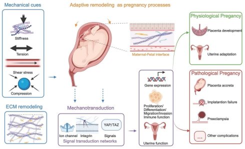
Pregnancy is a highly coordinated process that relies on the precise regulation of the extracellular matrix, cells, and various molecules at the maternal-fetal interface. As the fetus grows and amniotic fluid increases, the uterus undergoes adaptive remodeling, a process in which biomechanics plays an indispensable role. This review aims to systematically explore the biomechanical characteristics of the uterus and specifically elucidate the crucial regulatory roles of mechanical signals such as stiffness, shear stress, tension, and compression force in both physiological and pathological pregnancies. We focus on the remodeling of the extracellular matrix, the mechanical property changes of the maternal-fetal interface, and the associated mechanotransduction pathways, such as PIEZO1, integrins, and YAP/TAZ. Finally, we summarize the cutting-edge technologies used to investigate uterine biomechanics and discuss the clinical translational potential of targeting mechanical signaling pathways as novel diagnostic and therapeutic strategies for pregnancy complications. This review provides a comprehensive analysis of how biomechanical cues regulate uterine function at the molecular, cellular, and tissue levels.
Keywords: maternal-fetal interface, biomechanics, extracellular matrix, mechanotransduction, pregnancy complications
Introduction
Pregnancy is a highly coordinated biological process involving embryo implantation, placental development, and fetal growth within a dynamically changing maternal environment. The success of this process depends on the precise regulation of cellular, molecular, and tissue-level processes within the maternal-fetal microenvironment. Traditional research on pregnancy mainly focused on genetic and biochemical regulation. Hormonal regulation [1, 2], immune tolerance [3], and metabolic disorders [4] have been widely recognized as key contributors.
The maternal-fetal interface is a complex system composed of various components, including decidual stromal cells (dS), trophoblasts, immune cells, and the extracellular matrix (ECM) (5). These components do not exist independently but are involved in cell activities and intercellular communication through numerous biochemical signals. The role of biomechanics in this process has recently attracted attention. Biomechanics primarily applies mechanics principles to biological systems, providing a framework for understanding how mechanical properties influence cellular behavior and tissue function [5]. Animal and in vitro models have indicated that such mechanical cues may modulate processes such as vascular remodeling [6] and trophoblast behavior [7]. More importantly, mechanical abnormalities may lead to impaired placental development, contributing to complications such as placenta accreta spectrum (PAS) [8] and preeclampsia (PE) [9]. Nevertheless, the extent to which these mechanisms operate in human pregnancy and how they integrate with known biochemical pathways remains unclear.
Several reviews have explored uterine biomechanics [10] and mechanotransduction at the maternal-fetal interface in isolation [11]. Given the growing but heterogeneous body of evidence, a systematic and critical review is needed to clarify the mechanical characteristics of the maternal-fetal interface and its spatiotemporal dynamics. This review aims to follow the “mechanics-structure-signal-function-disease” axis as the main thread and to conduct a hierarchical, comprehensive analysis of the existing evidence. Specifically, we discuss how mechanical signals shape tissue organization, are perceived by cells, and are transformed into cellular responses, ultimately leading to physiological adaptation or pathological progression. We also evaluate current biomechanics-related tools based on purpose and clinical maturity. Moreover, we discuss their translational potential, as well as the prospects and limitations of targeted mechanical drug application in pregnancy complications.
Uterine Biomechanical Framework: From Tissue Mechanics to Mechanotransduction
Uterine Structure and Biomechanical Properties
The uterus is an organ composed of muscle tissue. The uterine wall is composed of the endometrium, the myometrium, and the perimetrium. Each layer contributes distinct mechanical functions. The endometrium can provide a contact interface for embryo implantation, the myometrium can generate contractile forces, and the perimetrium can preserve structural integrity. This functional differentiation suggests that pregnancy-adaptive remodeling may exhibit layer-specific characteristics. As pregnancy progresses, the uterine cavity expands significantly by 500 to 1000 times [12], accompanied by pronounced structural remodeling of the uterine wall. Early pregnancy is dominated by tissue growth, whereas later stages involve progressive stretching and thinning of the uterine wall [13]. Imaging-based studies on humans have demonstrated specific changes in myometrium thickness across different regions. For example, the thickness of the myometrium in the lower uterine segment decreases significantly with advancing gestational age, whereas the myometrium at the placental attachment site becomes markedly thicker [14]. During the second and third trimesters, the fundal myometrium is the thinnest [15]. In the intrapartum period, the myometrium first thins symmetrically and then undergoes a rebound thickening [16]. These spatial differences are not randomly distributed. Instead, they may reflect the structural adaptation of the uterus to meet region-specific mechanical load requirements, thereby influencing the load distribution and contractile function.
Uterine tissue exhibits typical biomechanical characteristics of soft biological materials, including anisotropy, nonlinearity, frequency dependence, viscoelasticity, and strain-dependent stiffening (Figure 1). These properties collectively reflect the way the uterus responds to mechanical loads to accommodate the dynamic demands of pregnancy. Specifically, the diffusion anisotropy of the uterus is related to its ordered fiber structure [17, 18]. Experimental studies have characterized uterine mechanical behavior under both compressive and tensile loads. The uterine tissue displays relatively low resistance to compression but exhibits strain-stiffening behavior under tension, in which its resistance to deformation increases with increasing strain [19, 20]. Within the frequency range of 0.1 to 100 Hz, the modulus of uterine tissue shows greater frequency dependence, consistent with its viscoelastic behavior [21]. Although these observations have been primarily from in vitro mechanical tests, they provide key parameters for quantifying the integrity and functional reserve of the uterine structure.
Structure and mechanical characteristics of the uterus. The uterus is a complex hollow organ whose wall is mainly composed of three layers: the endometrium, myometrium, and perimetrium. The myometrium consists of smooth muscle, with an outer longitudinal layer ensuring contractile function and an inner circular layer primarily responsible for integrity. During the menstrual cycle, the mechanical properties of the uterus change periodically, exhibiting decreased stiffness during the secretory phase. Furthermore, the uterus is continuously subjected to external multiscale forces, including uterine wall tension caused by fetal growth, compressive forces from villous growth, and shear stress. Changes in extracellular matrix (ECM) composition and structure confer typical soft tissue mechanical properties on the uterus: strain-dependent stiffening, viscoelasticity, anisotropy, and nonlinear characteristics.
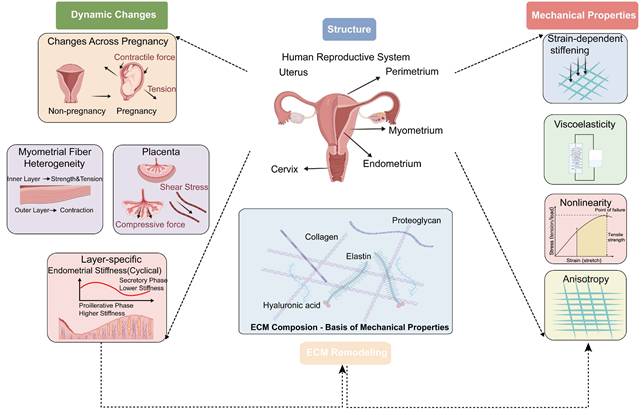
Of note, uterine biomechanical properties are not constant, as shown in Table 1. Ex vivo studies suggest that during pregnancy, the passive compliance and extensibility of uterine tissue gradually increase to accommodate the growth needs of the fetus. One study found no effect of parity, age, or menstrual cycle stage on uterine mechanical properties [22]. In contrast, in vivo imaging studies indicate that endometrial stiffness is higher during the proliferative phase than during the secretory phase [23]. These inconsistent findings may be attributed to methodological heterogeneity and structural heterogeneity across the uterine layers (periodic hormonal effects may act on the endometrium rather than the myometrium), so these results are not always directly comparable. Near the implantation window, the uterus adjusts its mechanical properties to prepare for subsequent embryo implantation. Understandably, the tension in the uterine wall increases as pregnancy progresses, consistent with the continuously increasing mechanical demands imposed by fetal development [24]. During delivery, the mechanical behavior of the uterus is characterized by nonlinear stress-strain responses and coordinated contraction activity, which may be influenced by tissue structure and loading conditions [25]. On one hand, uterine fibers exhibit structural heterogeneity. Dense muscle fibers are arranged to withstand tension, while the longer outer muscle layer fibers are responsible for contraction [26]. On the other hand, the uterine smooth muscle undergoes passive stress relaxation at a slower rate, which further maintains the stability of uterine wall tension and promotes the formation of the lower uterine segment [27]. Due to ethical and technical limitations in obtaining human samples throughout pregnancy, animal models provide valuable complementary insights. Studies in rhesus monkeys have suggested the presence of spatial gradients in mechanical properties across uterine layers. Specifically, the stiffness from the endometrium to the perimetrium increases gradually. The viscoelastic ratio of the endometrium is higher, while its permeability and diffusivity are lower than those of the myometrium and perimetrium [10]. This is slightly different from the increase in uterine decidua stiffness observed during human pregnancy, possibly due to species differences [28]. Additionally, the stiffness of uterine tissues under pathological conditions also varies. Uterine fibroids are stiffer than normal tissues, while malignant tissues are the opposite. This suggests that there may also be biomechanical differences between different uterine pathologies [29]. Interspecies differences should be considered when extrapolating these findings to human pregnancy.
Representative Biomechanical Properties of the Uterus
| Tissue Region | Mechanical Property* | Measurement Methods (Quantitative Metric) | Species | Findings |
|---|---|---|---|---|
| The entire uterine wall | Tensile Strength a | Ex vivo Dynamic Uniaxial tension to failure (Peak true tensile stress, kPa; Elongation at break, dimensionless/%) | Human (Fresh tissue) | Pregnant lower uterine segment: Nonlinear stress-strain behavior b; Peak true tensile stress: 656.3±483.9kPa; Elongation at break: 32.7%±11.2% (range: 14-45%) [25] |
| Stiffness c Viscoelasticity d | In vivo and Ex vivo IFEA (Model-derived parameter, N/m2; Maximum elongation ratio, dimensionless) | Human | Non-pregnant uterus: Regional stiffness-related parameter: Fundus (3477 N/m2) > Dorsal region (3044 N/m2) > Ventral region (1239 N/m2); Apparent modulus decreased markedly ex vivo; Maximum elongation ratio e: 1.1-1.3 in vivo, 1.2-1.45 ex vivo [18] | |
| Uterine Layers | Stiffness c Viscoelasticity d | Ex vivo Microindentation and PVE model (Elastic modulus, Pa; Viscoelastic ratio, dimensionless) | Rhesus macaque (Frozen tissue) | Layer-specific stiffness: Endometrium / Decidua < Myometrium < Perimetrium, (range 10-104 Pa); Viscoelastic Ratio: Endometrium/Decidua < Myometrium, Perimetrium (range 0.3-0.6); The endometrium and pregnant decidua are the least stiff, most viscous, least diffusible, and most hydrated layers; No significant gestational changes in elastic modulus, viscoelastic ratio, permeability, or diffusivity were observed [10] |
| Stiffness c | Ex vivo AFM indentation (Apparent elastic modulus, Pa) | Human (Fresh tissue) | Region-dependent stiffness: Decidua Basalis (1250Pa) > Endometrium (250Pa) > Placenta (232Pa) > Decidua Parietalis (171Pa) [66] | |
| Viscoelasticity d Anisotropy f | Ex vivo Dynamic Uniaxial cyclic compression (Complex modulus, kPa) | Human (Fresh tissue) | Myometrium, non-pregnant uterus: Frequency-dependent Viscoelasticity; Complex modulus increased with frequency: 28 -35 kPa at 0.1 Hz and 78-95 kPa at 100 Hz; Parallel direction > Perpendicular direction [21] | |
| Stiffness c Compliance g Tensile Strength a | Ex vivo Uniaxial tensile and compression test (Elastic modulus, kPa/psi; Tensile strength, MPa/psi) | Human (Fresh tissue) | Myometrium: Stiffness: Non-pregnancy-965kPa (140psi) > Pregnancy-586kPa (85psi); Tensile strength: 0.55-2.07MPa (80-300psi); The outer layer is softer in compression, stiffer in tension [19] | |
| Compliance g Stress Relaxation h | Ex vivo Passive stretch test (Passive length-tension behavior; Stress relaxation rate, %) | Human (Fresh tissue) | Myometrium Pregnancy increased passive compliance, as shown by flatter passive length-tension curves [20, 22]; Stress Relaxation Rate: Non-pregnancy (27.24%) > Pregnancy (22.67%)[27] | |
| Stiffness c Viscoelasticity d | In vivo 3DMMRE (Magnitude of complex shear modulus, kPa; Phase angle, dimensionless) | Human | Myometrium, non-pregnant uterus: Magnitude of complex shear modulus: Uterine corpus (2.58±0.52kPa) > Cervix (2.00±0.34kPa); Magnitude of shear modulus: Endometrium-Proliferative phase (3.34±0.42kPa) > Secretory phase (1.97±0.34kPa); Myometrium-Proliferative phase (3.01±0.26kPa) > Secretory phase (2.22±0.26kPa); Phase angle: Myometrium (0.57±0.09) > Endometrium (0.32±0.10) [23] | |
| Equilibrium stress I Anisotropy f | Ex vivo Spherical indentation, Uniaxial tensile test, IFEA (Equilibrium stress, kPa) | Human (Fresh tissue) | Equilibrium stress: Non-pregnant (7.69±5.89kPa)> Pregnant (3.04±0.80kPa) [37] | |
| Stiffness c Viscoelasticity d | Ex vivo nanoindentation, PVE model (Elastic modulus, kPa; Viscoelastic ratio, dimensionless) | Human (Frozen tissue) | Elastic modulus: Endometrium (0.143±0.04kPa) < Decidua (0.352±0.07kPa) < Perimetrium (0.365±0.17kPa) < Non-pregnant myometrium (0.738±0.45kPa); Viscoelastic Ratio: Endometrium (0.430±0.02) < Decidua (0.485±0.03) [28] |
*Definition and units of mechanical parameters
a Tensile strength, kPa/MPa/psi: The maximum tensile stress sustained by tissue before rupture; b Stress-strain behavior, stress in kPa and strain dimensionless or %: The relationship between applied stress and tissue deformation during loading; nonlinear stress-strain behavior indicates that tissue stiffness changes with increasing deformation; c Stiffness-related metrics, N/m2 or Pa/kPa/MPa/psi: The resistance of tissue to deformation. In this table, stiffness-related behavior is reported according to the original studies as model-derived stiffness-related parameter (N/m2), elastic modulus (Pa/kPa/MPa), apparent elastic modulus (Pa), complex modulus (kPa), or magnitude of complex shear modulus (kPa). d Viscoelasticity-related metrics, unit depends on the reported metric: Time- or frequency- dependent mechanical behavior combining elastic and viscous responses. In this table, viscoelasticity is reported as complex modulus (kPa), magnitude of complex shear modulus (kPa), viscoelastic ratio (dimensionless), phase angle (dimensionless); e Maximum elongation ratio, dimensionless: The maximum relative extension ratio achieved by tissue under loading; f Anisotropy, dimensionless or direction-specific modulus in Pa/kPa: direction-dependent mechanical behavior in which tissue properties differ along different fiber orientations; g Compliance, depending on the test: the extent of deformation or change produced by a given load. Higher compliance indicates greater deformability; h Stress relaxation, %: Time-dependent decrease in stress under a constant imposed strain; I Equilibrium stress, Pa/kPa: the steady-state stress reached after stress relaxation under a constant strain, reflecting passive resistance after transient viscoelastic effects have dissipated.
3DMMRE: Three-dimensional multi-frequency magnetic resonance elastography; IFEA: Inverse finite element modeling; AFM: Atomic force microscopy; PVE: Poro-viscoelastic model
At the organ level, the uterus does not function in isolation, but forms a mechanically coupled system with the cervix and fetal membranes. Its moderate stiffness and ability to expand are crucial for accommodating fetal growth and transmitting mechanical loads [30]. Finite element models indicate that the cervix, characterized by a complex three-dimensional stress state, contributes to load redistribution among the uterus, fetal membranes, and itself, with its stretching depending on geometric morphology and material properties [31, 32]. These biomechanical properties ensure the uterine structural stability and adaptability throughout gestation.
Mechanotransduction Pathways in the Uterus
At the microscopic level, the mechanical behavior of the uterus is related to the composition of the ECM. The uterine ECM is primarily composed of type I, III, and V collagens, laminin, fibronectin, and proteoglycans, which together form a fibrous network that supports the tissue structure [33]. Importantly, beyond composition, the organization, arrangement, and cross-linking density of collagen fibers also affect the mechanical properties of the tissue [34]. These structural features are not static but are constantly reshaped. As decidualization progresses, decidual fibroblasts secrete type IV collagen and laminin, forming a transient basement membrane-like structure [35, 36]. The fibrous network becomes more extensible and less densely arranged, while matrix stiffness remains unchanged [37]. It is worth noting that similar changes also occur in cervical tissue. Before delivery, the collagen arrangement becomes looser, the water content increases, and the content of sulfated glycosaminoglycans rises [38-40]. These observations indicate that the remodeling of the uterine ECM may be a common biomechanical adaptation strategy during pregnancy.
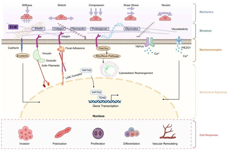
Mechanical cues within the uterine microenvironment are transmitted to cells through interactions with the ECM. Cells sense mechanical signals through mechanosensitive ion channels or integrin-based adhesion complexes. Among them, Piezo1 and Piezo2 are well-studied mechanosensitive ion channels [41]. Experimental studies have shown that Piezo1 is mainly expressed in uterine smooth muscle cells and microvascular endothelial cells, and its expression is upregulated during term labor and downregulated during preterm birth [42]. In contrast, Piezo2 is expressed in sensory neurons of the lower reproductive tract. Moreover, impairment of channel activity may be related to reduced contractility and altered labor progression [43]. Mechanosensitive ion channels mediate the remote activation effect of stretch, thereby enabling synchronized contraction of the entire uterus [44]. Integrins activate downstream signaling cascades, including focal adhesion kinase (FAK), which, in turn, regulates cytoskeletal organization and cell fate [45, 46]. The intracellular mechanical transduction pathways, especially the Hippo signaling pathway and its effectors YAP/TAZ, act as a downstream pathway. They are the mediators through which cells respond to mechanical stimuli. Their activity is regulated by cytoskeletal tension and can be transferred to the nucleus upon activation, driving transcriptional programs related to cell proliferation and differentiation [47]. In vitro studies have shown that ECM stiffness may affect the proliferation of endometrial stromal cells via the YAP signaling axis and promote uterine repair [48]. These mechanical transduction mechanisms jointly ensure the foundation for uterine function during pregnancy (Figure 2 and Table 2).
Overview of biomechanical cues, mechanosensors, and pregnancy outcomes
| Biomechanical Cues | Mechanosensors | Mechanisms | Main Tissue/ Cell Type | Physiological Process | Pathological Associations | Ref. |
|---|---|---|---|---|---|---|
| Stiffness | Myosin-II | Myosin-II | STB | Trophoblast Fusion, Morphological, and Hormone release | Not reported | [77] |
| Piezo1 | Piezo1-Glycolysis-Ca2+-PKC -IL8/G-CSF | dESF | Trophoblast invasion | PAS | [8] | |
| Not reported | Not Reported | Early Embryo | Early embryo development | Embryo Arrest | [56] | |
| Fibrillin1 | Feedback Positive | EVT | EVT invasion | Not Reported | [66] | |
| Not reported | LINC00458-SMAD2/3 | hPSCs | Endodermal lineage differentiation | Not Reported | [59] | |
| Shear Stress | Piezo1/YAP | Piezo1-Ca2+-YAP-PFKFB3 Piezo1-eNOS | EPCs | Glycolysis Vascularization | PE | [154] |
| Piezo1 TRPV4 Glycocalyx | PGE2/PGI2-paracrine Piezo1-eNOS-NO TRPV4- Ca2+ Glycocalyx-NO | Endothelial | Decidualization Vasodilation | SGA PE | [69, 84-86, 147-149] | |
| Piezo1 | Piezo1-Ca2+-TMEM16F cAMP-PKA-PIGF | Trophoblasts (STB) | Trophoblast fusion and migration Vascularization Metabolism | Embryo death PE | [79, 89-91] | |
| Focal adhesion | Focal adhesion assembly and actin polymerization | Fibroblasts Stroma cells | Pig placenta sculpt folds | Not reported | [87] | |
| Tension | Not reported | IL-8/MCP3 | Fibroblasts | Cervical Remodeling | Not reported | [115] |
| Piezo1 | Piezo1-Inflammatory | Myometrium | Uterine Contraction | PTB | [166, 167] | |
| Vinculin | Vinculin-occludin/ZO-1-ppMRLC/Myh9 | Blastomeres | Embryo compaction ICM/TE specification | Not reported | [53] | |
| Compression | Syndecan1 | Syndecan1- E-cadherin | STB | Trophoblast fusion | Implantation failure | [80] |
| Traction force/ Contractility | YAP Actomyosin | Actomyosin /YAP | Blastomeres | Embryo Compaction ICM/TE Specification Implantation pattern | Embryo quality Implantation failure | [51, 52, 61] |
| Oscillation | YAP | PDGFRα/YAP | Early embryo | ICM formation | Not reported | [54] |
| ECM (Composition /Arrangement) | Adhesion molecule | Actin cytoskeleton | Trophoblast (CTB) | Endometrial-trophoblast adhesion Trophoblast invasion | RPL RIF | [67, 75] |
| Collagen | Collagen-LAIR1 NKp30/Perforin Type I/V collagen | Macrophage dNK Th17/Treg | Immune tolerance | RPL | [97, 98, 127, 131] | |
| α-actinin-1 | ACTN1-NEBL-F-actin | Epithelial | Endometrial receptivity | RIF | [124] | |
| Collagen | SAA1-TypeI collagen | Fetal Membrane | Rupture of fetal membrane | PPROM | [160] | |
| Versican | Not reported | Stromal cells | Spiral artery dilation | Not reported | [99] |
Note: The biomechanical terms listed in this table refer to biomechanical cues.
Stiffness mainly refers to ECM or substrate stiffness. Shear Stress mainly refers to blood flow-induced wall shear stress acting on cells. Tension refers to tensile forces generated between neighboring cells or across cell-cell junctions. Compression refers to compressive forces generated between adjacent cells or within confined tissue spaces. Traction force refers to the force exerted by cells on the adhesive substrate. Contractility refers to active force generation by cells, often mediated by actomyosin activity. Oscillation refers here mainly to blastocyst cavity oscillation or periodic biomechanical fluctuation during early embryo development.
STB: syncytiotrophoblast; PAS: placenta accreta spectrum; dESF: decidual endometrial stroma fibroblasts; EVT: extravillous trophoblast; EPCs: Endothelial progenitor cells; hPSCs: human pluripotent stem cells; SGA: small for gestational age; PE: preeclampsia; RPL: recurrent pregnancy loss; RIF: recurrent implantation failure; CTB: Cytotrophoblast; PGE2: Prostaglandin E2; PGI2: Prostacyclin PGI2; ICM: Inner cell mass; TE: trophectoderm; PPROM: preterm premature rupture of membranes; ECM: Extracellular Matrix.
The mechanics-structure-mechanoreceptor-mechano-signaling-cell response axis at the maternal-fetal interface. This axis elucidates the relationship between the biomechanical properties of the maternal-fetal interface microenvironment and cellular function. Mechanical forces (extracellular matrix stiffness, compression, tensile force, tension, and shear stress) act on each cell at the maternal-fetal interface, triggering a mechanotransduction cascade. Alterations in extracellular matrix remodeling are sensed by mechanosensitive ion channels on the cell membrane (e.g., Piezo1, TRPV4), integrin receptors, and cell adhesion molecules, which transduce mechanical signals via calcium ion influx or conformational changes, activating cytoskeletal rearrangement and downstream pathways such as YAP/TAZ, leading to changes in gene transcription. Ultimately, these changes result in altered cell behaviors, including trophoblast invasion and fusion, morphological changes in stromal cells, shifts in immune cell polarization, and participation in spiral artery remodeling.
Biomechanics Across Pregnancy: From Implantation to Parturition
Embryo Implantation: Mechanical Dialogue Between Embryo and Endometrium
Embryo implantation is a dynamic mechanical interaction process involving forces generated by the embryo and physical constraints imposed by the uterus. Before implantation, the uterine contractions and ciliary movements propel the embryo through the reproductive tract. During the early stages of mammalian embryo development, the preimplantation embryo is encapsulated by the zona pellucida, which prevents it from directly sensing matrix stiffness or shear stress through integrins. In vitro models have shown that the zona pellucida in mouse stiffens after fertilization [49]. And the mechanical properties of the zona pellucida may affect the development and positioning of early mouse embryos [50]. As the embryo undergoes division and enters the maturation stage, this process is mainly driven by actin-myosin-dependent pulsed contractions. During this process, CDH1 regulates contractile force at cell-cell contact sites [51]. Subsequently, the embryo undergoes asymmetric division, producing blastomeres with distinct contractile capabilities, thereby separating the fates of the inner cell mass (ICM) and the trophoblast cells (TE). In TE, YAP localization can enhance its mechanical sensing [52]. During cleavage and differentiation into the blastocyst, the increase in intracavitary pressure promotes the formation of tight junctions through tension-cytoskeletal adhesion protein positive feedback loop [53]. However, pressure above the threshold may lead to TE rupture, which might represent a protective mechanism. Periodic mechanical oscillations within the mouse blastocyst cavity could shape the spatial structure of the ICM lineage through fluctuations in cell-cell contact, cell movement, and PDGFRα/YAP signaling [54]. In parallel, blastocyst expansion generates hydrostatic pressure, which may serve as a potential mechanical signal contributing to endometrial receptivity [55]. The embryo is sensitive to the mechanical properties of the surrounding microenvironment. Three-dimensional in vitro studies have shown that culturing mouse embryos on a soft hydrogel mimicking uterine stiffness significantly increases blastocyst formation rate and hatching efficiency, while also increasing the number of trophoblasts [56]. While, a stiffer matrix tends to inhibit development or promote osteogenic differentiation [57]. This difference may be due to the embryonic stage. Mouse ESCs only acquire mechanical responsiveness after exiting the ground-state pluripotent state [58]. Moreover, experimental studies using human pluripotent stem cells have shown that the soft matrix may regulate SMAD2/3 by upregulating the lncRNA LINC00458 to contribute to embryo development [59]. Emerging research suggests that stress relaxation rate may be a more crucial parameter regulating implantation [60]. When comparing across species, imaging platforms have also revealed specific differences in implantation modes: mouse embryos tend to exhibit expansive growth, whereas human embryos display more directional extension and adopt an embedded morphology [61]. These observations highlight the diversity of implantation strategies across species and suggest that mechanical interactions between the embryo and uterine environment may vary accordingly.
At the maternal level, the endometrium undergoes significant structural and mechanical remodeling during the implantation window. It has been reported that ECM composition changes, including alterations in collagen subtypes in the endometrium during implantation and enrichment of type IV collagen in the embryonic basement membrane [62]. The ECM can promote physical interactions between the embryo and the endometrium, influencing spatial positioning. Decidual stromal cells (dS) exhibit dual characteristics. On the one hand, they undergo matrix degradation to accommodate trophoblast invasion while maintaining an anti-matrix state to prevent excessive invasion [63]. At the epithelial level, endometrial barrier properties are dynamically regulated. Redistribution of adherens junctions and desmosomes in endometrial epithelial cells shifts from apical to lateral, potentially reducing intercellular adhesion strength and facilitating trophoblast invasion [64, 65].
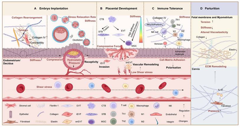
Embryonic and maternal cells jointly regulate and respond to the mechanical microenvironment at the maternal-fetal interface. During trophoblast invasion, dynamic changes in type IV collagen and fibronectin-1, as well as vascular remodeling, have been reported. These changes may be related to the regional differences in human decidual stiffness (basal decidua vs. parietal decidua: 1250 Pa vs. 171 Pa) [66]. Trophoblasts exhibit elongated pseudopodia and increased physical stiffness to reduce fluid shear-induced damage and enhance interaction with the surrounding matrix. In parallel, endometrial epithelial cells undergo changes in non-polarized actin cytoskeletal organization, apically localized integrins, and calcium signaling, which may affect tight junctions at the maternal-fetal interface [67, 68]. Microfluidic models further demonstrate that shear stress and trace forces may contribute to decidualization by regulating cytoskeletal rearrangement [69]. Endometrial epithelial cells express PIEZO1 and epithelial sodium channels (ENaC), converting mechanical stimuli into biochemical signals, including calcium ion influx, to regulate prostaglandin E2 production during early implantation [65, 70]. While deletion of Piezo1 in mice may even result in embryonic death. After implantation, the mechanical constraints imposed by the maternal uterus may affect the formation of the body axis in early mouse embryos by limiting developmental space [71]. Most of these findings come from animal models or in vitro systems, while human data are relatively sparse. Nevertheless, the overall situation indicates that ECM properties and intercellular connections affect the balance of local forces and regulate decidualization and blastocyst implantation (Figure 3A).
Mechanical changes and mechanisms during normal pregnancy. This figure illustrates the key mechanical events and potential mechanisms during the process from embryo implantation to delivery. A. The endometrium undergoes structural and mechanical remodeling. The collagen subtypes in the extracellular matrix change, and decidual stromal cells combine with this changed ECM, resulting in morphological changes. Before embryo implantation, the periodic mechanical oscillations of the blastocyst form the inner cell mass. The intracavitary hydrostatic pressure of the blastocyst increases, serving as a mechanical signal that promotes endometrial receptivity. B. Shear stress, stiffness gradients, and villous compressive forces regulate trophoblast differentiation, migration, and vascular remodeling processes. C. The stiffness and composition of the extracellular matrix regulate the polarization and cytokine secretion of immune cells, such as macrophages and NK cells, through adhesion molecules. D. The tension exerted on the myometrium and fetal membranes, along with their intrinsic stiffness and viscoelasticity, as well as the mechanical compression on the cervix, participate in the process of parturition by modulating extracellular matrix remodeling through mechano-immunological mechanisms.
Placental Development: Forces in Trophoblast Invasion and Vascular Remodeling
The placenta is a critical organ during pregnancy, exhibiting compressive symmetry, nonlinear viscoelasticity, and strain-rate dependence [72, 73]. These properties ensure the structural integrity under physiological loads. During placental development, cytotrophoblast (CTB) gradually fuses into syncytiotrophoblast (STB) or differentiates into extravillous trophoblast (EVT), and EVT further differentiates into endovascular (enEVT) and interstitial (iEVT) subtypes. These cells invade the decidua and maternal vasculature, remodeling spiral arteries to establish an efficient maternal-fetal circulation [11, 74]. During this process, trophoblasts appear to sense and respond to mechanical signals in the surrounding microenvironment. Cell surface integrins and intracellular focal adhesion complexes play a role and exhibit lineage-specific expression patterns. For example, VCT preferentially expresses α4β6, whereas EVT expresses α5β1, which is associated with the invasive phenotype [75]. In addition, ECM stiffness has been reported to modulate the morphology and invasion ability of trophoblasts. A certain high stiffness can promote the directional behavior of EVT [76]. While upon encountering regions of higher stiffness, such as the uterine myometrium, EVT may exhibit inhibited migration and fuse into multinucleated giant cells. Similarly, matrix stiffness can regulate the fusion and morphological transformation to STB form and hormone release through myosin-II [77]. STB simultaneously expresses mechanosensitive ion channels (Piezo 1, Polycystin 2, TRPV6) and motility proteins related to primary cilia (Dynein 1, IFT88, Kinesin 2) [78, 79]. These molecules may be crucial for the key mechanical transduction. Moreover, experimental and computational studies have shown that directional compressive forces could enhance trophoblast fusion efficiency and villous formation by syndecan1 and E-cadherin in vitro [80]. This indicates a close connection between multiple mechanical signals and the behaviors of different trophoblast types.
Hemodynamic factors provide another perspective on mechanical regulation during placental development. During pregnancy, the uterine spiral arteries undergo significant remodeling. Incomplete vascular circumferential adaptation in mice, potentially alters stress distribution within the uteroplacental circulation [81]. In human, structural changes, including the replacement of vascular smooth muscle with fibrinoid material and the formation of the Nitabuch layer, are associated with increased local tissue stiffness [82, 83]. Shear stress, arising from increased maternal blood flow, modulates cellular functions through distinct mechanisms in a cell-type-specific manner. In endothelial cells, Piezo1 mediates shear stress-induced calcium influx, thereby promoting nitric oxide production and vascular adaptation in rats [84]. The same holds true in human placental vascular endothelial cells, and may be associated with small for gestational age babies [85, 86]. Pig models suggested that mechanical forces generated by increased blood flow at the uteroplacental interface may trigger focal adhesion assembly and actin polymerization, thereby forming sculp folds [87]. A computational model suggested that the optimal morphology of human terminal villi is also regulated by the pressure gradient and changes in blood flow velocity, and driven by shear forces and deformation [88]. In the intervillous space, shear stress upregulates angiogenic factors such as PlGF via the cAMP-PKA pathway, thereby participating in the differentiation and functional maturation of human STB [7, 89]. Additionally, shear stress reprograms STB metabolism by inhibiting glycolysis and activating GOT-mediated amino acid metabolism, an effect dysregulated in fetal growth restriction (FGR) [90]. enEVT may respond to low shear stress by inhibiting directed migration while enhancing endothelium-derived chemotactic gradients during vascular remodeling [11, 91]. In contrast, elevated shear stress may act as a biomechanical signal to terminate spiral artery remodeling [6]. These mechanical-biological feedback mechanisms ensure the effective establishment and development of the placental structure and maternal-fetal circulation (Figure 3B).
Pregnancy Maintenance: Biomechanic-Immune Crosstalk
Immune tolerance at the maternal-fetal interface is essential for maintaining pregnancy, allowing the semi-allogeneic fetus to coexist with the maternal immune system. This process involves decidual natural killer (dNK) cells, macrophages, and T cells, forming an immunosuppressive microenvironment [92]. Emerging evidence suggests that immune cells are sensitive to mechanical cues, including stiffness, shear stress, spatial confinement, and tension. Insights from other fields, such as tumor immunology and computational modeling, suggest that these factors influence immune cell migration, activation, and polarization through mechanotransduction pathways [93]. However, their relevance to pregnancy requires further investigation. At the maternal-fetal interface, immune cells are embedded within a dynamically remodeling ECM, which may provide both structural support and mechanical signals. Decidual macrophages predominantly exhibit an M2-like anti-inflammatory phenotype, which has been associated with placental development and immune regulation during early pregnancy [94]. Ex vivo studies suggest that matrix physical properties may influence macrophage polarization and cytokine secretion by the Piezo1-YAP axis [95, 96]. A pregnancy-related study further indicates that decidual stromal cells may influence CD16+ decidual macrophages through ECM-adhesion molecule interactions, promoting an immunoregulatory phenotype during early pregnancy [97]. ECM components may also regulate immune cell function. Collagen has been reported to reduce dNK cytotoxicity by downregulating molecules such as NKp30 and perforin, while influencing cytokine production, such as IFN-γ and TNF-α [98]. Proteoglycans such as versican, secreted by endometrial stromal cells, have been implicated in regulating the proliferation of tissue-resident NK cells and spiral artery remodeling [99]. The ECM and its physical properties seem to be a key regulator hub for immune tolerance in pregnancy. Different responses of distinct immune cells to the same mechanical signal enable fine-tuned regulation of immune homeostasis (Figure 3C).
Parturition: Mechanical Remodeling of Cervix and Fetal Membranes
The cervix, the fetal membranes, and the uterus form a complete mechanical coupling system. This system can maintain the integrity of pregnancy and coordinate the delivery process. The cervix provides structural support, the fetal membranes help to bear the load, and the uterus generates contraction force.
The fetal membrane is a thin layer of tissue composed of the inner amnion and the outer chorion. There is a movable interface between the two, allowing stress distribution during pregnancy. Among them, the amnion is the main load-bearing layer, and the chorion serves as the buffering layer [88]. The structural integrity of both depends on their tensile strength, extensibility, and viscoelasticity, which are jointly regulated by the contents of elastin and collagen [100, 101]. As amniotic fluid volume increases and the fetus grows, the fetal membranes must resist the pressure within the uterus and adapt to the gradually increasing mechanical stretching. In vitro experiments have shown that before labor, the intermediate layer of the amnion undergoes collagen degradation and softening, with a significant reduction in fiber density, resulting in decreased stiffness, which is the basis for labor initiation [101]. At full term, the rupture of the amnion is related to the physiological weakening of its mechanical properties.
Similarly, during normal pregnancy, the cervix remains closed and firm, and its mechanical strength is mainly maintained by type I and type III collagen [102]. During late pregnancy and delivery, the cervix undergoes adaptive remodeling. It goes through the stages of softening, maturation, dilation, and postpartum repair, characterized by decreased tissue stiffness, increased viscoelasticity, increased compliance, and decreased load-bearing capacity [103, 104]. This behavior is driven by time-dependent matrix reorganization [105, 106]. Collagen transforms from a highly ordered to a more disordered network, with increased fiber dispersion and solubility. At the same time, EP4 regulates the stretching strength of the rat cervix to adapt to the stress required for delivery by modulating collagen rearrangement and microstructural changes [107]. At the molecular level, it is driven by increased matrix metalloproteinase activity and decreased tissue inhibitor expression [108, 109]. The expression of proteoglycans and collagen fibers in the rat cervix increases during the delivery period, and their interaction promotes cervical dilation, rapidly decreasing to the non-pregnant level one day after delivery [110]. Cervical shortening is most significant in the later stages of pregnancy, reflecting the cumulative effects of mechanical load and tissue remodeling [111]. Mechanical force participates in cervical remodeling through feedback mechanisms. The gradual increase in uterine volume and intrauterine pressure increases the pressure on the cervix, affecting the coupling of uterine contractions and cervical dilation [112, 113]. Mouse models indicate that the cervical tissue is anisotropic, and its axial contraction force and circumferential stiffness play a key role in maintaining homeostasis [114]. An increase in intrauterine pressure and mechanical stimulation of the cervix by the fetal head can activate mechanosensitive pathways in cervical fibroblasts, regulate IL-8 release, and thereby modulate ECM synthesis and degradation [115]. This mechanical-biochemical feedback loop coordinates uterine contractions, fetal membrane tension, and cervical remodeling during delivery. Before full-term delivery, there is a clear "weak zone" above the cervix in the area of the fetal membranes, which is the starting point of physiological rupture [116]. The cervix exhibits regional-specific structural and functional differences. The upper part may participate in the regulation of labor through contraction activities [117]. The three-dimensional finite element model in the late pregnancy further confirms that intrauterine pressure acts on both the fetal membranes and the cervix, and cervical shortening reversely affects the stress distribution in the weak zone of the fetal membranes, suggesting that studying a single tissue in isolation cannot reflect the true interactive nature of the prenatal mechanical environment [108].
Mechanical forces have been suggested to interact with electrophysiological pathways in regulating uterine contraction. For example, multiscale modeling studies have indicated that tensile forces may contribute to synchronized whole-uterine contraction [44]. Clinical findings have indirectly validated this theory, showing that mechanical stimulation (e.g., abdominal pressure) can directly induce bioelectrical activity, confirming the mechanosensitivity of the uterus [118]. These findings suggest the presence of a potential feedback system in which mechanical forces, cellular responses, and electrophysiological signals are dynamically integrated (Figure 3D).
Biomechanical Dysregulation in Pregnancy Complications: From Adaptation to Maladaptation
Implantation Failure
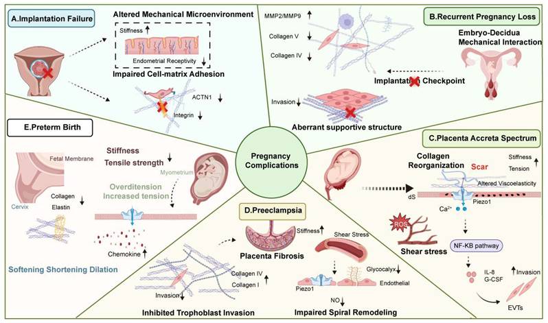
Embryo implantation requires a coordinated interaction between the embryo and the endometrium. Changes in uterine mechanical activity, including uterine contractility and endometrial stiffness, may be associated with implantation outcome. Fanchin [119] and Chung's team [120] suggested that a high-frequency contraction pattern may lead to implantation failure. In contrast, Blank [121] proposed that high-frequency, low-amplitude uterine activity is beneficial for implantation. The inconsistency may be due to differences in observation time and measurement methods. In addition, mechanical stimulation of the endometrium has been suggested to improve endometrial receptivity to reduce implantation failure [122]. At the cellular level, cell-matrix interactions play a critical role in mediating embryo attachment. Studies in mouse models have revealed that preimplantation embryo movement comprises three stages: the entry phase, the unidirectional clustering movement phase, and the bidirectional dispersal phase. Among these, embryo clustering movement depends on myometrial contractions, whereas dispersal relies on the LPAR3 signaling pathway. While disruption of integrin-mediated force transmission impairs implantation in mouse models [123]. In RIF patients, it has been further found that upregulation of anti-adhesion proteins PDX and α-actinin-1 impairs endometrial epithelial cell adhesion [124, 125]. Collectively, these findings suggest that RIF may be associated with alterations in the mechanical microenvironment and impaired cell-matrix interactions, although the causal relationships remain to be fully established (Figure 4A and Table 2).
Mechanical mechanisms underlying pregnancy complications. This figure summarizes the mechanisms associated with several pregnancy complications related to biomechanical abnormalities. A. Alterations in the mechanical microenvironment and impaired cell-matrix interactions are associated with implantation failure; B. Abnormal extracellular matrix remodeling and aberrant supportive structures disrupt the mechanical homeostasis of the maternal-fetal interface, contributing to recurrent pregnancy loss; C. Altered mechanical properties of scar tissue, manifested as increased stiffness, reduced tension, and disorganized collagen architecture, activate Piezo1 ion channels, leading to the release of pro-inflammatory factors, regulating abnormal trophoblast invasion, and contributing to placenta accreta spectrum (PAS); D. Shear stress resulting from abnormal vascular remodeling and increased stiffness from aberrant extracellular matrix remodeling inhibit trophoblast invasion and spiral artery remodeling, contributing to preeclampsia (PE); E. Preterm birth is associated with weakened mechanical properties of the fetal membranes, cervical remodeling, and uterine contractions induced by abnormal tension.
Recurrent Pregnancy Loss
Following successful implantation, maintenance of early pregnancy requires coordinated interactions between the embryo and the maternal decidua. If this interaction is abnormal, it will lead to recurrent pregnancy loss (RPL). RPL is a multifactorial condition, with a substantial proportion of cases remaining unclear. Congenital and acquired uterine morphology abnormalities are associated with RPL. Changes in morphology may affect the uneven distribution of tension within the uterine cavity, influencing uterine cavity mechanics and, in turn, embryo implantation and stability [126]. Emerging evidence suggests that alterations in ECM composition may be associated with RPL. Reduced expression of type IV and type V collagen has been observed in the decidua of RPL patients, indicating potential changes in tissue structure and mechanical properties [127-129]. While upregulation of MMP-9 in RPL suggests that an imbalance between ECM degradation and synthesis may weaken the ECM and interfere with the embedding processes [130]. In the RPL mouse, supplementation with recombinant human collagen can regulate the Th17/Treg balance, reduce the incidence of retained fetuses, reshape the immune microenvironment at the maternal-fetal interface, and improve reproductive capacity [131]. The endometrium functions as a selective interface that integrates biochemical and mechanical signals during implantation. It has been proposed that this “implantation checkpoint” may involve not only molecular signaling but also mechanical interactions between the embryo and the endometrium [132]. While alterations in tissue structure and cell-matrix interactions may contribute to RPL, direct evidence linking specific mechanical parameters to RPL remains limited. We hypothesize that the mechanism of selective elimination of low-quality embryos may also involve abnormal mechanical interactions between the embryo and the endometrium (Figure 4B and Table 2).
Placenta Accreta Spectrum (PAS)
Normal placental development requires tightly regulated trophoblast invasion and coordinated remodeling of the maternal vasculature. Under physiological conditions, EVT invade the decidua and remodel spiral arteries to establish a low-resistance, high-capacity circulation.
Cesarean Scar pregnancy (CSP) and placental accreta spectrum (PAS) are associated with abnormal implantation and excessive trophoblast invasion [133]. Scar has been identified as a common risk factor for both conditions [134]. In vitro and in vivo experiments have demonstrated that scars exhibit altered mechanical properties, including increased stiffness and changes in collagen organization [135-137]. While no impact of CS scars on uterine motility patterns has been identified, regional anterior-posterior wall motion asymmetry and abnormal viscoelasticity of the lower uterine segment muscle exist, potentially leading to subsequent abnormal embryo implantation [138, 139]. These changes may influence the local microenvironment at the implantation site. Alterations in tissue mechanics may affect trophoblast behavior. Experimental observations suggest that there may be differences in the invasive capacity of trophoblast cells between scar and non-scar tissue. Compared with non-scar regions, the invasive ability of trophoblasts in scar tissue is enhanced, and restoring normal tissue structure appears to reverse this behavior [140]. In vitro scar models have shown that Piezo1 participates in trophoblast cell responses to mechanical stimuli by secreting IL-8 and G-CSF [8]. In addition to local tissue properties, abnormal vascular remodeling contributes to altered hemodynamic conditions in PAS. Impaired spiral artery remodeling may result in increased blood flow velocity and altered shear stress within the intervillous space [141]. On one hand, reactive oxygen species (ROS) are generated, disrupting ECM structure and altering EVT invasion signals [142]. On the other hand, increased shear stress and mechanical injury from unreconstructed arteries induce lacunar formation and thrombosis. These hemodynamic disturbances may impair placental function and are associated with an increased risk of adverse pregnancy outcomes [143].
Collectively, PAS may involve complex interactions between altered ECM structure, abnormal tissue mechanics, and dysregulated hemodynamic forces. These findings support a conceptual framework in which the local microenvironment (“soil”), trophoblasts (“seeds”), vascular flow (“flow”), and ECM structure (“scaffold”) interact to shape placental implantation. Within this context, a mechanical-biological imbalance of the endometrium may influence trophoblast behavior, contributing to disease. Therefore, in clinical practice, we need to assess the mechanical state of the uterine scar to determine the risks of PAS or uterine rupture (Figure 4C and Table 2).
Preeclampsia
Preeclampsia (PE) is associated with impaired placental vascular remodeling and alterations in the mechanical properties of the uteroplacental circulation. Clinical and experimental studies have reported reduced uterine artery dilation, increased vascular stiffness, and altered pulse wave transmission in early PE pregnancies [81, 144, 145]. In addition, abnormal shear stress patterns have been associated with PE and fetal growth restriction, suggesting that hemodynamic forces may be altered in this condition [146]. At the mechanistic level, endothelial cells are known to respond to altered mechanical cues. Piezo1 has been implicated in shear stress-induced calcium signaling and nitric oxide (NO) production via the Ca2+/eNOS pathway, which is involved in vascular function. Rat models suggest that disruption of the Piezo1-NO axis may be associated with impaired vasodilation in PE [9, 147]. Other mechanosensitive channels, such as TRPV4, have also been reported to participate in NO-mediated vascular responses, and their altered activity has been associated with early-onset PE [148]. Furthermore, the structural components of the vascular microenvironment may also affect mechanical signal transmission. In PE, an abnormal glycocalyx may impair endothelial cells' ability to sense shear stress and regulate NO production [149]. Additionally, ECM remodeling and fibrosis were observed in PE placentas, manifested as increased collagen deposition and tissue stiffness. Increased COL4A1 and COL4A2 expression has been associated with impaired vascular remodeling [150, 151]. In vitro studies suggest that hypoxia may promote ECM deposition via TGF-β1 signaling, contributing to placental fibrosis [152, 153]. The mechanically sensitive signaling pathway can also regulate cellular metabolism. Recent studies have shown that the Piezo1/YAP/PFKFB3 axis may regulate the glycolysis of endothelial progenitor cells (EPCs) and promote the formation of new blood vessels in the placenta during PE [154]. However, these findings are primarily derived from experimental systems and require further validation in humans. Beyond the uteroplacental circulation, mechanical alterations were also observed in fetal tissues. Increased collagen fiber density and stiffness in the umbilical artery, along with reduced Wharton's jelly area and elasticity, have been observed in PE, which may have implications for fetal cardiovascular development [155]. The abnormality in the above-mentioned behavior may interfere with normal placental development, leading to abnormal mechanical properties. This also explains why PE presents abnormal mechanical properties of the placenta [88]. The abnormality of the placenta, as a bridge for transporting nutrients and connecting the mother and the fetus, reflects the mechanism of abnormal fetal outcomes. For example, the stiffness and viscosity of the placenta in cases of fetal growth restriction decrease [156, 157].
Collectively, these findings suggest that PE may involve interactions between altered ECM remodeling, abnormal vascular mechanical properties, and dysregulated mechanosensory pathways. However, whether these mechanical alterations represent primary drivers of disease or secondary responses remains unclear. Therefore, mechanical parameters are currently better considered as potential contributors or biomarkers (Figure 4D and Table 2).
Preterm Premature Rupture of Membranes and Preterm Birth
Before delivery, a series of coordinated mechanical and biochemical changes occur, including fetal membrane relaxation, cervix remodeling, and uterine contractions. These processes are tightly regulated to ensure term delivery. Preterm birth (PTB) is associated with conditions involving increased mechanical load, such as multiple pregnancies and polyhydramnios, suggesting that uterine overdistension may alter the mechanical environment of pregnancy [158]. One cause of PTB is preterm premature rupture of membranes (PPROM). The high tensile stiffness and rupture critical tension of the fetal membranes depend on the content and cross-linking structure of the mature collagen fiber network. When these two are abnormal, the tensile strength and stiffness of the fetal membranes decrease, causing physiological pressure-induced membrane rupture [159]. In the tissues obtained from vaginal delivery with spontaneous rupture of the fetal membranes, COL1A1 and COL1A2 expression were significantly lower than those from elective cesarean section without fetal membrane rupture [160]. Computational biomechanical modeling has further revealed that lubrication at the chorion-amnion interface decreases, leading to chorion thinning and potentially increasing fetal membrane stress and rupture [161]. This suggests the importance of tissue structure and mechanical properties in maintaining the integrity of the fetal membranes. The amnion has a certain degree of toughness, and a macroscopic mechanical defect of the amnion alone cannot cause rupture [162]. Therefore, PPROM is more likely to result from a biochemical imbalance accompanied by increased mechanical load. Another cause of PTB is cervical factors. Alterations in the collagen and elastin networks of fetal membranes may also lead to premature softening, shortening, and dilation of the cervix [101]. A clinical trial found that the collagen concentration in the cervix of women with cervical insufficiency (CI) was lower, and the tissue strength was reduced [163]. Perhaps the abnormal mechanical properties triggered premature contractions of the uterus, resulting in miscarriage and PTB. In addition, the uterine myometrium is not a passive load-bearing structure that bears tension; rather, it actively senses and responds to mechanical stretch [164]. At the cellular level, mechanical stretch has been reported to regulate chemokine expression and inflammatory signaling pathways, which may contribute to the initiation of uterine contractions [165, 166]. In addition, stretch-induced electromechanical coupling may activate calcium channels in uterine smooth muscle cells, contributing to contractile activity [37]. Experimental studies suggest that Piezo1 activation modulates uterine contractility and inflammatory signaling [154, 167]. Preterm birth is likely the result of immune activation under mechanical load (Figure 4E and Table 2).
Translational Research and Future Directions
Biomechanical Monitoring
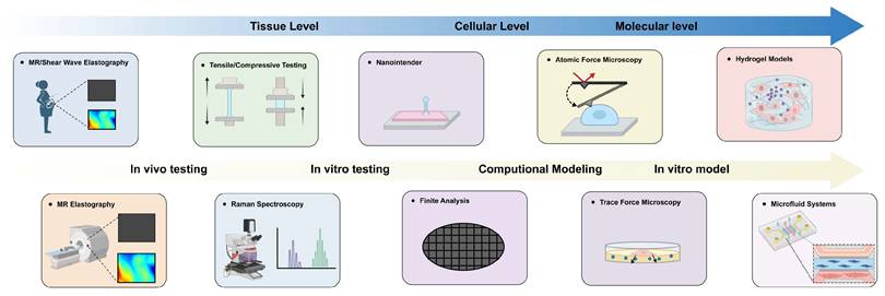
Currently, certain biophysical imaging techniques have enabled non-invasive assessment of tissue mechanical properties during pregnancy (Figure 5), but their clinical maturity varies considerably. Strain and shear wave elastography have been applied in several preliminary clinical exploratory studies to evaluate cervical softening and predict preterm birth risk [168-170]. Ex vivo experiments have further characterized the spatial variation in cervical shear wave velocity and proposed it as a potential quantitative indicator of cervical softening [171]. In addition, ultrasound imaging has also been used to detect changes in stiffness at cesarean section scars [172]. In vivo measurements of cervical stiffness using ultrasound-based maximal deformation and the aspiration method have also been used to predict preterm birth, and they may perform better than elastography [173]. However, most of these applications are currently based on small sample studies. Raman spectroscopy has been shown to capture changes in collagen structure, water content, and lipid signals in the pregnant mouse cervix, and these changes correlate with decreases in cervical stiffness during pregnancy [174]. An MRI-based parametric solid model can reconstruct a three-dimensional uterine structure during late pregnancy to simulate tissue deformation and mechanical heterogeneity. Still, some deviation between the models and the actual situation remains [175]. A dual-arm nanorobot system enables non-invasive measurement of embryo mechanics, but it is still at an early experimental stage [176]. Tensile tests and nanoindentation mainly measure the material properties of each layer of the uterus in vitro [28]. The future applications of these technologies may enable in vivo dynamic mechanical monitoring of the uterus during pregnancy. Moreover, they can provide key biomechanical indicators to determine the optimal time of delivery and prevent complications (e.g., cervical stiffness and placental elasticity). Given the differences in mechanical properties between in vivo and ex vivo conditions, future work should focus on standardizing operating protocols, establishing quantitative thresholds, and basing them on human samples to explore the feasibility of mechanical monitoring.
Multi-scale biomechanical tools for mechanical properties. This figure provides an overview of the tools that can be applied to monitor the mechanical properties of the maternal-fetal interface and explore mechanical mechanisms at the tissue level, cell level, and molecular level. It covers macroscopic elastic imaging, microscopic atomic force microscopy, and hydrogels and microfluidic models for dynamic regulation of mechanical properties.
Experimental Models and Mechanobiology Tools
At the microscale, some experimental tools have helped us explore microscopic mechanics (Figure 5). Atomic force microscopy (AFM) and traction force microscopy are measurement tools; the former directly measures local stiffness and adhesion forces of individual cells or the ECM, while the latter can visualize the forces exerted by cells during migration or contraction on the substrate [61, 177]. Additionally, a microfluidic system can simulate cells under dynamic mechanical conditions, such as shear stress and concentration gradients, to explore changes in vascular remodeling during pregnancy [178]. The combination of organoid and hydrogel models enables the adjustment of parameters, such as stiffness and viscoelasticity, to simulate the regulatory role of cell behavior under various mechanical conditions [60, 179]. Currently, these methods are mainly used to explore the mechanism of the mechanical signal axis. Nevertheless, in vitro models cannot fully capture the spatiotemporal complexity in vivo, making it difficult to recapitulate physiological phenomena and limiting the extrapolation of research results to humans.
Therapeutic Strategies Targeting Mechanics
Some scholars have begun to attach importance to biomechanics in clinical practice during pregnancy [180]. Physical biomechanical interventions have already been preliminarily applied clinically. For example, the silicone double-balloon catheter promotes cervical ripening through mechanical cervical dilation [181]. In contrast, drugs targeting mechanotransduction signaling pathways remain largely at the concept stage. The Piezo1 agonist Yoda1 can restore mechanosensitivity of Piezo1 mutants, suggesting its potential therapeutic effect on hydrops fetalis [182]. Vedolizumab targets integrin α4β7 and blocks MAdCAM-1-mediated cell adhesion and invasion [183]. In vitro experiments have shown that it inhibits trophoblast invasion, but its safety in vivo has not been validated. In the future, perhaps targeted drugs targeting the ECM, or drugs targeting the core mechanical transduction elements, could be considered to intervene in the mechanical characteristics and in cells' perception and response to the mechanical environment. Physical interventions target the upstream source directly and act quickly. Molecular interventions focus on sensing and transducing mechanical signals. Of note, we have to carefully monitor the safety of applying mechanically targeted drugs during pregnancy.
Modeling and Future Directions
Computational modeling offers a unique perspective for research in pregnancy biomechanics. The microstructural fiber network model of fetal membrane rupture shows that the high toughness of the amnion arises from non-affine deformation and crack deflection of its collagen fibers. The multiscale model of the maternal-fetal interface simulates the process by which trophoblasts gradually remodel blood vessels [184]. However, current models remain partially dependent on parameter selection, which limits their physiological relevance and extrapolation capability. Future efforts should focus on constructing molecular-cellular-tissue system models that comprehensively reveal the biomechanical behavior of the uterus under physiological and pathological conditions.
Conclusions
Overall, biomechanics is not merely a passive factor in pregnancy. On the contrary, it is involved in regulating key events throughout the entire pregnancy process. Currently, most evidence on the correlation between mechanics and pregnancy comes from animal models and in vitro experiments. Nevertheless, these studies have identified several possible mechanical mechanisms, including PIEZO1, integrins, and YAP/TAZ, which seem to play important roles in trophoblast cell invasion, vascular remodeling, immune tolerance, and cervical maturation. Moreover, abnormalities in stiffness, shear stress, and tension may be related to the occurrence of pregnancy complications. Future research should combine advanced biomechanical modeling with validation using real human samples to confirm the role of mechanical forces. Additionally, the feasibility of targeting mechanosensors such as PIEZO1 and YAP/TAZ for the treatment of pregnancy complications should be explored.
Abbreviations
ECM: Extracellular Matrix; FAK: Focal Adhesion Kinase; ICM: Inner Cell Mass; TE: Trophectoderm; ENaC: Epithelial Sodium Channels; dS: Decidual Stromal Cells; CTB: Cytotrophoblast; STB: Syncytiotrophoblast; EVT: Extravillous Trophoblast; enEVT: Endovascular Extravillous Trophoblast; iEVT: Interstitial Extravillous Trophoblast; FGR: Fetal Growth Restriction; dNK: decidual natural killer; RPL: Recurrent pregnancy loss; CSP: Cesarean Scar pregnancy; PAS: Placental accrete spectrum; ROS: reactive oxygen species; PE: Preeclampsia; NO: nitric oxide; PPROM: preterm premature rupture of membranes; CI: cervical insufficiency; EPCs: endothelial progenitor cells; PTB: Preterm birth; AFM: Atomic force microscopy.
Acknowledgements
All the figures were created in https://BioRender.com.
Funding
This work was supported by the National Natural Science Foundation of China (82371688) and the Ningxia Natural Science Foundation (2025AAV030082).
Authorship Contributions
Tingting Wang: Writing - original draft, Writing - review & editing, Visualization. Wanru Chen: Writing - revise & editing. Huixia Yang: Funding acquisition, Conceptualization, Supervision. Jie Yan: Writing - review & editing, Conceptualization, Funding acquisition, Supervision. All authors have read and approved the final manuscript.
AI usage statement
The conception of this study, literature search, logical structure, and initial draft were independently completed by the authors. ChatGPT was used solely for language polishing, including correction of grammar, sentence structure, and readability. This tool was not used for study design, statistical analysis, result interpretation, image creation, or formulation of any conclusions. All AI-polished edits were carefully reviewed and revised by the authors. The authors take full responsibility for the accuracy, completeness, and final content of the manuscript.
Competing Interests
The authors have declared that no competing interest exists.
References
1. Cooke PS, Buchanan DL, Young P, Setiawan T, Brody J, Korach KS. et al. Stromal estrogen receptors mediate mitogenic effects of estradiol on uterine epithelium. Proc Natl Acad Sci U S A. 1997;94:6535-40
2. Wetendorf M, DeMayo FJ. The progesterone receptor regulates implantation, decidualization, and glandular development via a complex paracrine signaling network. Mol Cell Endocrinol. 2012;357:108-18
3. Qiu Y, Chen M, Lin R, Chen X, He L, Yin H. et al. Crosstalk between decidual natural killer cells and extravillous trophoblasts at the maternal-fetal interface: current status and future perspectives. Front Immunol. 2025;16:1703156
4. Brown K, Heller DS, Zamudio S, Illsley NP. Glucose transporter 3 (GLUT3) protein expression in human placenta across gestation. Placenta. 2011;32:1041-9
5. Myers KM, Elad D. Biomechanics of the human uterus. Wiley Interdiscip Rev Syst Biol Med. 2017 9
6. James JL, Whitley GS, Cartwright JE. Shear stress and spiral artery remodelling: the effects of low shear stress on trophoblast-induced endothelial cell apoptosis. Cardiovasc Res. 2011;90:130-9
7. Inohaya A, Chigusa Y, Takakura M, Io S, Kim MA, Matsuzaka Y. et al. Shear stress in the intervillous space promotes syncytial formation of iPS cells-derived trophoblasts†. Biol Reprod. 2024;110:300-9
8. Wenqiang D, Novin A, Liu Y, Afzal J, Suhail Y, Liu S. et al. Scar matrix drives Piezo1 mediated stromal inflammation leading to placenta accreta spectrum. Nat Commun. 2024;15:8379
9. Arishe OO, Ebeigbe AB, Webb RC. Mechanotransduction and Uterine Blood Flow in Preeclampsia: The Role of Mechanosensing Piezo 1 Ion Channels. Am J Hypertens. 2020;33:1-9
10. Fodera DM, Xu EZ, Duarte CA, M HW, Fang S, Chen X. et al. Time-dependent material properties and composition of the nonhuman primate uterine layers through gestation. Sci Rep. 2025;15:23194
11. Szukiewicz D, Trojanowski S, Wróbel E, Wojdasiewicz P, Szewczyk G. Mechanosensing of Shear Stress and Uterine Spiral Artery Remodeling by Invasive Trophoblasts in Early Pregnancy. Int J Mol Sci. 2025 26
12. Cunningham FG LK, Bloom SL, et al. Williams Obstetrics.; 2022
13. Gillespie EC. Principles of uterine growth in pregnancy. Am J Obstet Gynecol. 1950;59:949-59
14. Degani S, Leibovitz Z, Shapiro I, Gonen R, Ohel G. Myometrial thickness in pregnancy: longitudinal sonographic study. J Ultrasound Med. 1998;17:661-5
15. Durnwald CP, Mercer BM. Myometrial thickness according to uterine site, gestational age and prior cesarean delivery. J Matern Fetal Neonatal Med. 2008;21:247-50
16. Buhimschi CS, Buhimschi IA, Malinow AM, Weiner CP. Myometrial thickness during human labor and immediately post partum. Am J Obstet Gynecol. 2003;188:553-9
17. Weiss S, Jaermann T, Schmid P, Staempfli P, Boesiger P, Niederer P. et al. Three-dimensional fiber architecture of the nonpregnant human uterus determined ex vivo using magnetic resonance diffusion tensor imaging. Anat Rec A Discov Mol Cell Evol Biol. 2006;288:84-90
18. Kauer M, Vuskovic V, Dual J, Szekely G, Bajka M. Inverse finite element characterization of soft tissues. Med Image Anal. 2002;6:275-87
19. Pearsall GW, Roberts VL. Passive mechanical properties of uterine muscle (myometrium) tested in vitro. J Biomech. 1978;11:167-76
20. Conrad JT, Kuhn W. The active length-tension relationship in human uterine muscle. Am J Obstet Gynecol. 1967;97:154-60
21. Kiss MZ, Hobson MA, Varghese T, Harter J, Kliewer MA, Hartenbach EM. et al. Frequency-dependent complex modulus of the uterus: preliminary results. Phys Med Biol. 2006;51:3683-95
22. Conrad JT, Johnson WL, Kuhn WK, Hunter CA Jr. Passive stretch relationships in human uterine muscle. Am J Obstet Gynecol. 1966;96:1055-9
23. Jiang X, Asbach P, Streitberger KJ, Thomas A, Hamm B, Braun J. et al. In vivo high-resolution magnetic resonance elastography of the uterine corpus and cervix. Eur Radiol. 2014;24:3025-33
24. Sokolowski P, Saison F, Giles W, McGrath S, Smith D, Smith J. et al. Human uterine wall tension trajectories and the onset of parturition. PLoS One. 2010;5:e11037
25. Manoogian SJ, Bisplinghoff JA, Kemper AR, Duma SM. Dynamic material properties of the pregnant human uterus. J Biomech. 2012;45:1724-7
26. Fujimoto K, Kido A, Okada T, Uchikoshi M, Togashi K. Diffusion tensor imaging (DTI) of the normal human uterus in vivo at 3 tesla: comparison of DTI parameters in the different uterine layers. J Magn Reson Imaging. 2013;38:1494-500
27. Conrad JT, Kuhn WK, Johnson WL. Stress relaxation in human uterine muscle. Am J Obstet Gynecol. 1966;95:254-65
28. Fodera DM, Russell SR, Jackson JLL, Fang S, Chen X, Vink J. et al. Material properties of nonpregnant and pregnant human uterine layers. J Mech Behav Biomed Mater. 2024;151:106348
29. Omari EA, Varghese T, Kliewer MA, Harter J, Hartenbach EM. Dynamic and quasi-static mechanical testing for characterization of the viscoelastic properties of human uterine tissue. J Biomech. 2015;48:1730-6
30. Mahmoud H, Wagoner Johnson A, Chien EK, Poellmann MJ, McFarlin B. System-level biomechanical approach for the evaluation of term and preterm pregnancy maintenance. J Biomech Eng. 2013;135:021009
31. Westervelt AR, Fernandez M, House M, Vink J, Nhan-Chang CL, Wapner R. et al. A Parameterized Ultrasound-Based Finite Element Analysis of the Mechanical Environment of Pregnancy. J Biomech Eng. 2017;139:0510041-05100411
32. Fernandez M, House M, Jambawalikar S, Zork N, Vink J, Wapner R. et al. Investigating the mechanical function of the cervix during pregnancy using finite element models derived from high-resolution 3D MRI. Comput Methods Biomech Biomed Engin. 2016;19:404-17
33. Aplin JD, Charlton AK, Ayad S. An immunohistochemical study of human endometrial extracellular matrix during the menstrual cycle and first trimester of pregnancy. Cell Tissue Res. 1988;253:231-40
34. Kurt I, Kulhan M, AlAshqar A, Borahay MA. Uterine Collagen Cross-Linking: Biology, Role in Disorders, and Therapeutic Implications. Reprod Sci. 2024;31:645-60
35. Iwahashi M, Muragaki Y, Ooshima A, Yamoto M, Nakano R. Alterations in distribution and composition of the extracellular matrix during decidualization of the human endometrium. J Reprod Fertil. 1996;108:147-55
36. Tong J, Lv S, Yang J, Li H, Li W, Zhang C. Decidualization and Related Pregnancy Complications. Matern Fetal Med. 2022;4:24-35
37. Fang S, Duarte CA, Fodera DM, Shi L, Chen X, Advincula AP. et al. Equilibrium mechanical properties of the human uterus in tension and compression. Acta Biomater. 2025;194:219-32
38. Myers KM, Paskaleva AP, House M, Socrate S. Mechanical and biochemical properties of human cervical tissue. Acta Biomater. 2008;4:104-16
39. Myers K, Socrate S, Tzeranis D, House M. Changes in the biochemical constituents and morphologic appearance of the human cervical stroma during pregnancy. Eur J Obstet Gynecol Reprod Biol. 2009;144(Suppl 1):S82-9
40. Gan Y, Yao W, Myers KM, Vink JY, Wapner RJ, Hendon CP. Analyzing three-dimensional ultrastructure of human cervical tissue using optical coherence tomography. Biomed Opt Express. 2015;6:1090-108
41. Coste B, Mathur J, Schmidt M, Earley TJ, Ranade S, Petrus MJ. et al. Piezo1 and Piezo2 are essential components of distinct mechanically activated cation channels. Science. 2010;330:55-60
42. Barnett SD, Asif H, Buxton ILO. Novel identification and modulation of the mechanosensitive Piezo1 channel in human myometrium. J Physiol. 2023;601:1675-90
43. Zhang Y, Kini SA, Mishkanian SA, Yarishkin O, Luo R, Seradj SH. et al. PIEZO channels link mechanical forces to uterine contractions in parturition. Science. 2025;390:eady3045
44. Yochum M, Laforêt J, Marque C. Multi-scale and multi-physics model of the uterine smooth muscle with mechanotransduction. Comput Biol Med. 2018;93:17-30
45. Katoh K. Signal Transduction Mechanisms of Focal Adhesions: Src and FAK-Mediated Cell Response. Front Biosci (Landmark Ed). 2024;29:392
46. Benn MC, Pot SA, Moeller J, Yamashita T, Fonta CM, Orend G. et al. How the mechanobiology orchestrates the iterative and reciprocal ECM-cell cross-talk that drives microtissue growth. Sci Adv. 2023;9:eadd9275
47. Aragona M, Panciera T, Manfrin A, Giulitti S, Michielin F, Elvassore N. et al. A mechanical checkpoint controls multicellular growth through YAP/TAZ regulation by actin-processing factors. Cell. 2013;154:1047-59
48. Zhang T, Hu R, Wang Y, Guo S, Wu Z, Liu J. et al. Extracellular matrix stiffness mediates uterine repair via the Rap1a/ARHGAP35/RhoA/F-actin/YAP axis. Cell Commun Signal. 2023;21:22
49. Sun Y, Wan KT, Roberts KP, Bischof JC, Nelson BJ. Mechanical property characterization of mouse zona pellucida. IEEE Trans Nanobioscience. 2003;2:279-86
50. Elad D, Jaffa AJ, Grisaru D. Biomechanics of Early Life in the Female Reproductive Tract. Physiology (Bethesda). 2020;35:134-43
51. Maître JL, Niwayama R, Turlier H, Nédélec F, Hiiragi T. Pulsatile cell-autonomous contractility drives compaction in the mouse embryo. Nat Cell Biol. 2015;17:849-55
52. Maître JL, Turlier H, Illukkumbura R, Eismann B, Niwayama R, Nédélec F. et al. Asymmetric division of contractile domains couples cell positioning and fate specification. Nature. 2016;536:344-8
53. Chan CJ, Costanzo M, Ruiz-Herrero T, Mönke G, Petrie RJ, Bergert M. et al. Hydraulic control of mammalian embryo size and cell fate. Nature. 2019;571:112-6
54. Guo Z, Yao J, Zheng X, Cao J, Lv X, Gao Z. et al. Cavity oscillation drives pattern formation in early mammalian embryos. Cell Rep. 2025;44:115342
55. Enders AC. Anatomical aspects of implantation. J Reprod Fertil Suppl. 1976:1-15
56. Kolahi KS, Donjacour A, Liu X, Lin W, Simbulan RK, Bloise E. et al. Effect of substrate stiffness on early mouse embryo development. PLoS One. 2012;7:e41717
57. Evans ND, Minelli C, Gentleman E, LaPointe V, Patankar SN, Kallivretaki M. et al. Substrate stiffness affects early differentiation events in embryonic stem cells. Eur Cell Mater. 2009;18:1-14
58. Verstreken CM, Labouesse C, Agley CC, Chalut KJ. Embryonic stem cells become mechanoresponsive upon exit from ground state of pluripotency. Open Biol. 2019;9:180203
59. Chen YF, Li YJ, Chou CH, Chiew MY, Huang HD, Ho JH. et al. Control of matrix stiffness promotes endodermal lineage specification by regulating SMAD2/3 via lncRNA LINC00458. Sci Adv. 2020;6:eaay0264
60. Guo J, Lyu J, Gao Z, Jia T, Wang L, Dong J. et al. 3D biomimetic niche modulates embryo development in vitro. Nat Commun. 2026;17:1279
61. Godeau AL, Seriola A, Tchaicheeyan O, Casals M, Denkova D, Aroca E. et al. Traction force and mechanosensitivity mediate species-specific implantation patterns in human and mouse embryos. Sci Adv. 2025;11:eadr5199
62. Diao H, Aplin JD, Xiao S, Chun J, Li Z, Chen S. et al. Altered spatiotemporal expression of collagen types I, III, IV, and VI in Lpar3-deficient peri-implantation mouse uterus. Biol Reprod. 2011;84:255-65
63. Afzal J, Suhail Y, Du W, Liu Y, Ramasamy R, Liu Z. et al. Evidence for coopetition at the maternal-fetal interface shaping placental invasion. Proc Natl Acad Sci U S A. 2025;122:e2323038122
64. Buck VU, Windoffer R, Leube RE, Classen-Linke I. Redistribution of adhering junctions in human endometrial epithelial cells during the implantation window of the menstrual cycle. Histochem Cell Biol. 2012;137:777-90
65. Sternberg AK, Buck VU, Classen-Linke I, Leube RE. How Mechanical Forces Change the Human Endometrium during the Menstrual Cycle in Preparation for Embryo Implantation. Cells. 2021 10
66. Abbas Y, Carnicer-Lombarte A, Gardner L, Thomas J, Brosens JJ, Moffett A. et al. Tissue stiffness at the human maternal-fetal interface. Hum Reprod. 2019;34:1999-2008
67. Thie M, Herter P, Pommerenke H, Dürr F, Sieckmann F, Nebe B. et al. Adhesiveness of the free surface of a human endometrial monolayer for trophoblast as related to actin cytoskeleton. Mol Hum Reprod. 1997;3:275-83
68. Thie M, Röspel R, Dettmann W, Benoit M, Ludwig M, Gaub HE. et al. Interactions between trophoblast and uterine epithelium: monitoring of adhesive forces. Hum Reprod. 1998;13:3211-9
69. Gnecco JS, Ding T, Smith C, Lu J, Bruner-Tran KL, Osteen KG. Hemodynamic forces enhance decidualization via endothelial-derived prostaglandin E2 and prostacyclin in a microfluidic model of the human endometrium. Hum Reprod. 2019;34:702-14
70. Hennes A, Held K, Boretto M, De Clercq K, Van den Eynde C, Vanhie A. et al. Functional expression of the mechanosensitive PIEZO1 channel in primary endometrial epithelial cells and endometrial organoids. Sci Rep. 2019;9:1779
71. Hiramatsu R, Matsuoka T, Kimura-Yoshida C, Han SW, Mochida K, Adachi T. et al. External mechanical cues trigger the establishment of the anterior-posterior axis in early mouse embryos. Dev Cell. 2013;27:131-44
72. Pérès J, Thollon L, Delotte J, Tillier Y, Brunet C, Kayvantash K. et al. Material properties of the placenta under dynamic loading conditions. Comput Methods Biomech Biomed Engin. 2014;17:958-64
73. Weed BC, Borazjani A, Patnaik SS, Prabhu R, Horstemeyer MF, Ryan PL. et al. Stress state and strain rate dependence of the human placenta. Ann Biomed Eng. 2012;40:2255-65
74. Sheldon RE, Shmygol A, van den Berg HA, Blanks AM. Functional and morphological development of the womb throughout life. Sci Prog. 2015;98:103-27
75. Damsky CH, Fitzgerald ML, Fisher SJ. Distribution patterns of extracellular matrix components and adhesion receptors are intricately modulated during first trimester cytotrophoblast differentiation along the invasive pathway, in vivo. J Clin Invest. 1992;89:210-22
76. Cao J, Li H, Tang H, Gu X, Wang Y, Guan D. et al. Stiff Extracellular Matrix Promotes Invasive Behaviors of Trophoblast Cells. Bioengineering (Basel). 2023 10
77. Ma Z, Sagrillo-Fagundes L, Mok S, Vaillancourt C, Moraes C. Mechanobiological regulation of placental trophoblast fusion and function through extracellular matrix rigidity. Sci Rep. 2020;10:5837
78. Lee TC, Moulvi A, James JL, Clark AR. Multi-scale Modelling of Shear Stress on the Syncytiotrophoblast: Could Maternal Blood Flow Impact Placental Function Across Gestation? Ann Biomed Eng. 2023;51:1256-69
79. Zhang Y, Shan KZ, Liang P, Lowry AJ, Feng L, Yang H. PIEZO1 drives trophoblast fusion and placental development. Nat Commun. 2025;16:6895
80. Parameshwar PK, Li C, Arnauts K, Jiang J, Rostami S, Campbell BE. et al. Directed biomechanical compressive forces enhance fusion efficiency in model placental trophoblast cultures. Sci Rep. 2024;14:11312
81. Murtada SI, Latorre M, Humphrey JD. Remodeling of the uterine artery during and early after pregnancy in the mouse. Biomech Model Mechanobiol. 2023;22:1531-40
82. Pijnenborg R, Vercruysse L, Hanssens M. The uterine spiral arteries in human pregnancy: facts and controversies. Placenta. 2006;27:939-58
83. Pijnenborg R, Dixon G, Robertson WB, Brosens I. Trophoblastic invasion of human decidua from 8 to 18 weeks of pregnancy. Placenta. 1980;1:3-19
84. John L, Ko NL, Gokin A, Gokina N, Mandalà M, Osol G. The Piezo1 cation channel mediates uterine artery shear stress mechanotransduction and vasodilation during rat pregnancy. Am J Physiol Heart Circ Physiol. 2018;315:H1019-H1026
85. Morley LC, Shi J, Gaunt HJ, Hyman AJ, Webster PJ, Williams C. et al. Piezo1 channels are mechanosensors in human fetoplacental endothelial cells. Mol Hum Reprod. 2018;24:510-20
86. Morley LC, Debant M, Gaunt HJ, Simpson NAB, Beech DJ. Nitric oxide synthase phosphorylation in fetoplacental endothelium is enhanced by agonism of Piezo1 mechanosensor in small for gestational age babies. Reprod Fertil. 2023 4
87. Seo H, Li X, Wu G, Bazer FW, Burghardt RC, Bayless KJ. et al. Mechanotransduction drives morphogenesis to develop folding during placental development in pigs. Placenta. 2020;90:62-70
88. Plitman Mayo R. Advances in Human Placental Biomechanics. Comput Struct Biotechnol J. 2018;16:298-306
89. Lecarpentier E, Atallah A, Guibourdenche J, Hebert-Schuster M, Vieillefosse S, Chissey A. et al. Fluid Shear Stress Promotes Placental Growth Factor Upregulation in Human Syncytiotrophoblast Through the cAMP-PKA Signaling Pathway. Hypertension. 2016;68:1438-46
90. Brugger BA, Neuper L, Guettler J, Forstner D, Wernitznig S, Kummer D. et al. Fluid shear stress induces a shift from glycolytic to amino acid pathway in human trophoblasts. Cell Biosci. 2023;13:163
91. James JL, Cartwright JE, Whitley GS, Greenhill DR, Hoppe A. The regulation of trophoblast migration across endothelial cells by low shear stress: consequences for vascular remodelling in pregnancy. Cardiovasc Res. 2012;93:152-61
92. Alippe Y, Hatterschide J, Coyne CB, Diamond MS. Innate immune responses to pathogens at the maternal-fetal interface. Nat Rev Immunol. 2025;25:869-84
93. Zhang X, Kim TH, Thauland TJ, Li H, Majedi FS, Ly C. et al. Unraveling the mechanobiology of immune cells. Curr Opin Biotechnol. 2020;66:236-45
94. Wang H, He M, Hou Y, Chen S, Zhang X, Zhang M. et al. Role of decidual CD14(+) macrophages in the homeostasis of maternal-fetal interface and the differentiation capacity of the cells during pregnancy and parturition. Placenta. 2016;38:76-83
95. Derisoud E, Jiang H, Zhao A, Chavatte-Palmer P, Deng Q. Revealing the molecular landscape of human placenta: a systematic review and meta-analysis of single-cell RNA sequencing studies. Hum Reprod Update. 2024;30:410-41
96. Mei F, Guo Y, Wang Y, Zhou Y, Heng BC, Xie M. et al. Matrix stiffness regulates macrophage polarisation via the Piezo1-YAP signalling axis. Cell Prolif. 2024;57:e13640
97. Yang HL, Shi JW, Lai ZZ, Li X, Gu CJ, Zheng ZM. et al. Decidual stromal cells drive CD16(+) macrophages towards an immunoregulatory phenotype via extracellular matrix-adhesion molecule interaction during early pregnancy. Front Immunol. 2025;16:1747323
98. Fu Q, Tao Y, Piao H, Du MR, Li DJ. Trophoblasts and decidual stromal cells regulate decidual NK cell functions via interaction between collagen and LAIR-1. Am J Reprod Immunol. 2014;71:368-78
99. Sagae Y, Horie A, Yanai A, Ohara T, Nakakita B, Kitawaki Y. et al. Versican provides the provisional matrix for uterine spiral artery dilation and fetal growth. Matrix Biol. 2023;115:16-31
100. Grémare A, Jean-Gilles S, Musqui P, Magnan L, Torres Y, Fénelon M. et al. Cartography of the mechanical properties of the human amniotic membrane. J Mech Behav Biomed Mater. 2019;99:18-26
101. Jabareen M, Mallik AS, Bilic G, Zisch AH, Mazza E. Relation between mechanical properties and microstructure of human fetal membranes: an attempt towards a quantitative analysis. Eur J Obstet Gynecol Reprod Biol. 2009;144(Suppl 1):S134-41
102. Akins ML, Luby-Phelps K, Bank RA, Mahendroo M. Cervical softening during pregnancy: regulated changes in collagen cross-linking and composition of matricellular proteins in the mouse. Biol Reprod. 2011;84:1053-62
103. Read CP, Word RA, Ruscheinsky MA, Timmons BC, Mahendroo MS. Cervical remodeling during pregnancy and parturition: molecular characterization of the softening phase in mice. Reproduction. 2007;134:327-40
104. Yoshida K, Jayyosi C, Lee N, Mahendroo M, Myers KM. Mechanics of cervical remodelling: insights from rodent models of pregnancy. Interface Focus. 2019;9:20190026
105. Myers KM, Hendon CP, Gan Y, Yao W, Yoshida K, Fernandez M. et al. A continuous fiber distribution material model for human cervical tissue. J Biomech. 2015;48:1533-40
106. Duarte CA, Fang S, Rosado Mendez IM, Ateshian DGA, Hall T, Feltovich H. et al. An Anisotropic Reactive Viscoelastic Model of the Rhesus Macaque Cervix for Studying Cervical Remodeling. J Biomech Eng. 2025:1-37
107. Feltovich H, Ji H, Janowski JW, Delance NC, Moran CC, Chien EK. Effects of selective and nonselective PGE2 receptor agonists on cervical tensile strength and collagen organization and microstructure in the pregnant rat at term. Am J Obstet Gynecol. 2005;192:753-60
108. Fidalgo DS, Jorge RMN, Parente MPL, Louwagie EM, Malanowska E, Myers KM. et al. Pregnancy state before the onset of labor: a holistic mechanical perspective. Biomech Model Mechanobiol. 2024;23:1531-50
109. Mahendroo M. Cervical remodeling in term and preterm birth: insights from an animal model. Reproduction. 2012;143:429-38
110. Kokenyesi R, Woessner JF Jr. Relationship between dilatation of the rat uterine cervix and a small dermatan sulfate proteoglycan. Biol Reprod. 1990;42:87-97
111. Louwagie EM, Russell SR, Hairston JC, Nottman C, Nhan-Chang CL, Fuchs K. et al. The biomechanical evolution of the uterus and cervix and fetal growth in human pregnancy. NPJ Womens Health. 2024 2
112. House M, Paskaleva A, Myers K, Craigo S, Socrate S. The effect of uterine volume, uterine pressure and maternal position on cervical stress: The biomechanics of common static loads. American Journal of Obstetrics & Gynecology. 2005;193:S112
113. House M, Paskaleva A, Myers K, Craigo S, Socrate S. The connection between uterine contractions and cervical dilation: The biomechanics of cervical deformation. American Journal of Obstetrics & Gynecology. 2005;193:S55
114. Conway CK, Varghese A, Mahendroo M, Miller KS. The Role of Biaxial Loading on Smooth Muscle Contractility in the Nulliparous Murine Cervix. Ann Biomed Eng. 2021;49:1874-87
115. Takemura M, Itoh H, Sagawa N, Yura S, Korita D, Kakui K. et al. Cyclic mechanical stretch augments both interleukin-8 and monocyte chemotactic protein-3 production in the cultured human uterine cervical fibroblast cells. Mol Hum Reprod. 2004;10:573-80
116. Joyce EM, Moore JJ, Sacks MS. Biomechanics of the fetal membrane prior to mechanical failure: review and implications. Eur J Obstet Gynecol Reprod Biol. 2009;144(Suppl 1):S121-7
117. Hansen CJ, Rogers JH, Brown AJ, Boatwright N, Siricilla S, O'Brien CM. et al. Regional differences in three-dimensional fiber organization, smooth muscle cell phenotype, and contractility in the pregnant mouse cervix. Sci Adv. 2024;10:eadr3530
118. Garfield RE, Murphy L, Gray K, Towe B. Review and Study of Uterine Bioelectrical Waveforms and Vector Analysis to Identify Electrical and Mechanosensitive Transduction Control Mechanisms During Labor in Pregnant Patients. Reprod Sci. 2021;28:838-56
119. Fanchin R, Righini C, Olivennes F, Taylor S, de Ziegler D, Frydman R. Uterine contractions at the time of embryo transfer alter pregnancy rates after in-vitro fertilization. Hum Reprod. 1998;13:1968-74
120. Chung CH, Wong AW, Chan CP, Saravelos SH, Kong GW, Cheung LP. et al. The changing pattern of uterine contractions before and after fresh embryo transfer and its relation to clinical outcome. Reprod Biomed Online. 2017;34:240-7
121. Blank C, Sammali F, Kuijsters N, Huang Y, Rabotti C, de Sutter P. et al. Assessment of uterine activity during IVF by quantitative ultrasound imaging: a pilot study. Reprod Biomed Online. 2020;41:1045-53
122. Zhang XL, Fu YL, Kang Y, Qi C, Zhang QH, Kuang YP. Clinical observations of sequential therapy with Chinese medicine and hysteroscopic mechanical stimulation of the endometrium in infertile patients with repeated implantation failure undergoing frozen-thawed embryo transfer. Chin J Integr Med. 2015;21:249-53
123. Flores D, Madhavan M, Wright S, Arora R. Mechanical and signaling mechanisms that guide pre-implantation embryo movement. Development. 2020 147
124. Zhou X, Duan J, Zhou W, Zhang A, Chen Q. Upregulated α-actinin-1 impairs endometrial epithelial cell adhesion by downregulating NEBL in recurrent implantation failure. iScience. 2024;27:109046
125. Tas M. Anti-Adhesive Podocalyxin Expression Is Disrupted in Recurrent Implantation Failure. Diagnostics (Basel). 2025 15
126. Carbonnel M, Pirtea P, de Ziegler D, Ayoubi JM. Uterine factors in recurrent pregnancy losses. Fertil Steril. 2021;115:538-45
127. Iwahashi M, Nakano R. Decreased type V collagen expression in human decidual tissues of spontaneous abortion during early pregnancy. J Clin Pathol. 1998;51:44-6
128. Iwahashi M, Muragaki Y, Ooshima A, Nakano R. Decreased type IV collagen expression by human decidual tissues in spontaneous abortion. J Clin Endocrinol Metab. 1996;81:2925-9
129. Shi JW, Lai ZZ, Yang HL, Yang SL, Wang CJ, Ao D. et al. Collagen at the maternal-fetal interface in human pregnancy. Int J Biol Sci. 2020;16:2220-34
130. Goto S, Ozaki Y, Mori R, Ozawa F, Obayashi Y, Kitaori T. et al. MMP2 and MMP9 are associated with the pathogenesis of recurrent pregnancy loss through protein expression rather than genetic polymorphism. J Reprod Immunol. 2024;164:104270
131. Wang L, Zeng H, Li H, Dai J, You S, Jiang H. et al. Recombinant humanized type I collagen remodels decidual immune microenvironment at maternal-fetal interface by modulating Th17/Treg imbalance. Int J Biol Macromol. 2024;276:133994
132. Brosens JJ, Bennett PR, Abrahams VM, Ramhorst R, Coomarasamy A, Quenby S. et al. Maternal selection of human embryos in early gestation: Insights from recurrent miscarriage. Semin Cell Dev Biol. 2022;131:14-24
133. D'Antonio F, Calì G, Palacios-Jaraquemada J, Khalil A, Timor-Tritsch IE. Cesarean scar pregnancy is associated with abnormal implantation but not macroscopic myometrial invasion in early first trimester of pregnancy. Ultrasound Obstet Gynecol. 2022;59:550-1
134. Naji O, Wynants L, Smith A, Abdallah Y, Saso S, Stalder C. et al. Does the presence of a Caesarean section scar affect implantation site and early pregnancy outcome in women attending an early pregnancy assessment unit? Hum Reprod. 2013;28:1489-96
135. Moretti L, Stalfort J, Barker TH, Abebayehu D. The interplay of fibroblasts, the extracellular matrix, and inflammation in scar formation. J Biol Chem. 2022;298:101530
136. di Pasquo E, Kiener AJO, DallAsta A, Commare A, Angeli L, Frusca T. et al. Evaluation of the uterine scar stiffness in women with previous Cesarean section by ultrasound elastography: A cohort study. Clin Imaging. 2020;64:53-6
137. Kashani Ligumsky L, Jeong A, Martinez G, Marks H, Shah S, Staniczek J. et al. Placenta accreta spectrum: disrupted collagen architecture at a previous scar is a defining characteristic of placental adherence. Am J Obstet Gynecol. 2026;233:S645-S661.e1
138. Gora S, Elad D, Jaffa AJ. Objective Analysis of Vaginal Ultrasound Video Clips for Exploring Uterine Peristalsis Post Vaginal and Cesarean Section Deliveries. Reprod Sci. 2018;25:899-908
139. Buhimschi CS, Buhimschi IA, Yu C, Wang H, Sharer DJ, Diamond MP. et al. The effect of dystocia and previous cesarean uterine scar on the tensile properties of the lower uterine segment. Am J Obstet Gynecol. 2006;194:873-83
140. Garmi G, Goldman S, Shalev E, Salim R. The effects of decidual injury on the invasion potential of trophoblastic cells. Obstet Gynecol. 2011;117:55-9
141. Pollheimer J, Vondra S, Baltayeva J, Beristain AG, Knöfler M. Regulation of Placental Extravillous Trophoblasts by the Maternal Uterine Environment. Front Immunol. 2018;9:2597
142. Jauniaux E, Hempstock J, Greenwold N, Burton GJ. Trophoblastic oxidative stress in relation to temporal and regional differences in maternal placental blood flow in normal and abnormal early pregnancies. Am J Pathol. 2003;162:115-25
143. Afshar Y, Kashani Ligumsky L, Bartels HC, Krakow D. Biology and Pathophysiology of Placenta Accreta Spectrum Disorder. Obstet Gynecol. 2025;145:611-20
144. Schliefsteiner C, Wadsack C, Allerkamp HH. Exploring the Lifeline: Unpacking the Complexities of Placental Vascular Function in Normal and Preeclamptic Pregnancies. Compr Physiol. 2024;14:5763-87
145. Sedaghati F, Gleason RL Jr. A mathematical model of vascular and hemodynamics changes in early and late forms of preeclampsia. Physiol Rep. 2023;11:e15661
146. Morley LC, Beech DJ, Walker JJ, Simpson NAB. Emerging concepts of shear stress in placental development and function. Mol Hum Reprod. 2019;25:329-39
147. Arishe OO, McKenzie J, Dela Justina V, Dos Anjos Moraes R, Webb RC, Priviero F. Piezo1 channels mediate vasorelaxation of uterine arteries from pseudopregnant rats. Front Physiol. 2023;14:1140989
148. Allerkamp HH, Bondarenko AI, Tawfik I, Kamali-Simsek N, Horvat Mercnik M, Madreiter-Sokolowski CT. et al. In vitro examination of Piezo1-TRPV4 dynamics: implications for placental endothelial function in normal and preeclamptic pregnancies. Am J Physiol Cell Physiol. 2025;328:C227-c44
149. Moore KH, Murphy HA, George EM. The glycocalyx: a central regulator of vascular function. Am J Physiol Regul Integr Comp Physiol. 2021;320:R508-R518
150. Li W, Mata KM, Mazzuca MQ, Khalil RA. Altered matrix metalloproteinase-2 and -9 expression/activity links placental ischemia and anti-angiogenic sFlt-1 to uteroplacental and vascular remodeling and collagen deposition in hypertensive pregnancy. Biochem Pharmacol. 2014;89:370-85
151. Yong HE, Murthi P, Borg A, Kalionis B, Moses EK, Brennecke SP. et al. Increased decidual mRNA expression levels of candidate maternal pre-eclampsia susceptibility genes are associated with clinical severity. Placenta. 2014;35:117-24
152. Semenza GL. Hypoxia-inducible factors in physiology and medicine. Cell. 2012;148:399-408
153. Ohmaru-Nakanishi T, Asanoma K, Fujikawa M, Fujita Y, Yagi H, Onoyama I. et al. Fibrosis in Preeclamptic Placentas Is Associated with Stromal Fibroblasts Activated by the Transforming Growth Factor-β1 Signaling Pathway. Am J Pathol. 2018;188:683-95
154. Chen Y, Wan G, Liu X, Li L, Zhang Y, Gao H. et al. Piezo1 modulates PFKFB3 during placental neovascularization in preeclampsia via YAP to regulate glycolysis in endothelial progenitor cells. Placenta. 2026;177:17-31
155. Dodson RB, Martin JT, Hunter KS, Ferguson VL. Determination of hyperelastic properties for umbilical artery in preeclampsia from uniaxial extension tests. Eur J Obstet Gynecol Reprod Biol. 2013;169:207-12
156. Lau JS, Saw SN, Buist ML, Biswas A, Zaini Mattar CN, Yap CH. Mechanical testing and non-linear viscoelastic modelling of the human placenta in normal and growth restricted pregnancies. J Biomech. 2016;49:173-84
157. Saw SN, Low JYR, Ong MHH, Poh YW, Mattar CNZ, Biswas A. et al. Hyperelastic Mechanical Properties of Ex Vivo Normal and Intrauterine Growth Restricted Placenta. Ann Biomed Eng. 2018;46:1066-77
158. Kirkinen P, Jouppila P. Polyhydramnion. A clinical study. Ann Chir Gynaecol. 1978;67:117-22
159. Buerzle W, Haller CM, Jabareen M, Egger J, Mallik AS, Ochsenbein-Koelble N. et al. Multiaxial mechanical behavior of human fetal membranes and its relationship to microstructure. Biomech Model Mechanobiol. 2013;12:747-62
160. Wang WS, Li WJ, Wang YW, Wang LY, Mi YB, Lu JW. et al. Involvement of serum amyloid A1 in the rupture of fetal membranes through induction of collagen I degradation. Clin Sci (Lond). 2019;133:515-30
161. Verbruggen SW, Oyen ML, Phillips AT, Nowlan NC. Function and failure of the fetal membrane: Modelling the mechanics of the chorion and amnion. PLoS One. 2017;12:e0171588
162. Koh CT, Tonsomboon K, Oyen ML. Fracture toughness of human amniotic membranes. Interface Focus. 2019;9:20190012
163. Oxlund BS, Ørtoft G, Brüel A, Danielsen CC, Oxlund H, Uldbjerg N. Cervical collagen and biomechanical strength in non-pregnant women with a history of cervical insufficiency. Reprod Biol Endocrinol. 2010;8:92
164. Douglas AJ, Clarke EW, Goldspink DF. Influence of mechanical stretch on growth and protein turnover of rat uterus. Am J Physiol. 1988;254:E543-8
165. Shynlova O, Lee YH, Srikhajon K, Lye SJ. Physiologic uterine inflammation and labor onset: integration of endocrine and mechanical signals. Reprod Sci. 2013;20:154-67
166. Adams Waldorf KM, Singh N, Mohan AR, Young RC, Ngo L, Das A. et al. Uterine overdistention induces preterm labor mediated by inflammation: observations in pregnant women and nonhuman primates. Am J Obstet Gynecol. 2015;213:830.e1-e19
167. Bi Y, Li H, Diao M, Liu Q, Huang L, Tao Y. et al. Piezo1 overexpression in the uterus contributes to myometrium contraction and inflammation-associated preterm birth. J Transl Med. 2024;22:1140
168. Fruscalzo A, Mazza E, Feltovich H, Schmitz R. Cervical elastography during pregnancy: a critical review of current approaches with a focus on controversies and limitations. J Med Ultrason (2001). 2016;43:493-504
169. Carlson LC, Romero ST, Palmeri ML, Muñoz Del Rio A, Esplin SM, Rotemberg VM. et al. Changes in shear wave speed pre- and post-induction of labor: a feasibility study. Ultrasound Obstet Gynecol. 2015;46:93-8
170. Donaldson K, Huntington A, De Vita R. Mechanics of Uterosacral Ligaments: Current Knowledge, Existing Gaps, and Future Directions. Ann Biomed Eng. 2021;49:1788-804
171. Carlson LC, Feltovich H, Palmeri ML, Dahl JJ, Munoz del Rio A, Hall TJ. Estimation of shear wave speed in the human uterine cervix. Ultrasound Obstet Gynecol. 2014;43:452-8
172. Nosiadek M, Marusiak J, Gamoń K, Bączkowicz D. Inter- and Intra-Rater Reliability of Myotonometric Assessment of the Mechanical Properties of Caesarean Section Scar Skin Using the MyotonPRO With an L-Shaped Probe. Skin Res Technol. 2026;32:e70315
173. Mazza E, Parra-Saavedra M, Bajka M, Gratacos E, Nicolaides K, Deprest J. In vivo assessment of the biomechanical properties of the uterine cervix in pregnancy. Prenat Diagn. 2014;34:33-41
174. O'Brien CM, Vargis E, Paria BC, Bennett KA, Mahadevan-Jansen A, Reese J. Raman spectroscopy provides a noninvasive approach for determining biochemical composition of the pregnant cervix in vivo. Acta Paediatr. 2014;103:715-21
175. Louwagie EM, Rajasekharan D, Feder A, Fang S, Nhan-Chang CL, Mourad M. et al. Parametric Solid Models of the At-Term Uterus From Magnetic Resonance Images. J Biomech Eng. 2024 146
176. Xi Y. et al. Dual-Arm Nanorobotic System for Measuring Embryo with Minimum Disturbances. 2023 IEEE 18TH INTERNATIONAL CONFERENCE ON NANO/MICRO. 2023
177. Arshee MR, Shukla R, Li J, Doha U, Bagchi IC, Ziv-Gal A. et al. Impact of paraben on uterine collagen: An integrated and targeted Correlative approach using second harmonic generation microscopy, nanoindentation, and atomic force microscopy. J Mech Behav Biomed Mater. 2025;165:106926
178. Abbas Y, Oefner CM, Polacheck WJ, Gardner L, Farrell L, Sharkey A. et al. A microfluidics assay to study invasion of human placental trophoblast cells. J R Soc Interface. 2017 14
179. Fujimura T, Tamura I, Yoshimura A, Yoneda T, Takasaki H, Shiroshita A. et al. Establishment of an in vitro implantation model using a newly developed mouse endometrial organoid. Development. 2025 152
180. Firmin J, Maître JL. Morphogenesis of the human preimplantation embryo: bringing mechanics to the clinics. Semin Cell Dev Biol. 2021;120:22-31
181. Diguisto C, Le Gouge A, Giraudeau B, Perrotin F. Mechanical cervicAl ripeninG for women with PrOlongedPregnancies (MAGPOP): protocol for a randomised controlled trial of a silicone double balloon catheter versus the Propess system for the slow release of dinoprostone for cervical ripening of prolonged pregnancies. BMJ Open. 2017;7:e016069
182. Jiang J, Guo W, Chen X, He Q, Wang Y, Zhu X. et al. The fetal hydrops-associated single-residue mutation L322P disrupts mechanical but not chemical activation of the PIEZO1 ion channel. Proc Natl Acad Sci U S A. 2025;122:e2503793122
183. Blaisdell A, Zhou Y, Kattah MG, Fisher SJ, Mahadevan U. Vedolizumab Antagonizes MAdCAM-1-Dependent Human Placental Cytotrophoblast Adhesion and Invasion In Vitro. Inflamm Bowel Dis. 2022;28:1219-28
184. Clark AR, Yoshida K, Oyen ML. Computational modeling in pregnancy biomechanics research. J Mech Behav Biomed Mater. 2022;128:105099
Author contact
Corresponding authors: Tel: +861083573456; Jie Yan (yanjieedu.cn), and Huixia Yang (yanghuixiaedu.cn).