Impact Factor ISSN: 1449-2288




Int J Biol Sci 2026; 22(13):7332-7348. doi:10.7150/ijbs.138435 This issue Cite
Review
Mitophagy and Cellular Homeostasis in Kidney Diseases: Mechanisms and Potential Therapeutics
Hao Lu1*, Shenyu Yan1*, Baoyi Shao1*, Jianbo Guo1, Yifan Wang1,2, Rui Hou1, Shen Chen1, Meicen Wu1, Biao Wei1, Fei Li3, Xiaoming Meng4  , Haiyong Chen1,5
1. School of Chinese Medicine, Li Ka Shing Faculty of Medicine, The University of Hong Kong, Hong Kong, China.
2. Department of Nephrology, The University of Hong Kong-Shenzhen Hospital, Shenzhen, China.
3. Department of Urology, Nanfang Hospital, Southern Medical University, Guangzhou, Guangdong, China.
4. Inflammation and Immune Mediated Diseases Laboratory of Anhui Province, The Key Laboratory of Anti-Inflammatory of Immune Medicines, Ministry of Education, Anhui Institute of Innovative Drugs, School of Pharmacy, Anhui Medical University, Hefei 230032, China.
5. Department of Chinese Medicine, The University of Hong Kong-Shenzhen Hospital, Shenzhen, China.
* These authors contributed equally to this work.
Received 2026-5-27; Accepted 2026-7-21; Published 2026-8-12
Abstract
Mitochondrial homeostasis has attracted increasing interest and is now recognized as playing a significant role in both kidney development and the progression of kidney disease. Among these, the latest approach, mitophagy, has been shown to be activated dynamically and reversibly under various physiological conditions, including reactive oxygen stress, nutrient deficiency, and cellular senescence, to maintain mitochondrial homeostasis and function. Moreover, findings indicate that mitophagy can also maintain mitochondrial quality through interactions and mutual regulation with mitochondrial dynamics. Crucially, a growing number of kidney diseases, such as acute kidney injury, diabetic kidney disease, and other chronic kidney diseases, are linked to abnormal levels of mitophagy. In this review, we comprehensively examined the vital role of mitophagy in kidney diseases, discussed the potential of mitophagy-targeted therapies, and described the detailed alterations in specific mitophagy-related proteins associated with kidney diseases.
Keywords: mitochondrial quality control, mitophagy, kidney disease, targeted therapy
1. Introduction
The global number of people suffering from chronic kidney disease is estimated to be approximately 850 million, and about 4 million people require kidney replacement therapy due to kidney failure. By 2050, chronic kidney disease is projected to rank as the fifth leading cause of death globally [1]. The age-standardized prevalence rate of chronic kidney disease among adults worldwide is 14.2% (ranging from 13.4% to 15.2%) [2]. Consequently, there is an urgent need to enhance understanding of the mechanisms underlying kidney disease and to develop effective preventive and therapeutic strategies. Recently, studies have provided valuable new insights into various aspects of kidney pathology and treatments [3]. Therefore, it is essential to synthesize these findings and to explore innovative approaches for the prevention and treatment of these conditions.
Mitochondria are double-membrane organelles found in most eukaryotic cells. As the main energy generators and the primary sites of aerobic respiration, they provide most of the energy required for organ physiology. The kidney has the second-highest mitochondrial content and oxygen consumption in the human body, and mitochondrial function has been shown to play a crucial role in kidney development and disease progression [4]. To maintain mitochondrial integrity and homeostasis, cells have evolved a variety of quality-control mechanisms, including mitochondrial DNA (mtDNA) repair, mitochondrial dynamics, mitophagy, and mitochondrial biogenesis [5]. Among these, mitophagy, a highly conserved process in eukaryotic cells that selectively eliminates damaged or superfluous mitochondria via autophagy, has attracted increasing research attention due to its essential role in maintaining mitochondrial homeostasis [6]. Consistent with the vital role of mitochondria in renal physiology, dysregulated mitophagy has been implicated in a growing number of kidney diseases, including acute kidney injury (AKI), chronic kidney disease (CKD), and diabetic kidney disease (DKD) [7-9]. Despite this growing interest in investigating the mechanisms and potential targets of mitophagy in kidney diseases, the functions of many mitophagy-related targets remain incompletely understood.
In view of this, we provide a comprehensive review of recent findings on the role of mitophagy in kidney diseases and identify potential strategies to target mitophagy in their treatment. This review offers valuable insights into identifying potential targets in kidney disease, developing effective therapeutic strategies, and implementing them in clinical settings.
2. Mitophagy and mitochondrial homeostasis
Autophagy is an intracellular system responsible for waste disposal and recycling, playing a crucial role in maintaining cellular homeostasis. It facilitates the removal of pathological protein aggregates and helps cells cope with stresses [10]. Through autophagy, a broad range of cytoplasmic components, including nucleic acids, proteins, and organelles, are degraded and transported to lysosomes, which function in the removal of specific organelles [11].
The autophagy process comprises four distinct stages: initiation, formation of the isolating membrane and autophagosome, fusion of the autophagosome with the lysosome, and the degradation of the autophagosome contents. Autophagy-related proteins (ATGs) are involved at various stages of this process [12]. Mitophagy, as a selective form of autophagy, specifically targets damaged mitochondria exhibiting mtDNA injuries, lost membrane potential, and oxidative protein accumulation through various mitophagy pathways [13].
2.1. Classical mitophagy pathways
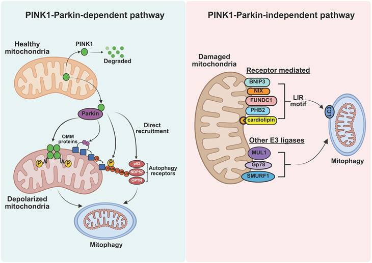
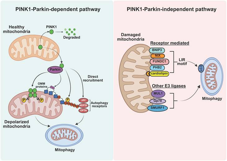
Mitophagy has attracted significant research interest since it was first proposed in 2005 [14]. The mechanisms of mitophagy can be broadly categorized into two pathways: the PINK1-Parkin-dependent pathway and the PINK1-Parkin-independent pathway [15] (Figure 1).
The mechanisms of mitophagy. Two main pathways: PINK1-Parkin-dependent pathway and PINK1-Parkin-independent pathway.
2.1.1 PINK1-Parkin-dependent pathway
The dependent pathway relies on extensive ubiquitination of mitochondrial surface proteins to facilitate mitophagy. The primary proteins involved are PTEN-induced putative kinase 1 (PINK1) and Parkin, forming the PINK1-Parkin-mediated mitophagy pathway [16]. PINK1 is a serine/threonine kinase located on depolarized mitochondria, and Parkin serves as an E3 ubiquitin ligase, facilitating the conjugation of ubiquitin to mitochondrial substrates [17]. Under normal conditions, the PINK1 precursor protein in the cytoplasm is imported into healthy mitochondria by its N-terminal mitochondrial-targeting sequences (MTSs), where it is cleaved by mitochondrial proteases in the matrix and inner membrane. The cleaved PINK1 is then degraded in the cytoplasm through the ubiquitin-proteasome system. However, when mitochondrial membrane potential (MMP) diminishes, the entry of PINK1 precursor protein into mitochondria is blocked, leading to its accumulation on the outer mitochondrial membrane (OMM). This accumulation triggers PINK1 dimerization and autophosphorylation, activating its kinase activity. Activated PINK1 recruits Parkin from the cytoplasm to the OMM and initiates its E3 ubiquitin ligase activity. Parkin then ubiquitinates various OMM proteins, and these ubiquitin chains are subsequently phosphorylated. The phosphorylated ubiquitin-modified proteins on the OMM are recognized by autophagy adaptor proteins, such as p62, nuclear dot protein 52 (NDP52), and optineurin (OPTN), which act as "eat me" signals to initiate autophagy [18-20]. Moreover, in addition to the PINK1-Parkin pathway, there are Parkin-independent ubiquitin-dependent pathways. It has been reported that PINK1 can directly recruit autophagy adaptor proteins to the mitochondria via ubiquitin phosphorylation, thereby promoting mitophagy [21]. Notably, PINK1 can amplify mitophagy by generating phosphorylated ubiquitin, which enhances the recruitment and activation of downstream effectors [22].
2.1.2 PINK1-Parkin-independent pathway
PINK1-Parkin-independent mitophagy is primarily mediated by mitophagy receptors that directly interact with autophagic machinery. Unlike the ubiquitin-dependent pathway, this process involves mitophagy receptors that contain a conserved microtubule-associated protein 1 light chain 3 (LC3)-interacting region (LIR). These receptors, including BCL2 interacting protein 3 (BNIP3), BCL2 interacting protein 3 like (BNIP3L, also known as NIX), FUN14 domain-containing 1 (FUNDC1), prohibitin 2 (PHB2), and cardiolipin, can directly bind to ATGs (e.g., LC3) via the LIR motif to initiate autophagosome formation [23, 24]. In addition, other E3 ligases involved in mitophagy include Smad ubiquitination regulatory factor 1 (SMURF1), mitochondrial E3 ubiquitin protein ligase 1 (MUL1), and Gp78 [25]. The main mitophagy receptors and E3 ligases identified so far are listed in the table below (Table 1). These mitophagy regulators sense mitochondrial damage and facilitate mitophagy independently of PINK1-Parkin, often responding to specific signals or stress conditions.
An overview of main PINK1-Parkin-independent mitophagy regulators
| Protein or lipid | Property | Localization | Inducers | Regulators | Autophagic interactors | References |
|---|---|---|---|---|---|---|
| BNIP3 | Receptor | OMM | Hypoxia | FOXO3↑ HIF1α↑ MA-5↑ | LC3B Atg8 | [56, 68] [142-144] |
| BNIP3L/NIX | Receptor | OMM | Hypoxia High OXPHOS activity | HIF1α↑ FBXL4↓ | GABARAPL1 | [145-148] |
| FUNDC1 | Receptor | OMM | Hypoxia FCCP | ULK1↑ PGAM5↑ CK2↓ SRC↓ | LC3B | [149-153] |
| FKBP8 | Receptor | OMM | Starvation | Rheb↓ | LC3A | [154-156] |
| BCL2L13 | Receptor | OMM | CCCP | - | LC3B | [157] |
| AMBRA1 | Receptor | OMM | FCCP | MCL-1↓ IKKα↑ SRC↓ HUWE1↑ | LC3 Beclin1 | [92, 158-162] |
| NLRX1 | Receptor | mtMatrix | CCCP | TRMT10C↓ | LC3 | [163-166] |
| PHB2 | Receptor | IMM | CCCP oligomycin + antimycin | TIPE1↓ miR-24-3p↓ ALDH2↓ | LC3B | [81, 104, 167, 168] |
| Cardiolipin | Receptor | IMM OMM | FCCP Rotenone Staurosporine 6-hydroxydopamine | CRLS1↑ SNCA↑ PLSCR3↑ | LC3 Beclin1 | [169-171] |
| NIPSNAP1/2 | Receptor | mtMatrix | Hypoxia CCCP oligomycin + antimycin | ATMLP↓ | LC3B Atg8 | [172, 173] |
| SMURF1 | E3-ubiquitin ligase | OMM | Hypoxia | TRIB3↓ | LC3 | [174, 175] |
| MUL1 | E3-ubiquitin ligase | OMM | Selenite | miR-135b-5p↑ | GABARAPL1 | [176, 177] |
| Gp78 | E3-ubiquitin ligase | ER membrane | CCCP | MGRN1↓ | LC3 | [178, 179] |
| SIAH1 | E3-ubiquitin ligase | Cytoplasm Nuclear | - | PINK1↑ | LC3 | [180] |
| ARIH1 | E3-ubiquitin ligase | Cytoplasm Nuclear | CCCP | PINK1↑ | - | [181] |
Abbreviations: AMBRA1: autophagy and beclin 1 regulator 1; ATMLP: lncRNA AFAP1-AS1 translated mitochondrial-localized peptide; ARIH1: ariadne RBR E3 ubiquitin protein ligase 1; AURKA: aurora kinase A; BCL2L13: BCL2 like 13; CCCP: carbonyl cyanide 3-chlorophenylhydrazone; CK2: casein kinase 2; CRLS1: cardiolipin synthase 1; FBXL4: F-box and leucine-rich repeat protein 4; FCCP: carbonyl cyanide 4-(trifluoromethoxy)phenylhydrazone; GABARAPL1: GABA type A receptor associated protein like 1; HUWE1: HECT, UBA and WWE domain containing E3 ubiquitin protein ligase 1; IKKα: inhibitory kappa B kinase α; LC3B: microtubule associated protein 1 light chain 3 beta; MA-5: mitochonic acid 5; MGRN1: mahogunin, ring finger 1; NIPSNAP1/2: nitrophenylphosphatase domain and non-neuronal SNAP25-like protein homolog 1/2; NLRX1: NLR family member X1; PLSCR3: phospholipid scramblase 3; Rheb: ras homolog, mTORC1 binding; SIAH1: siah E3 ubiquitin protein ligase 1; SNCA: alpha-synuclein; SRC: SRC proto-oncogene, non-receptor tyrosine kinase; TRIB3: tribbles pseudokinase 3; TRMT10C: tRNA methyltransferase 10C.
2.2. Mitophagy in renal physiology
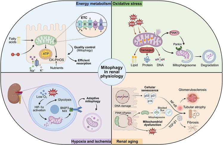
The kidney is a vital organ within the human urinary system, responsible for filtering metabolic waste products and reabsorbing essential nutrients [26]. This active excretion and reabsorption require substantial energy, primarily supplied by mitochondrial oxidative metabolism [27]. Consequently, mitophagy, a key component of mitochondrial quality control (MQC), plays an important role in maintaining renal physiological functions (Figure 2).
Mitophagy in renal physiology. Mitophagy is involved in various physiological processes of the kidney, such as energy metabolism, oxidative stress, hypoxia-ischemia, and aging.
2.2.1 Energy metabolism and homeostasis
Mitochondria power renal proximal tubules, which require high energy for nutrients and ion reabsorption [28]. These cells primarily rely on fatty acid oxidation (FAO) and oxidative phosphorylation (OX-PHOS) to produce ATP via the electron transport chain (ETC) [29]. However, in renal medullary regions, particularly within distal tubules and collecting ducts where oxygen tension is relatively low, glycolysis serves as the predominant energy-producing pathway, ensuring cellular function under hypoxic conditions [30].
FAO and OX-PHOS are tightly linked mitochondrial energy metabolism processes. Long-chain fatty acids are transported into mitochondria through the carnitine transport system, where they undergo β-oxidation in the mitochondrial matrix to generate acetyl coenzyme A (acetyl-CoA), as well as the reduced electron carriers-nicotinamide adenine dinucleotide (NADH) and flavin adenine dinucleotide (FADH₂). Acetyl-CoA then enters the tricarboxylic acid cycle, leading to further production of NADH, FADH₂, and GTP/ATP. Subsequently, NADH donates electrons to complex I of the electron transport chain, whereas FADH₂-derived electrons enter through complex II or related flavoprotein-dependent pathways. Electron transfer through the respiratory chain, including complexes I-IV, drives proton pumping across the inner mitochondrial membrane (IMM), thereby generating an electrochemical proton gradient. Finally, ATP synthase, also known as complex V, uses this gradient to synthesize ATP [31].
Mitophagy plays a critical role in MQC during these metabolic processes. In response to mitochondrial damage, such as a decline of MMP, or an excessive production of reactive oxygen species (ROS), mitophagy-related signaling pathways including PINK1/Parkin, BNIP3, and FUNDC1 are activated to facilitate the selective removal of damaged mitochondria [32].
This targeted clearance prevents dysfunctional mitochondria from impairing the efficiency of FAO and OXPHOS, thereby preserving a healthy mitochondrial population, optimizing ATP synthesis, and maintaining the homeostasis of proximal tubule cells [33].
2.2.2 Oxidative stress
Oxidative stress occurs when ROS production exceeds the capacity of antioxidant defenses. At physiological levels, ROS participate in the regulation of cellular signaling, metabolism, and immune responses, but excessive ROS can induce lipid peroxidation and damage cellular membranes, proteins, and DNA, thereby contributing to kidney injury [34]. To maintain mitochondrial homeostasis, mitophagy is activated under oxidative stress to remove damaged mitochondria. Excessive ROS can also impair mitochondrial function and reduce MMP, triggering PINK1 accumulation on the outer mitochondrial membrane. Activated PINK1 subsequently phosphorylates ubiquitin and Parkin, promoting Parkin recruitment and activation, and promoting the ubiquitination of outer mitochondrial membrane proteins, thereby initiating the PINK1-Parkin-mediated mitophagy [35, 36]. This process is essential for MQC and plays a protective role in kidney diseases, such as AKI and CKD.
2.2.3 Response to hypoxia and ischemia
Hypoxia increases mitochondrial ROS (mtROS) production, in part due to electron leakage from respiratory chain complexes I and III and the accumulation of reducing equivalents such as NADH [37]. Elevated ROS levels can disrupt redox hemostasis and contribute to the activation of hypoxia-responsive signaling pathways. Hypoxia-inducible factor (HIF) is primarily driven by oxygen limitation through stabilization of hypoxia-inducible factor 1, alpha subunit (HIF-1α), while mtROS may further modulate this response. Activated HIF promotes adaptive metabolic and quality-control programs including enhanced glycolysis and mitophagy [38]. In renal cells, hypoxia or ischemia-reperfusion injury causes oxidative damage to proteins, lipids, and mitochondrial components, impairing ETC activity and membrane integrity [39]. Under these conditions, mitophagy is primarily mediated by HIF-inducible receptors such as BNIP3 and NIX, which act as ubiquitin-independent mitophagy receptors by linking damaged mitochondria to LC3/GABARAP-positive autophagosomes for lysosomal degradation, thereby maintaining MQC and supporting cellular homeostasis [40, 41].
2.2.4 Renal aging
The kidney is highly susceptible to aging, characterized by structural and functional changes such as glomerulosclerosis, tubular atrophy, and interstitial fibrosis [42]. Cellular senescence drives these changes through DNA damage responses, p53/p21 and p16INK4a/Rb signaling, NF-κB-mediated inflammation, and transforming growth factor β1 (TGF-β1)-induced fibrosis [43]. Mitochondrial dysfunction is a hallmark of aging, involving impaired mitophagy, mtDNA damage, disrupted dynamics, and metabolic imbalance [44]. According to the mitochondrial free radical theory of aging (MFRTA), the accumulation of oxidative stress impairs mitochondrial function and mitophagy, leading to decreased efficiency of OX-PHOS and elevated production of ROS [45]. Although aging can activate mitophagy through pathways such as PINK1-Parkin, the concomitant blockade of autophagic flux prevents the clearance of damaged mitochondria. As a result, dysfunctional mitochondria accumulate, perpetuating oxidative stress and metabolic imbalance, thereby accelerating renal aging and functional decline [46].
3. Mitophagy and acute kidney injuries
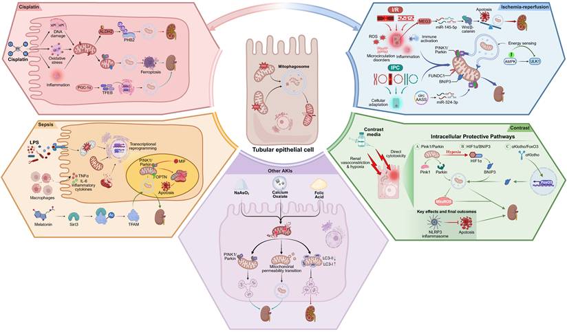
Acute kidney injury is a clinical syndrome characterized by a rapid deterioration of renal function over a short period, caused by a variety of etiological factors. It manifests as a reduction in glomerular filtration rate, accumulation of creatinine and urea nitrogen, and disturbances in water, electrolyte, and acid-base homeostasis [47]. Factors such as ischemia/reperfusion, exposure to surgical contrast agents, improper medication use, rhabdomyolysis, and infections can lead to AKI, many of which are known to stimulate mitophagy. Accumulating evidence suggests that mitophagy exerts a protective function, notably through the PINK1-Parkin and the BNIP3-mediated pathways [48, 49]. Interestingly, some studies have also reported that mitophagy aggravates AKI [50]. The discrepancy indicates the multiple roles of mitophagy in AKIs. Here, we review the role of mitophagy in different types of AKI and elucidate the mechanism underlying its activation in the kidney (Figure 3).
Mitophagy in AKI. AKI profoundly impairs mitochondrial homeostasis, leading to energy metabolism disorders, oxidative stress, and apoptosis. Concurrently, AKI induces activation of mitophagy to eliminate damaged mitochondria, and insufficient or excessive mitophagy further aggravates kidney injury.
3.1. Ischemia-reperfusion-related AKI
Ischemia-reperfusion injury (IRI) denotes the exacerbation of tissue following the restoration of blood flow after a period of ischemia. In clinical settings, AKI caused by IRI is associated with a high mortality rate [51]. The underlying mechanisms are intricate, including oxidative stress, endothelial and microcirculatory disorders, inflammation and immune activation, and various forms of controlled cell death (apoptosis, necroptosis, pyroptosis, and ferroptosis) [52]. Studies show that ischemia/reperfusion (I/R) significantly induces oxidative damage in the kidney, resulting in renal tubular injury and alterations in mitochondrial ultrastructure [53]. Growing research highlights the pivotal role of mitophagy in I/R-induced AKI. For example, renal IRI is exacerbated in mice with both single and double knockout of Pink1 and Parkin, indicating that PINK1-Parkin-mediated mitophagy plays a protective role in ischemic AKI [54]. Additionally, knockout of Bnip3, a specific mitophagy receptor, is shown to exacerbate injury during I/R-induced AKI in vivo [55]. Other researchers found that overexpression of BNIP3 restored hypoxia/reoxygenation (H/R)- induced reductions in mitophagy and mitigated cellular damage in vitro. These findings support the protective role of BNIP3-mediated mitophagy in renal IRI [56]. Moreover, a recent study showed that circAASS alleviates I/R-induced AKI by improving mitochondrial homeostasis and inhibiting apoptosis and inflammatory response of renal tubular epithelial cells (RTECs). Specifically, the circAASS in the cytoplasm acts as a competitive endogenous RNA (ceRNA) by binding to MIR324-3p, thereby promoting PINK1 expression and enhancing mitophagy [57].
Ischemic preconditioning (IPC) involves a brief, intermittent period of I/R that induces adaptive responses in affected tissues, such as alterations in energy metabolism, reduced free radical production, and decreased inflammation and apoptosis [58]. Studies have demonstrated that IPC confers protection against subsequent renal I/R in mice, primarily through PINK1-Parkin-dependent mitophagy [59]. Furthermore, the study found that the renal protective effects of IPC against renal injury, inflammation, and tubular cell death are abolished in proximal tubule-specific Fundc1 (a mitophagy receptor) knockout mice, highlighting the essential protective role of mitophagy in IPC [60]. Additional studies have linked energy-sensing pathways to the regulation of mitophagy. In both in vivo and in vitro experiments, the phosphorylation level of Thr172- AMP-activated protein kinase (AMPK) α decreases rapidly after I/R. Mice with tubular epithelial cell-specific AMPKα deficiency exhibit more severe renal injury and increased apoptosis under I/R, partly because reduced AMPK activity inhibits UNC51-like kinase 1 (ULK1)-mediated autophagy, impairing the clearance of dysfunctional mitochondria [61]. Contradictory findings also exist. c-MYC-activated maternally expressed 3 (MEG3) exacerbates renal IRI by upregulating rhotekin (RTKN) to induce mitophagy and trigger the Wnt/β-catenin pathway [62]. The discrepancy may be explained by the fact that Wnt/β-catenin pathway activation can also promote apoptosis of tubular epithelial cells during AKI, with the pro-apoptotic effects potentially outweighing the protective benefits of mitophagy [63].
3.2. Contrast-induced AKI
Contrast media have been widely used to enhance the visibility of internal structures, organs, and tissues in medical imaging. Contrast-induced AKI (CI-AKI) occurs in more than 30% of patients following intravenous iodine administration, and currently, there are no effective targeted therapies [64]. Contrast media have direct cytotoxicity leading to cellular death and tubular injury, while contrast media also alter renal hemodynamics, leading to renal vasoconstriction and renal hypoxia [65]. Studies demonstrated that PINK1-Parkin-mediated mitophagy plays a crucial role in preventing apoptosis of RTECs by decreasing mtROS levels, which in turn reduces the activation of the NOD-like receptor thermal protein domain associated protein 3 (NLRP3) inflammasome in CI-AKI [66]. The research group also revealed that upregulation of HIF-1α-BNIP3-mediated mitophagy attenuates NLRP3 inflammasome-mediated apoptosis in CI-AKI [67]. Similar results were observed in that αKlotho promotes BNIP3-mediated mitophagy by activating forkhead box O3 (FOXO3), consequently reducing mtROS, cell apoptosis, and renal injury in CI-AKI [68]. Collectively, these findings suggest that mitophagy forms a positive feedback loop in protecting against CI-AKI, highlighting its potential as a therapeutic target in drug development.
3.3. Mitophagy in sepsis-associated AKI
Sepsis-associated AKI (SA-AKI) is a sudden loss of kidney function due to the body's extreme response to infection, common in critically ill patients [69, 70]. Sepsis-associated AKI progresses through four stages: intense inflammation (initiated by innate immune response), self-amplification (to exert antiviral response), maladaptive metabolic shutdown (transcriptional reprogramming), and recovery (metabolic adaptation) [71]. Ultrastructural mitochondrial alterations, such as cristae disruption and mitochondrial swelling, are frequently observed during SA-AKI. In response, mitophagy is activated to minimize cellular damage and promote recovery [72]. Mitophagy is activated, as demonstrated by the upregulation of PINK1-Parkin in mouse models of SA-AKI induced by lipopolysaccharide (LPS) or cecal ligation and puncture (CLP), as well as in human renal proximal tubular epithelial cells (HK-2) exposed to LPS. Nonetheless, knockout of Pink1 or Park2 impairs mitophagy by blocking mitochondrial accumulation of the autophagy receptor OPTN, resulting in exacerbated kidney injury and increased apoptosis [73]. Alternatively, macrophage migration inhibitory factor (MIF) interferes with PINK1-Parkin interaction, affecting Parkin's mitochondrial recruitment and thereby impairing mitophagy [74]. Researchers analyzed 48 patients with clinical sepsis and found that the extent of mitochondrial damage in renal tubular epithelial cells inversely correlates with recovery from AKI. They further found that melatonin, a potent SIRT3 (sirtuin 3) agonist, alleviates SA-AKI by promoting mitophagy via SIRT3-mediated deacetylation of transcription factor A, mitochondrial (TFAM), offering promising therapeutic insights [75].
3.4. Cisplatin-induced AKI
Cisplatin is an effective chemotherapeutic agent used to treat various solid tumors. However, its clinical application is limited by significant nephrotoxicity [76]. Cisplatin has been shown to cause DNA/mitochondrial damage, oxidative stress, endoplasmic reticulum (ER) stress, inflammation (cytokines/immune cells), mitochondrial dysfunction (ATP loss, calcium imbalance), metabolic reprogramming (glycolysis, impaired FAO), and cell death (apoptosis, necroptosis, ferroptosis, etc) [77]. Evidence indicates that mitophagy exerts protective effects in cisplatin-induced AKI [78]. For instance, BNIP3 and PINK1-Parkin-mediated mitophagy can protect renal tubular epithelial cells from ferroptosis induced by cisplatin, primarily via the ROS/HO1/GPX4 signaling pathway [79]. However, another study has shown that the deficiency of Pink1 can improve acute kidney injury induced by cisplatin, possibly by inhibiting mitochondrial fission mediated by dynamin-related protein 1 (DRP1) and excessive mitochondrial phagocytosis [80]. In addition, a recent study found markedly elevated levels of lactylation, a post-translational modification (PTM), in kidneys of AKI patients and in cisplatin-induced mice. Further studies showed that aldehyde dehydrogenase 2 (ALDH2) lactylation facilitates the ubiquitination and proteasome degradation of PHB2, a vital mitophagy receptor, thereby suppressing mitophagy and aggravating mitochondrial dysfunction [81]. Conversely, a separate study reported that cisplatin inhibits mitophagy in renal tubule cells. However, peroxisome proliferator-activated receptor γ coactivator-1α (PGC-1α) reactivated mitophagy and lysosomal biogenesis via the transcription factor EB (TFEB), ultimately ameliorating kidney injury [82]. These conflicting findings may suggest that mitophagy plays distinct, context-dependent roles at different stages of AKI.
3.5. Other AKIs
In clinical practice, several less common factors can induce AKI, including arsenic exposure, folic acid (FA) toxicity, and calcium oxalate crystal deposition. The role of mitophagy in AKI triggered by these factors is not fully understood. Exposure to NaAsO2 remarkably exacerbates renal tubular injury and mitochondrial dysfunction and increases the expression of mitophagy-related proteins. This study also demonstrated that PINK1-Parkin-dependent mitophagy exerts a protective effect against NaAsO2-induced AKI, but the precise underlying mechanism has yet to be elucidated [83]. In a rat model of FA-induced AKI, the expression of PINK1 and p62 increases, while LC-3II/I decreases, suggesting that mitophagy is impaired [84]. Additionally, AKI caused by calcium oxalate crystal deposition in distal renal tubular epithelial cells is associated with mitochondrial alterations characterized by mitochondrial permeability transition, which ultimately triggers mitophagy [85].
4. Mitophagy in chronic kidney disease
Chronic kidney disease (CKD) is characterized by persistent structural and functional renal abnormalities over three months [86]. Common causes include diabetes mellitus, glomerulonephritis, and polycystic kidney disease, among others [87]. Despite extensive research, the precise mechanisms underlying CKD development remain incompletely understood. Renal inflammation and fibrosis are common pathogenic mechanisms in CKD [88]. Accumulating evidence indicates that mitochondrial dysfunction in renal tubular epithelial cells plays a crucial role in CKD progression and is closely linked to renal fibrosis [89]. Studies have shown that the deficiency of acyl-CoA synthetase family 3 (Acsm3), involved in the metabolism of medium-chain fatty acids (MCFA), leads to metabolic disorders, which in turn disrupt mitochondrial homeostasis and ultimately aggravate unilateral ureteral obstruction (UUO)-induced renal fibrosis [90]. Studies have demonstrated that mitophagy is induced in tubular epithelial cells during renal fibrosis. Under UUO or hypoxic conditions, deletion or silencing of Bnip3 resulted in significantly increased mtROS production, mitochondrial damage, NLRP3 inflammasome activation, and elevated levels of α-smooth muscle actin (α-SMA) and TGF-β1, indicating that BNIP3-mediated mitophagy exhibits a protective effect against fibrosis [91]. Furthermore, the knockout of Parkin inhibits mitophagy in endothelial cells, leading to an increase in interleukin-6 production, thereby inducing epithelial-to-mesenchymal transition (EndoMT), and ultimately exacerbating interstitial fibrosis in renal allografts of rats subjected to allogeneic kidney transplantation [92]. Nevertheless, another study indirectly indicates that overactivation of mitophagy might cause a buildup of autophagosomes, hindering the efficient degradation of damaged mitochondria. This process instead results in the release of more mtDNA, triggers the stimulator of interferon genes (STING) pathway, and worsens inflammation and fibrosis [93]. Of particular interest, an earlier study reported reduced levels of Parkin and autophagy in the renal tissues of UUO mice, in macrophages treated with TGF-β1, and in kidney tissues from adenine diet-induced CKD models [94, 95]. The discrepancy suggests that alterations in mitophagy may differ across various cell types and disease stages. Nonetheless, all the evidence indicates that the dysfunction of mitophagy is an important factor contributing to the acceleration of renal fibrosis.
In summary, the prevailing view is that mitophagy plays a protective role in renal fibrosis. By reducing mitochondrial damage, limiting epithelial cell apoptosis, and decreasing inflammatory infiltration, mitophagy can mitigate tubulointerstitial fibrosis. However, it is important to recognize that models such as UUO, which induce rapid and irreversible fibrosis, may not fully capture the complexities of renal repair processes.
5. Mitophagy and diabetic kidney disease
Diabetic kidney disease (DKD) is characterized by persistent proteinuria and a gradual decline in glomerular filtration rate (GFR) attributable to prolonged diabetes, representing one of the most common microvascular complications of diabetes [96]. Hyperglycemia is the primary driving pathogenesis in diabetic kidney disease, including hypertension, glomerular cell injury, disrupted tubular feedback mechanisms, renal hypoxia, inflammation, mitochondrial dysfunction, and impaired autophagy. These disturbances collectively promote podocyte loss, extracellular matrix expansion, glomerular sclerosis, and progressive renal function decline [97].
Bulk gene expression analyses have revealed several major dysregulated pathways in DKD, notably alterations in FAO and OXPHOS [98]. Mitochondrial abnormalities are increasingly recognized as the central contributors to these changes. Studies have shown that mitophagy in the podocytes of patients with DKD is severely inhibited [99]. Similarly, this phenomenon has also been observed in animal models of glomerular disease induced by high-fat diet (HFD) and streptozotocin (STZ) injection [100, 101]. Besides, impaired mitophagy has been observed in the kidneys of diabetic patients and in RTECs derived from mice exposed to high glucose (HG), suggesting that mitochondrial dysfunction plays a pivotal role in the onset and progression of DKD [102].
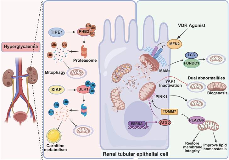
One study revealed that the deficiency of Ulk1 exacerbates kidney damage in diabetic mouse models. From a mechanistic perspective, the upregulation of X-linked inhibitor of apoptosis (XIAP) leads to the degradation of ULK1 through K48-linked polyubiquitination, thereby inhibiting mitophagy and disrupting carnitine metabolism. Restoring ULK1 protein expression by administering the ULK1 agonist echinacoside and supplementing with L-carnitine improves mitophagy and carnitine homeostasis, thereby alleviating DKD [103]. Furthermore, tumor necrosis factor alpha-induced protein 8-like 1 (TIPE1) has been shown to mediate ubiquitination and subsequent proteasomal degradation of PHB2, disrupting mitophagy in RTECs under hyperglycemic conditions, consequently culminating in tubular injury and exacerbating DKD progression [104]. Another study indicated that disrupted mitophagy and mitochondrial fission and fusion were associated with mtROS overproduction, MMP reduction, and renal interstitial fibrosis in diabetic rats. Notably, a Vitamin D receptor (VDR) agonist partially restored impaired mitophagy in diabetic rats via the mitofusin 2 (Mfn2)-mitochondria-associated ER membranes (MAMs) pathway, as evidenced by elevated levels of Pink1 and Parkin. From a mechanistic standpoint, VDR upregulates the expression of Mfn2, thereby reinstating the structural integrity of MAMs. This restoration of MAMs integrity facilitates the interaction between Fundc1 and LC3, which in turn triggers the activation of mitophagy [105]. Studies have shown that inactivation of yes-associated protein 1 (YAP1) results in dysregulation of MQC and triggers abnormalities in mitochondrial biogenesis and mitophagy in renal tubular cells, ultimately leading to tubular damage in DKD [106]. Additionally, Wang et al identified translocase of outer mitochondrial membrane 7 (TOMM7) as a critical regulator of mitophagy within renal tubules, contributing to the mitigation of DKD. Mechanistically, TOMM7 facilitates the recruitment of PLA2G6 to mitochondria. This process is accompanied by the restoration of mitochondrial membrane integrity, improved lipid homeostasis, and reduced ROS levels. Concurrently, TOMM7 promotes the stable accumulation of PINK1 on the OMM, thereby initiating the activation of mitophagy [107]. Recent studies also have demonstrated that in HK-2 cells under HG/palmitic acid (PA) conditions, the nuclear receptor estrogen-related receptor alpha (ESRRA) transcriptionally activates autophagy-related protein 5 (ATG5) to support PINK1-dependent mitophagy and maintain tubular homeostasis [108].
In summary, impairment of mitophagy and associated pathways, including ubiquitin-dependent and independent, is involved in ensuring MQC and sustaining the physiological function of mitophagy throughout DKD development (Figure 4). Nevertheless, the mechanisms by which impaired mitophagy contribute to glomerular and tubular injury in DKD remain incompletely understood, necessitating further mechanistic and translational research.
Mitophagy in DKD. In DKD, mitophagy is often inhibited, resulting in the accumulation of dysfunctional mitochondria. Conversely, proteins such as TOMM7, YAP1, and MFN2 promote mitophagy and improve mitochondrial function through distinct molecular pathways.
At this point, we have summarized the targets studied so far that mediate mitophagy in various kidney diseases, along with their potential functions and possible mechanisms (Table 2).
Regulatory targets and potential functions of mitophagy in different types of kidney diseases
| Kidney disease | Cause of disease | Regulatory target | Cell lines | Potential function | Possible mechanism | Reference |
|---|---|---|---|---|---|---|
| AKI | I/R | PINK1-Parkin | HK-2 | Anti-AKI | ROS production↓ Inflammation↓ | [54] |
| BNIP3 | BUMPT | Anti-AKI | Inflammatory cell infiltration↓ Release of proinflammatory cytokines↓ | [55] | ||
| BNIP3 | HK-2 | Anti-AKI | Cell apoptosis↓ ROS production↓ | [56] | ||
| Atg7 | RPTCs | Anti-AKI | Susceptibility to damage↓ Tubular cell necrosis↓ | [59] | ||
| FUNDC1 | PRTCs | Anti-AKI | Mitochondrial quality↑ Drp1-dependent mitochondrial fission↑ | [60] | ||
| ULK1 | PRTCs | Anti-AKI | Fatty acid oxidation↓ Tubular cell apoptosis↓ | [61] | ||
| AKI | Contrast | PINK1-Parkin | HK-2 | Anti-AKI | mtROS production↓ NLRP3 inflammasome activation↓ Cell apoptosis and tissue damage↓ | [66] |
| BNIP3 | HK-2 | Anti-AKI | mtROS production↓ Apoptosis↓ | [67, 68] | ||
| Spesis | PINK1-Parkin | RPTCs | Anti-AKI | Inflammation↓ Apoptosis↓ | [73] | |
| TFAM | HK-2 | Anti-AKI | Mitochondrial respiratory chain↑ NAD/NADH balance↑ | [75] | ||
| Cisplatin | PINK1-Parkin | BUMPT | Anti-AKI | Renal functional loss↓ Tissue damage↓ Apoptosis↓ | [78] | |
| PHB2 | HK-2 | Anti-AKI | Mitochondrial morphology/function↑ Mitochondrial apoptosis↓ | [81] | ||
| TFEB | mIMCD-3 | Anti-AKI | mtROS production↓ | [82] | ||
| AKI | Arsenic | PINK1-Parkin | HK-2 | Anti-AKI | Mitochondrial biogenesis and fission↑ Inflammation↓ | [83] |
| Folic acid | PINK1-Parkin | LLC-PK1 | Anti-AKI | Oxidative stress↓ Tubular injury and death↓ Inflammation and fibrosis↓ | [84] | |
| Calcium oxalate | PINK1-Parkin | HK-2 | Anti-AKI | Necroptosis↓ Interstitial inflammation↓ | [85] | |
| CKD | UUO H/R | BNIP3 | HK-2 | Anti-fibrotic | mtROS production↓ NLRP3 inflammasome↓ | [91] |
| UUO AD TGF-β1 | PINK1-Parkin | BMDMs THP-1 | Anti-fibrotic | mtROS production↓ Macrophage-polarization toward M2↑ | [94] | |
| DKD | HG | OPTN | mPTECs | Anti-DKD | mtROS production↓ Cellular senescence↓ | [102] |
| Single-side Nx+STZ | PHB2 | HK-2 | Anti-DKD | Mitochondrial morphology and MMP↑ EMT↓ Tubular cell death↓ | [104] | |
| DKD | db/db HFD+STZ HG/ TGF-β1 | ULK1 | mPTECs HK-2 | Anti-DKD | mtROS production↓ MMP↑ Carnitine homeostasis↑ | [103] |
| db/db HG/PA | PINK1-Parkin | HK-2 | Anti-DKD | mtROS production↓ MMP↑ Inflammation↓ | [182] |
Abbreviations: AD: adenine diet; Atg7: autophagy-related protein 7; BMDMs: bone marrow-derived macrophages; BUMPT: Boston University mouse proximal tubule; EMT: epithelial-mesenchymal transition; LLC-PK1: proximal tubular cell line derived from porcine kidney; mIMCD-3: mouse inner medullary collecting duct cell line; Nx: nephrectomy; PRTC: primary renal tubular epithelial cells; RPTC: renal proximal tubular cells; THP-1: human acute monocytic leukemia cells.
6. Targeting mitophagy for kidney diseases
6.1. Selective regulators of mitophagy
Numerous non-selective and selective mitophagy regulators have been identified, which modulate mitochondrial biological processes, such as FAO, OX-PHOS, ETC complexes, and mitophagy pathways [25]. Metformin is a non-selective regulator that promotes mitophagy in peripheral blood mononuclear cells from patients with type 2 diabetes, primarily through the activation of AMPK [109]. Several selective modulators have been developed to target the PINK1-Parkin signaling pathway. (Table 3). However, the druggability, clinical efficacy, and safety profiles of these modulators remain largely unknown.
Mitophagy-specific activators and modulators
| Effect | Drug name | Drug type | Target | Mechanisms | Research progress | Reference |
|---|---|---|---|---|---|---|
| Activators | FB231 | Small molecule | Parkin | Directly enhancing PINK1-Parkin-dependent mitophagy | Cell + animal | [119] |
| MTK458 | Small molecule | PINK1 | Lowering the threshold for cells to respond to mitochondrial damage | Cell + animal | [119] | |
| T0467 | Small molecule | Parkin | Promoting the translocation of Parkin to the damaged mitochondria | Cell-based | [183] | |
| PARL-IN-1/2 | Small molecule | Parkin | Promoting the translocation of Parkin to the damaged mitochondria | Cell-based | [183] | |
| KTP | Small molecule | PINK1 | Enhancing the kinase activity of PINK1 | Cell-based | [184] | |
| Modulators | BIO-2007817 | Molecular glue | PINK1 | Promoting the accumulation of PINK1 outside the damaged OMM and enhancing the phosphorylation of PINK1 by ubiquitin | Cell-based | [185] |
| LCL768 | Molecular glue | Parkin | Attenuating Parkin succination to promote Parkin activation | Cell-based | [120] |
6.2. Potential application for kidney diseases
Recently, mitophagy therapies targeting the kidneys have shown promise in cellular and animal models of kidney diseases.
6.2.1 PINK1-Parkin-dependent pathway
The small-molecule compound UMI-77, identified by high-throughput screening, is a novel mitophagy inducer that enhances the interaction between myeloid cell leukemia 1 (MCL-1) and LC3A on the mitochondrial surface. Studies using UMI-77 to activate mitophagy in RTECs demonstrate that mitophagy activation mitigates mitochondrial dysfunction, inhibits the TGF-β/Smad signaling pathway, and alleviates inflammation, thereby delaying the development of renal fibrosis in UUO mice [110]. Although UMI-77 has shown remarkable efficacy in preclinical animal studies, several potential obstacles remain before its clinical application can be realized. For example, MCL-1, the main target of UMI-77, not only regulates mitophagy but also plays critical roles in apoptosis, cell cycle progression, immune cell survival, and tumor biology. Therefore, pharmacological modulation of MCL-1 may produce unintended effects such as altered cell survival, immune dysregulation, or even oncogenic risk, which must be carefully evaluated in future studies [111]. Additionally, as a BH3 mimetic, UMI-77 may induce cell apoptosis at high doses, thereby increasing the risk of tissue injury [112]. Currently, systematic pharmacokinetic and toxicological data are still lacking, and further studies are needed to comprehensively evaluate its safety and translational potential. Furthermore, studies have found that the AMPK agonist metformin improves renal oxidative stress and tubulointerstitial fibrosis in diabetic mice by activating the p-AMPK-PINK1-Parkin mitophagy pathway [113]. Lycopene inhibits the activation of the protein kinase B (also known as AKT) signal, activates mitophagy, and alleviates renal fibrosis [114]. WJ-39, the novel aldo-keto reductase inhibitor, has been discovered to preserve the structural and functional integrity of renal tubules in diabetic nephropathy by activating PINK1-Parkin-mediated mitophagy [115]. Furthermore, Urolithin A (Uro A) suppresses the inflammatory response mediated by STING-NLRP3 through inducing PINK1-Parkin-dependent mitophagy, thereby alleviating fructose-induced hypouricemic nephropathy [116]. Uro A has advanced to the Phase I clinical trial, where oral administration in healthy adults improves immune function by modulating immune cell mitochondrial activity [117]. However, the efficacy of Uro A in treating kidney diseases remains to be verified through large-scale clinical studies. Its clinical translation also faces several challenges, including limited tissue specificity, significant interindividual variability in bioavailability, and a lack of long-term safety data. These limitations may hinder its further clinical application. Ruxolitinib has been demonstrated to ameliorate renal fibrosis by activating PINK1-Parkin-mediated mitophagy [118]. Recently, several other compounds, including MTK458, LCL768, and MTX115325, have been shown to modulate mitophagy, though their effects and mechanisms in kidney disease remain to be fully elucidated [119-121]. The potential therapeutic application of these compounds in kidney diseases warrants further investigation.
Chinese medicine and phytochemicals also have great potential to modulate mitophagy in kidney disease. For example, Huangkui capsule induces mitophagy in renal tubular cells through upregulating PINK1 expression, thereby alleviating diabetic nephropathy [122]. Similarly, swietenine has been reported to improve renal function in diabetic mice by enhancing mitophagy via the activation of the Acsf2/PHB2/PINK1 signaling pathway [123]. In addition, naringenin promotes mitophagy by directly binding to PINK1 and enhancing its stability, thereby alleviating LPS-induced renal structural damage and oxidative damage [124].
6.2.2 Receptor-mediated pathway
Resveratrol alleviates sepsis-induced acute kidney injury by activating SIRT1, promoting the upregulation of PGC-1α, and enhancing BNIP3/NIX-mediated mitophagy [125]. Cobalt oxide-polyethylene glycol-triphenylphosphine (COPT) nanoparticles have been reported to ameliorate H/R-induced mitochondrial damage by enhancing BNIP3-mediated mitophagy both in vitro and in vivo. These nanoparticles also alleviate ischemic AKI in a mouse model and gentamicin-induced AKI in a zebrafish model [126]. Additionally, a recent study suggests that melatonin can protect kidney function by regulating FUNDC1-mediated mitophagy [127].
6.3. Challenges in clinical application
Despite its therapeutic potential, the clinical application of mitophagy-targeted strategies in kidney disease still faces several challenges. First, mitophagy exhibits substantial cell-type and disease-subtype specificity, which complicates the development of personalized therapeutic approaches. For instance, activation of mitophagy in proximal tubular cells may exert protective effects, whereas excessive or maladaptive mitophagy in distal tubules or podocytes may contribute to cellular dysfunction, making it difficult to design uniform interventions. Second, variations in the agents, dosages, and delivery systems used to modulate mitophagy across different studies have resulted in inconsistent outcomes. This heterogeneity limits reproducibility and hinders the establishment of standardized treatment guidelines. More importantly, the long-term safety of mitophagy-targeted strategies remains to be fully determined, particularly in patients with severe kidney disease or immunosuppression. In addition, systemic delivery of mitophagy modulators raises concerns about off-target effects in non-renal tissues, such as the heart and liver [128]. Finally, the potential for drug resistance or maladaptive cellular adaptation should not be overlooked. If cells adapt to chronic mitophagy stimulation, therapeutic effect may diminish over time, while persistent mitochondrial stress could create latent health risks.
7. Discussion and Conclusion
The role of mitophagy in kidney diseases is complex and context-dependent, exhibiting a bidirectional effect influenced by the specific pathological environment. In AKI, mitophagy protects renal tubular cells by eliminating damaged mitochondria and reducing ROS accumulation [50]. In renal fibrosis, mitophagy suppresses EndoMT and thereby delays fibrotic progression; however, when excessively activated, mitophagy may promote mtDNA release and inflammatory responses, and the excessive clearance of mitochondria can further cause energy deficiency and structural abnormalities in renal tubular epithelial cells, ultimately leading to tubular atrophy and fibrosis [93, 114, 129]. In DKD, podocytes and renal tubular cells highly rely on mitochondrial homeostasis, and enhancement of mitophagy, such as through AMPK activation, appears to exert protective effects [113]. These findings highlight that future treatment strategies need to precisely regulate mitophagy levels based on the underlying disease mechanisms, rather than broadly enhancing or suppressing the process.
Furthermore, accumulating evidence indicates that the role of mitophagy varies across different kidney intrinsic cell types, including podocytes and endothelial cells [130]. For instance, podocytes are particularly reliant on mitochondrial homeostasis, and deficiency in PINK1-Parkin-mediated mitophagy has been shown to exacerbate proteinuria and podocyte injury [131]. By contrast, in human umbilical vein endothelial cells (HUVECs) induced by tumor necrosis factor-alpha (TNFα), suppression of the Rictor/mTORC2 signaling pathway enhances BNIP3-dependent mitophagy, thereby alleviating EndoMT and interstitial fibrosis in renal transplantation [132]. Beyond intrinsic renal cells, immune populations such as macrophages and T lymphocytes also play pivotal roles in kidney pathogenesis, with emerging evidence suggesting that mitophagy regulates macrophage polarization and T cell activation [133-135]. Collectively, these findings raise a compelling research question: Does mitophagy operate through distinct molecular pathways in podocytes, renal vascular endothelial cells, and immune cells, and do these cell-specific regulatory mechanisms ultimately shape the pathological classifications and therapeutic outcomes of kidney diseases? Addressing this question will require future investigations that focus on cell-type-specific mechanisms, integrating genetic models, single-cell approaches, and targeted delivery strategies to delineate the precise role of mitophagy in renal pathophysiology.
Emerging evidence indicates that mitophagy interacts intricately with multiple forms of cell death, thereby shaping the outcome of kidney injury [136]. On the one hand, mitophagy mitigates apoptosis by removing damaged mitochondria, reducing ROS accumulation and preventing cytochrome c release. Conversely, insufficient mitophagy accelerates apoptotic signaling [50]. In the context of necrosis, excessive or dysregulated mitophagy may precipitate energy collapse and membrane rupture, while necrotic inflammation can further impair autophagic flux [137]. Mitophagy also intersects with pyroptosis, where suppression of mtROS and mtDNA release attenuates NLRP3 inflammasome activation, reducing inflammatory cell death. Moreover, links between mitophagy and ferroptosis have been highlighted, as mitochondrial iron handling and lipid peroxidation can couple mitophagy imbalance to ferroptotic injury [138]. Taken together, these interactions underscore mitophagy as a central regulator of cellular fate in kidney diseases, functioning as either a protective mechanism or a potential pathological driver depending on the context.
Given that mitophagy is a ubiquitous cellular process, systemic administration of modulators may lead to widespread, non-targeted effects in other organs. Therefore, the targeted delivery strategy for the kidneys is critical for advancing therapeutic applications. Recent studies have demonstrated that nanoparticles encapsulating Parkin mRNA can promote synchronous regulation of functional and dysfunctional mitochondria, showing promise for treating pulmonary fibrosis [139]. Simultaneously, researchers have recently developed a biomimetic high-density lipoprotein (bHDL) lipid nanoparticle that exhibits excellent targeting ability for damaged renal tubular epithelial cells through endocytosis mediated by kidney injury molecule-1 (KIM-1) [140]. Based on these findings, we propose a testable hypothesis: The bHDL lipid nanoparticles that encapsulate Parkin mRNA could specifically target renal tissues and induce mitophagy, thus providing a potential strategy for the treatment of selected kidney diseases. Moreover, mitochondrial transplantation, an emerging cell therapy strategy, has been applied to treat experimental cerebellar degenerative diseases, in which mitochondria transplanted from the liver into the cerebellum of Drp1 conditional knockout mice improve mitochondrial function, delay neuronal apoptosis, and alleviate cerebellar ataxia [141]. These strategies offer valuable insights for developing targeted mitochondrial therapies in kidney diseases.
Additionally, some clinically used drugs indirectly modulate mitophagy. For instance, Metformin and other AMPK agonists help maintain mitochondrial homeostasis and have demonstrated protective effects in animal models of kidney diseases [109, 113]. Future research should explore the potential of these drugs to improve mitophagy-related kidney disease, with a systematic evaluation of their efficacy, safety, and dosimetric characteristics to facilitate clinical translation.
In conclusion, mitophagy plays a multifaceted role in kidney diseases, exerting both protective and detrimental effects depending on disease type, cellular context, and immune involvement. Moving forward, therapeutic strategies should focus on precise regulation and targeted delivery, combined with mechanism research and translational exploration, ultimately opening new avenues for the treatment of various kidney diseases.
Abbreviations
AKI: acute kidney injury; CKD: chronic kidney disease; DKD: diabetic kidney disease; MQC: mitochondrial quality control; ATGs: autophagy-related proteins; PINK1: PTEN-induced putative kinase 1; MMP: mitochondrial membrane potential; FAO: fatty acid oxidation; OX-PHOS: oxidative phosphorylation; ROS: reactive oxygen species; I/R: ischemia/reperfusion; OMM: outer mitochondrial membrane; NDP52: nuclear dot protein 52; OPTN: optineurin; ETC: electron transport chain; MFRTA: mitochondrial free radical theory of aging; RTECs: renal tubular epithelial cells; ULK1: UNC51-like kinase 1.
Acknowledgements
The figures were created using the BioRender platform (https://www.biorender.com).
Funding
This study was supported by grants from the Hong Kong Research Grants Council (17125323, 17125524, and 17125925) and the HKU Seed Funds (2502251990 and 2402101703).
Author contributions
Hao Lu, Shenyu Yan and Baoyi Shao read a large number of relevant literature and drafted the manuscript. Jianbo Guo, Yifan Wang and Rui Hou participated in the review and revision of the manuscript. Shen Chen and Meicen Wu eventually edited the manuscript. Biao Wei and Fei Li assisted in data extraction. Xiaoming Meng and Haiyong Chen supervised and advanced the process. All authors contributed to the final approval of the version to be published.
Competing Interests
The authors have declared that no competing interest exists.
References
1. Herrington WG, Judge PK, Grams ME, Wanner C. Chronic kidney disease. Lancet. 2026;407:90-104
2. Global regional, national burden of chronic kidney disease in adults 1990-2023, its attributable risk factors. a systematic analysis for the Global Burden of Disease Study 2023. Lancet. 2025;406:2461-82
3. Francis A, Harhay MN, Ong ACM, Tummalapalli SL, Ortiz A, Fogo AB. et al. Chronic kidney disease and the global public health agenda: an international consensus. Nat Rev Nephrol. 2024;20:473-85
4. Fu Y, Livingston MJ, Dong G, Chen A, Cai J, Dong Z. Selective autophagy in kidney health and disease. Nat Rev Nephrol. 2026
5. Tang C, Cai J, Yin XM, Weinberg JM, Venkatachalam MA, Dong Z. Mitochondrial quality control in kidney injury and repair. Nat Rev Nephrol. 2021;17:299-318
6. Picca A, Faitg J, Auwerx J, Ferrucci L, D'Amico D. Mitophagy in human health, ageing and disease. Nat Metab. 2023;5:2047-61
7. Wang Y, Cai J, Tang C, Dong Z. Mitophagy in Acute Kidney Injury and Kidney Repair. Cells. 2020 9
8. Li S, Lin Q, Shao X, Zhu X, Wu J, Wu B. et al. Drp1-regulated PARK2-dependent mitophagy protects against renal fibrosis in unilateral ureteral obstruction. Free Radic Biol Med. 2020;152:632-49
9. Yao L, Liang X, Qiao Y, Chen B, Wang P, Liu Z. Mitochondrial dysfunction in diabetic tubulopathy. Metabolism. 2022;131:155195
10. Vargas JNS, Hamasaki M, Kawabata T, Youle RJ, Yoshimori T. The mechanisms and roles of selective autophagy in mammals. Nat Rev Mol Cell Biol. 2023;24:167-85
11. Lamark T, Johansen T. Mechanisms of Selective Autophagy. Annu Rev Cell Dev Biol. 2021;37:143-69
12. Li X, He S, Ma B. Autophagy and autophagy-related proteins in cancer. Mol Cancer. 2020;19:12
13. Guo QQ, Wang SS, Jiang XY, Xie XC, Zou Y, Liu JW. et al. Mitochondrial ROS triggers mitophagy through activating the DNA damage response signaling pathway. Proc Natl Acad Sci U S A. 2025;122:e2502841122
14. Lemasters JJ. Selective mitochondrial autophagy, or mitophagy, as a targeted defense against oxidative stress, mitochondrial dysfunction, and aging. Rejuvenation Res. 2005;8:3-5
15. Wang S, Long H, Hou L, Feng B, Ma Z, Wu Y. et al. The mitophagy pathway and its implications in human diseases. Signal Transduct Target Ther. 2023;8:304
16. Han R, Liu Y, Li S, Li XJ, Yang W. PINK1-PRKN mediated mitophagy: differences between in vitro and in vivo models. Autophagy. 2023;19:1396-405
17. Valente EM, Abou-Sleiman PM, Caputo V, Muqit MM, Harvey K, Gispert S. et al. Hereditary early-onset Parkinson's disease caused by mutations in PINK1. Science. 2004;304:1158-60
18. Nguyen TN, Padman BS, Lazarou M. Deciphering the Molecular Signals of PINK1/Parkin Mitophagy. Trends Cell Biol. 2016;26:733-44
19. Pickrell AM, Youle RJ. The roles of PINK1, parkin, and mitochondrial fidelity in Parkinson's disease. Neuron. 2015;85:257-73
20. Narendra DP, Youle RJ. The role of PINK1-Parkin in mitochondrial quality control. Nat Cell Biol. 2024;26:1639-51
21. Lazarou M, Sliter DA, Kane LA, Sarraf SA, Wang C, Burman JL. et al. The ubiquitin kinase PINK1 recruits autophagy receptors to induce mitophagy. Nature. 2015;524:309-14
22. Lambourne OA, Bell S, Wilhelm LP, Yarbrough EB, Holly GG, Russell OM. et al. PINK1-Dependent Mitophagy Inhibits Elevated Ubiquitin Phosphorylation Caused by Mitochondrial Damage. J Med Chem. 2023;66:7645-56
23. Li Y, Zheng W, Lu Y, Zheng Y, Pan L, Wu X. et al. BNIP3L/NIX-mediated mitophagy: molecular mechanisms and implications for human disease. Cell Death Dis. 2021;13:14
24. Terešak P, Lapao A, Subic N, Boya P, Elazar Z, Simonsen A. Regulation of PRKN-independent mitophagy. Autophagy. 2022;18:24-39
25. Lu Y, Li Z, Zhang S, Zhang T, Liu Y, Zhang L. Cellular mitophagy: Mechanism, roles in diseases and small molecule pharmacological regulation. Theranostics. 2023;13:736-66
26. Wang Z, Ying Z, Bosy-Westphal A, Zhang J, Schautz B, Later W. et al. Specific metabolic rates of major organs and tissues across adulthood: evaluation by mechanistic model of resting energy expenditure. Am J Clin Nutr. 2010;92:1369-77
27. Bhargava P, Schnellmann RG. Mitochondrial energetics in the kidney. Nat Rev Nephrol. 2017;13:629-46
28. Picard M, Shirihai OS. Mitochondrial signal transduction. Cell Metab. 2022;34:1620-53
29. Kang HM, Ahn SH, Choi P, Ko YA, Han SH, Chinga F. et al. Defective fatty acid oxidation in renal tubular epithelial cells has a key role in kidney fibrosis development. Nat Med. 2015;21:37-46
30. Li H, Ren Q, Shi M, Ma L, Fu P. Lactate metabolism and acute kidney injury. Chin Med J (Engl). 2025;138:916-24
31. Liu H, Wang S, Wang J, Guo X, Song Y, Fu K. et al. Energy metabolism in health and diseases. Signal Transduct Target Ther. 2025;10:69
32. Liu L, Li Y, Chen G, Chen Q. Crosstalk between mitochondrial biogenesis and mitophagy to maintain mitochondrial homeostasis. J Biomed Sci. 2023;30:86
33. Liu BH, Xu CZ, Liu Y, Lu ZL, Fu TL, Li GR. et al. Mitochondrial quality control in human health and disease. Mil Med Res. 2024;11:32
34. Hoogstraten CA, Hoenderop JG, de Baaij JHF. Mitochondrial Dysfunction in Kidney Tubulopathies. Annu Rev Physiol. 2024;86:379-403
35. Xu X, Pang Y, Fan X. Mitochondria in oxidative stress, inflammation and aging: from mechanisms to therapeutic advances. Signal Transduct Target Ther. 2025;10:190
36. Zhao X, Wang Z, Wang L, Jiang T, Dong D, Sun M. The PINK1/Parkin signaling pathway-mediated mitophagy: a forgotten protagonist in myocardial ischemia/reperfusion injury. Pharmacol Res. 2024;209:107466
37. Wang B, Li ZL, Zhang YL, Wen Y, Gao YM, Liu BC. Hypoxia and chronic kidney disease. EBioMedicine. 2022;77:103942
38. Clague MJ, Urbé S. Diverse routes to mitophagy governed by ubiquitylation and mitochondrial import. Trends Cell Biol. 2025;35:527-38
39. Liu BC, Tang TT, Lv LL, Lan HY. Renal tubule injury: a driving force toward chronic kidney disease. Kidney Int. 2018;93:568-79
40. Sulkshane P, Ram J, Thakur A, Reis N, Kleifeld O, Glickman MH. Ubiquitination and receptor-mediated mitophagy converge to eliminate oxidation-damaged mitochondria during hypoxia. Redox Biol. 2021;45:102047
41. Li HS, Zhou YN, Li L, Li SF, Long D, Chen XL. et al. HIF-1α protects against oxidative stress by directly targeting mitochondria. Redox Biol. 2019;25:101109
42. Schmitt R, Melk A. Molecular mechanisms of renal aging. Kidney Int. 2017;92:569-79
43. Docherty MH, O'Sullivan ED, Bonventre JV, Ferenbach DA. Cellular Senescence in the Kidney. J Am Soc Nephrol. 2019;30:726-36
44. Miwa S, Kashyap S, Chini E, von Zglinicki T. Mitochondrial dysfunction in cell senescence and aging. J Clin Invest. 2022 132
45. Fang Y, Gong AY, Haller ST, Dworkin LD, Liu Z, Gong R. The ageing kidney: Molecular mechanisms and clinical implications. Ageing Res Rev. 2020;63:101151
46. Hruby AJ, Higuchi-Sanabria R. Mitochondrial dysfunction in cellular senescence: a bridge to neurodegenerative disease. NPJ Aging. 2025;11:99
47. Kellum JA, Romagnani P, Ashuntantang G, Ronco C, Zarbock A, Anders HJ. Acute kidney injury. Nat Rev Dis Primers. 2021;7:52
48. Zhao C, Chen Z, Xu X, An X, Duan S, Huang Z. et al. Pink1/Parkin-mediated mitophagy play a protective role in cisplatin induced renal tubular epithelial cells injury. Exp Cell Res. 2017;350:390-7
49. Ishihara M, Urushido M, Hamada K, Matsumoto T, Shimamura Y, Ogata K. et al. Sestrin-2 and BNIP3 regulate autophagy and mitophagy in renal tubular cells in acute kidney injury. Am J Physiol Renal Physiol. 2013;305:F495-509
50. Su L, Zhang J, Gomez H, Kellum JA, Peng Z. Mitochondria ROS and mitophagy in acute kidney injury. Autophagy. 2023;19:401-14
51. Li C, Yu Y, Zhu S, Hu Y, Ling X, Xu L. et al. The emerging role of regulated cell death in ischemia and reperfusion-induced acute kidney injury: current evidence and future perspectives. Cell Death Discov. 2024;10:216
52. Xu X, Zeng T, Chen S, Tian N, Zhang C, Chen Y. et al. Acute kidney injury: pathogenesis and therapeutic interventions. Mol Biomed. 2025;6:61
53. Hall AM, Rhodes GJ, Sandoval RM, Corridon PR, Molitoris BA. In vivo multiphoton imaging of mitochondrial structure and function during acute kidney injury. Kidney Int. 2013;83:72-83
54. Tang C, Han H, Yan M, Zhu S, Liu J, Liu Z. et al. PINK1-PRKN/PARK2 pathway of mitophagy is activated to protect against renal ischemia-reperfusion injury. Autophagy. 2018;14:880-97
55. Tang C, Han H, Liu Z, Liu Y, Yin L, Cai J. et al. Activation of BNIP3-mediated mitophagy protects against renal ischemia-reperfusion injury. Cell Death Dis. 2019;10:677
56. Fu ZJ, Wang ZY, Xu L, Chen XH, Li XX, Liao WT. et al. HIF-1α-BNIP3-mediated mitophagy in tubular cells protects against renal ischemia/reperfusion injury. Redox Biol. 2020;36:101671
57. Ma T, Yu Y, Luo H, Zhang Z, Wei M, Tian C. et al. CircAASS alleviates renal injury and fibrosis by regulating mitochondrial homeostasis in tubular epithelial cells. Autophagy. 2026;22:182-206
58. Pasupathy S, Homer-Vanniasinkam S. Ischaemic preconditioning protects against ischaemia/reperfusion injury: emerging concepts. Eur J Vasc Endovasc Surg. 2005;29:106-15
59. Livingston MJ, Wang J, Zhou J, Wu G, Ganley IG, Hill JA. et al. Clearance of damaged mitochondria via mitophagy is important to the protective effect of ischemic preconditioning in kidneys. Autophagy. 2019;15:2142-62
60. Wang J, Zhu P, Li R, Ren J, Zhou H. Fundc1-dependent mitophagy is obligatory to ischemic preconditioning-conferred renoprotection in ischemic AKI via suppression of Drp1-mediated mitochondrial fission. Redox Biol. 2020;30:101415
61. Ma H, Guo X, Cui S, Wu Y, Zhang Y, Shen X. et al. Dephosphorylation of AMP-activated protein kinase exacerbates ischemia/reperfusion-induced acute kidney injury via mitochondrial dysfunction. Kidney Int. 2022;101:315-30
62. Liu D, Liu Y, Zheng X, Liu N. c-MYC-induced long noncoding RNA MEG3 aggravates kidney ischemia-reperfusion injury through activating mitophagy by upregulation of RTKN to trigger the Wnt/β-catenin pathway. Cell Death Dis. 2021;12:191
63. Xiao L, Zhou D, Tan RJ, Fu H, Zhou L, Hou FF. et al. Sustained Activation of Wnt/β-Catenin Signaling Drives AKI to CKD Progression. J Am Soc Nephrol. 2016;27:1727-40
64. Fähling M, Seeliger E, Patzak A, Persson PB. Understanding and preventing contrast-induced acute kidney injury. Nat Rev Nephrol. 2017;13:169-80
65. Kusirisin P, Chattipakorn SC, Chattipakorn N. Contrast-induced nephropathy and oxidative stress: mechanistic insights for better interventional approaches. J Transl Med. 2020;18:400
66. Lin Q, Li S, Jiang N, Shao X, Zhang M, Jin H. et al. PINK1-parkin pathway of mitophagy protects against contrast-induced acute kidney injury via decreasing mitochondrial ROS and NLRP3 inflammasome activation. Redox Biol. 2019;26:101254
67. Lin Q, Li S, Jiang N, Jin H, Shao X, Zhu X. et al. Inhibiting NLRP3 inflammasome attenuates apoptosis in contrast-induced acute kidney injury through the upregulation of HIF1A and BNIP3-mediated mitophagy. Autophagy. 2021;17:2975-90
68. Zhu X, Lin Q, Yang Y, Li S, Shao X, Zhang W. et al. αKlotho modulates BNIP3-mediated mitophagy by regulating FoxO3 to decrease mitochondrial ROS and apoptosis in contrast-induced acute kidney injury. Cell Mol Life Sci. 2024;81:454
69. Poston JT, Koyner JL. Sepsis associated acute kidney injury. Bmj. 2019;364:k4891
70. Molema G, Zijlstra JG, van Meurs M, Kamps J. Renal microvascular endothelial cell responses in sepsis-induced acute kidney injury. Nat Rev Nephrol. 2022;18:95-112
71. Hato T, Dagher PC. Molecular Mechanisms of Sepsis-Associated Acute Kidney Injury. J Am Soc Nephrol. 2025;36:2259-68
72. Tran M, Tam D, Bardia A, Bhasin M, Rowe GC, Kher A. et al. PGC-1α promotes recovery after acute kidney injury during systemic inflammation in mice. J Clin Invest. 2011;121:4003-14
73. Wang Y, Zhu J, Liu Z, Shu S, Fu Y, Liu Y. et al. The PINK1/PARK2/optineurin pathway of mitophagy is activated for protection in septic acute kidney injury. Redox Biol. 2021;38:101767
74. Li T, Qu J, Hu C, Pang J, Qian Y, Li Y. et al. Macrophage migration inhibitory factor (MIF) suppresses mitophagy through disturbing the protein interaction of PINK1-Parkin in sepsis-associated acute kidney injury. Cell Death Dis. 2024;15:473
75. Deng Z, He M, Hu H, Zhang W, Zhang Y, Ge Y. et al. Melatonin attenuates sepsis-induced acute kidney injury by promoting mitophagy through SIRT3-mediated TFAM deacetylation. Autophagy. 2024;20:151-65
76. Tang C, Livingston MJ, Safirstein R, Dong Z. Cisplatin nephrotoxicity: new insights and therapeutic implications. Nat Rev Nephrol. 2023;19:53-72
77. Volarevic V, Djokovic B, Jankovic MG, Harrell CR, Fellabaum C, Djonov V. et al. Molecular mechanisms of cisplatin-induced nephrotoxicity: a balance on the knife edge between renoprotection and tumor toxicity. J Biomed Sci. 2019;26:25
78. Wang Y, Tang C, Cai J, Chen G, Zhang D, Zhang Z. et al. PINK1/Parkin-mediated mitophagy is activated in cisplatin nephrotoxicity to protect against kidney injury. Cell Death Dis. 2018;9:1113
79. Lin Q, Li S, Jin H, Cai H, Zhu X, Yang Y. et al. Mitophagy alleviates cisplatin-induced renal tubular epithelial cell ferroptosis through ROS/HO-1/GPX4 axis. Int J Biol Sci. 2023;19:1192-210
80. Zhou L, Zhang L, Zhang Y, Yu X, Sun X, Zhu T. et al. PINK1 Deficiency Ameliorates Cisplatin-Induced Acute Kidney Injury in Rats. Front Physiol. 2019;10:1225
81. Li J, Shi X, Xu J, Wang K, Hou F, Luan X. et al. Aldehyde Dehydrogenase 2 Lactylation Aggravates Mitochondrial Dysfunction by Disrupting PHB2 Mediated Mitophagy in Acute Kidney Injury. Adv Sci (Weinh). 2025;12:e2411943
82. Lynch MR, Tran MT, Ralto KM, Zsengeller ZK, Raman V, Bhasin SS. et al. TFEB-driven lysosomal biogenesis is pivotal for PGC1α-dependent renal stress resistance. JCI Insight. 2019 5
83. Liu S, Liu Y, Li J, Wang M, Chen X, Gan F. et al. Arsenic Exposure-Induced Acute Kidney Injury by Regulating SIRT1/PINK1/Mitophagy Axis in Mice and in HK-2 Cells. J Agric Food Chem. 2023;71:15809-20
84. Aparicio-Trejo OE, Reyes-Fermín LM, Briones-Herrera A, Tapia E, León-Contreras JC, Hernández-Pando R. et al. Protective effects of N-acetyl-cysteine in mitochondria bioenergetics, oxidative stress, dynamics and S-glutathionylation alterations in acute kidney damage induced by folic acid. Free Radic Biol Med. 2019;130:379-96
85. Mulay SR, Honarpisheh MM, Foresto-Neto O, Shi C, Desai J, Zhao ZB. et al. Mitochondria Permeability Transition versus Necroptosis in Oxalate-Induced AKI. J Am Soc Nephrol. 2019;30:1857-69
86. KDIGO 2024 Clinical Practice Guideline for the Evaluation and Management of Chronic Kidney Disease. Kidney Int. 2024; 105: S117-s314.
87. Kalantar-Zadeh K, Jafar TH, Nitsch D, Neuen BL, Perkovic V. Chronic kidney disease. Lancet. 2021;398:786-802
88. Liu Y. Cellular and molecular mechanisms of renal fibrosis. Nat Rev Nephrol. 2011;7:684-96
89. Ruiz-Ortega M, Rayego-Mateos S, Lamas S, Ortiz A, Rodrigues-Diez RR. Targeting the progression of chronic kidney disease. Nat Rev Nephrol. 2020;16:269-88
90. Li J, Xiang T, Zhang F, Feng L, Wu Y, Guo F. et al. Tubular ACSM3 deficiency impairs medium-chain fatty acid metabolism and aggravates kidney fibrosis. Proc Natl Acad Sci U S A. 2025;122:e2505752122
91. Li J, Lin Q, Shao X, Li S, Zhu X, Wu J. et al. HIF1α-BNIP3-mediated mitophagy protects against renal fibrosis by decreasing ROS and inhibiting activation of the NLRP3 inflammasome. Cell Death Dis. 2023;14:200
92. Gui Z, Liu X, Xu Z, Feng D, Hang Z, Zheng M. et al. Src inhibition modulates AMBRA1-mediated mitophagy to counteract endothelial-to-mesenchymal transition in renal allograft fibrosis. Cell Prolif. 2024;57:e13699
93. Chung KW, Dhillon P, Huang S, Sheng X, Shrestha R, Qiu C. et al. Mitochondrial Damage and Activation of the STING Pathway Lead to Renal Inflammation and Fibrosis. Cell Metab. 2019;30:784-99.e5
94. Bhatia D, Chung KP, Nakahira K, Patino E, Rice MC, Torres LK. et al. Mitophagy-dependent macrophage reprogramming protects against kidney fibrosis. JCI Insight. 2019 4
95. Bhatia D, Capili A, Nakahira K, Muthukumar T, Torres LK, Choi AMK. et al. Conditional deletion of myeloid-specific mitofusin 2 but not mitofusin 1 promotes kidney fibrosis. Kidney Int. 2022;101:963-86
96. Cole JB, Florez JC. Genetics of diabetes mellitus and diabetes complications. Nat Rev Nephrol. 2020;16:377-90
97. DeFronzo RA, Reeves WB, Awad AS. Pathophysiology of diabetic kidney disease: impact of SGLT2 inhibitors. Nat Rev Nephrol. 2021;17:319-34
98. Mohandes S, Doke T, Hu H, Mukhi D, Dhillon P, Susztak K. Molecular pathways that drive diabetic kidney disease. J Clin Invest. 2023 133
99. Guo Z, Gao S, Wang Z, Chen Z, Chen J, Duan A. et al. Engineered RGD-Treg-Exos Targeted Delivery of miR-218-5p to Activate Mitophagy and Attenuate Podocyte Injury in Diabetic Kidney Disease. Adv Sci (Weinh). 2025;12:e12034
100. Szeto HH, Liu S, Soong Y, Alam N, Prusky GT, Seshan SV. Protection of mitochondria prevents high-fat diet-induced glomerulopathy and proximal tubular injury. Kidney Int. 2016;90:997-1011
101. Qi W, Keenan HA, Li Q, Ishikado A, Kannt A, Sadowski T. et al. Pyruvate kinase M2 activation may protect against the progression of diabetic glomerular pathology and mitochondrial dysfunction. Nat Med. 2017;23:753-62
102. Chen K, Dai H, Yuan J, Chen J, Lin L, Zhang W. et al. Optineurin-mediated mitophagy protects renal tubular epithelial cells against accelerated senescence in diabetic nephropathy. Cell Death Dis. 2018;9:105
103. Hu H, Ji R, Hao Y, Liu Z, Yang J, Cao Y. et al. XIAP-ULK1-mediated mitophagy modulates carnitine metabolism to mitigate diabetic kidney disease. Autophagy. 2026;22:207-28
104. Liu L, Bai F, Song H, Xiao R, Wang Y, Yang H. et al. Upregulation of TIPE1 in tubular epithelial cell aggravates diabetic nephropathy by disrupting PHB2 mediated mitophagy. Redox Biol. 2022;50:102260
105. Chen H, Zhang H, Li AM, Liu YT, Liu Y, Zhang W. et al. VDR regulates mitochondrial function as a protective mechanism against renal tubular cell injury in diabetic rats. Redox Biol. 2024;70:103062
106. Ye S, Zhang M, Zheng X, Li S, Fan Y, Wang Y. et al. YAP1 preserves tubular mitochondrial quality control to mitigate diabetic kidney disease. Redox Biol. 2024;78:103435
107. Wang YH, Chang DY, Zhao YY, Tang SC, Zhao MH, Chen M. Mitochondrial protein TOMM7 alleviates diabetic kidney disease by regulating mitophagy via intracellular redistribution of phospholipase PLA2G6. Kidney Int. 2025
108. Hu H, Yang Q, Hu J, Fan Y, Zhang Z, Yang K. et al. ESRRA-ATG5-Mediated mitophagy enhances arginine metabolism to alleviate diabetic kidney disease. Autophagy. 2026;22:666-90
109. de Marañón AM, Díaz-Pozo P, Canet F, Díaz-Morales N, Abad-Jiménez Z, López-Domènech S. et al. Metformin modulates mitochondrial function and mitophagy in peripheral blood mononuclear cells from type 2 diabetic patients. Redox Biol. 2022;53:102342
110. Jin L, Yu B, Liu G, Nie W, Wang J, Chen J. et al. Mitophagy induced by UMI-77 preserves mitochondrial fitness in renal tubular epithelial cells and alleviates renal fibrosis. Faseb j. 2022;36:e22342
111. Yamaguchi R, Lartigue L, Perkins G. Targeting Mcl-1 and other Bcl-2 family member proteins in cancer therapy. Pharmacol Ther. 2019;195:13-20
112. Jia J, Ji W, Saliba AN, Csizmar CM, Ye K, Hu L. et al. AMPK inhibition sensitizes acute leukemia cells to BH3 mimetic-induced cell death. Cell Death Differ. 2024;31:405-16
113. Han YC, Tang SQ, Liu YT, Li AM, Zhan M, Yang M. et al. AMPK agonist alleviate renal tubulointerstitial fibrosis via activating mitophagy in high fat and streptozotocin induced diabetic mice. Cell Death Dis. 2021;12:925
114. Wang Y, Ping Z, Gao H, Liu Z, Xv Q, Jiang X. et al. LYC inhibits the AKT signaling pathway to activate autophagy and ameliorate TGFB-induced renal fibrosis. Autophagy. 2024;20:1114-33
115. Yang L, Xu L, Hao X, Song Z, Zhang X, Liu P. et al. An aldose reductase inhibitor, WJ-39, ameliorates renal tubular injury in diabetic nephropathy by activating PINK1/Parkin signaling. Eur J Pharmacol. 2024;967:176376
116. Zhang C, Song Y, Chen L, Chen P, Yuan M, Meng Y. et al. Urolithin A Attenuates Hyperuricemic Nephropathy in Fructose-Fed Mice by Impairing STING-NLRP3 Axis-Mediated Inflammatory Response via Restoration of Parkin-Dependent Mitophagy. Front Pharmacol. 2022;13:907209
117. Denk D, Singh A, Kasler HG, D'Amico D, Rey J, Alcober-Boquet L. et al. Effect of the mitophagy inducer urolithin A on age-related immune decline: a randomized, placebo-controlled trial. Nat Aging. 2025;5:2309-22
118. Li H, Meng P, Meng Y, Yang Y, Zheng W, Huang H. et al. Mechanism of ruxolitinib enhancing mitophagy against renal fibrosis via PINK1/Parkin pathway. Biochim Biophys Acta Mol Basis Dis. 2025;1871:167978
119. Rosencrans WM, Lee RW, McGraw L, Horsburgh I, Wang TY, Quan B. et al. Putative PINK1/Parkin activators lower the threshold for mitophagy by sensitizing cells to mitochondrial stress. Sci Adv. 2025;11:eady0240
120. Oleinik NV, Atilgan FC, Kassir MF, Lee HG, Janneh AH, Wofford W. et al. Ceramide-Induced Metabolic Stress Depletes Fumarate and Drives Mitophagy to Mediate Tumor Suppression. Cancer Res. 2025;85:3313-34
121. Fang TZ, Sun Y, Pearce AC, Eleuteri S, Kemp M, Luckhurst CA. et al. Knockout or inhibition of USP30 protects dopaminergic neurons in a Parkinson's disease mouse model. Nat Commun. 2023;14:7295
122. Zhu Z, Luan G, Peng S, Fang Y, Fang Q, Shen S. et al. Huangkui capsule attenuates diabetic kidney disease through the induction of mitophagy mediated by STING1/PINK1 signaling in tubular cells. Phytomedicine. 2023;119:154975
123. Pei F, Duan J, Tan L, Gu C, Miao J, Liu S. et al. Swietenine attenuates renal tubular injury by activating mitophagy in diabetic nephropathy. Phytomedicine. 2026;150:157668
124. Chen X, Shi X, Yang X, Gao M, Nie T, Xu S. Naringenin alleviates endotoxin-induced acute kidney injury in chicken by inhibiting pyroptosis through PINK1-dependent mitophagy. Phytomedicine. 2025;149:157429
125. Deng Z, Sun M, Wu J, Fang H, Cai S, An S. et al. SIRT1 attenuates sepsis-induced acute kidney injury via Beclin1 deacetylation-mediated autophagy activation. Cell Death Dis. 2021;12:217
126. Qin S, Liu C, Chen Y, Yao M, Liao S, Xin W. et al. Cobaltosic oxide-polyethylene glycol-triphenylphosphine nanoparticles ameliorate the acute-to-chronic kidney disease transition by inducing BNIP3-mediated mitophagy. Kidney Int. 2023;103:903-16
127. Li K, Xia X, Tong Y. Multiple roles of mitochondrial autophagy receptor FUNDC1 in mitochondrial events and kidney disease. Front Cell Dev Biol. 2024;12:1453365
128. Dong Y, Zhuang XX, Wang YT, Tan J, Feng D, Li M. et al. Chemical mitophagy modulators: Drug development strategies and novel regulatory mechanisms. Pharmacol Res. 2023;194:106835
129. Doke T, Susztak K. The multifaceted role of kidney tubule mitochondrial dysfunction in kidney disease development. Trends Cell Biol. 2022;32:841-53
130. Tang C, Livingston MJ, Liu Z, Dong Z. Autophagy in kidney homeostasis and disease. Nat Rev Nephrol. 2020;16:489-508
131. Zhou D, Zhou M, Wang Z, Fu Y, Jia M, Wang X. et al. PGRN acts as a novel regulator of mitochondrial homeostasis by facilitating mitophagy and mitochondrial biogenesis to prevent podocyte injury in diabetic nephropathy. Cell Death Dis. 2019;10:524
132. Feng D, Gui Z, Xu Z, Zhang J, Ni B, Wang Z. et al. Rictor/mTORC2 signalling contributes to renal vascular endothelial-to-mesenchymal transition and renal allograft interstitial fibrosis by regulating BNIP3-mediated mitophagy. Clin Transl Med. 2024;14:e1686
133. Kim S, Choi C, Son Y, Lee J, Joo S, Lee YH. BNIP3-mediated mitophagy in macrophages regulates obesity-induced adipose tissue metaflammation. Autophagy. 2025;21:2009-27
134. Franco F, Bevilacqua A, Wu RM, Kao KC, Lin CP, Rousseau L. et al. Regulatory circuits of mitophagy restrict distinct modes of cell death during memory CD8(+) T cell formation. Sci Immunol. 2023;8:eadf7579
135. Meng XM, Wang L, Nikolic-Paterson DJ, Lan HY. Innate immune cells in acute and chronic kidney disease. Nat Rev Nephrol. 2025;21:464-82
136. Yang M, Wei X, Yi X, Jiang DS. Mitophagy-related regulated cell death: molecular mechanisms and disease implications. Cell Death Dis. 2024;15:505
137. Nguyen TT, Wei S, Nguyen TH, Jo Y, Zhang Y, Park W. et al. Mitochondria-associated programmed cell death as a therapeutic target for age-related disease. Exp Mol Med. 2023;55:1595-619
138. Zhao P, Yin S, Qiu Y, Sun C, Yu H. Ferroptosis and pyroptosis are connected through autophagy: a new perspective of overcoming drug resistance. Mol Cancer. 2025;24:23
139. Wang Y, Hu LF, Liu NH, Yang JS, Xing L, Jeong JH. et al. Mitophagy-Enhanced Nanoparticle-Engineered Mitochondria Restore Homeostasis of Mitochondrial Pool for Alleviating Pulmonary Fibrosis. ACS Nano. 2024;18:32705-22
140. He S, Li X, He Y, Guo L, Dong Y, Wang L. et al. High-density lipoprotein nanoparticles spontaneously target to damaged renal tubules and alleviate renal fibrosis by remodeling the fibrotic niches. Nat Commun. 2025;16:1061
141. Li SJ, Zheng QW, Zheng J, Zhang JB, Liu H, Tie JJ. et al. Mitochondria transplantation transiently rescues cerebellar neurodegeneration improving mitochondrial function and reducing mitophagy in mice. Nat Commun. 2025;16:2839
142. Lei Q, Tan J, Yi S, Wu N, Wang Y, Wu H. Mitochonic acid 5 activates the MAPK-ERK-yap signaling pathways to protect mouse microglial BV-2 cells against TNFα-induced apoptosis via increased Bnip3-related mitophagy. Cell Mol Biol Lett. 2018;23:14
143. Zhu Y, Massen S, Terenzio M, Lang V, Chen-Lindner S, Eils R. et al. Modulation of serines 17 and 24 in the LC3-interacting region of Bnip3 determines pro-survival mitophagy versus apoptosis. J Biol Chem. 2013;288:1099-113
144. Bruick RK. Expression of the gene encoding the proapoptotic Nip3 protein is induced by hypoxia. Proc Natl Acad Sci U S A. 2000;97:9082-7
145. Esteban-Martínez L, Boya P. BNIP3L/NIX-dependent mitophagy regulates cell differentiation via metabolic reprogramming. Autophagy. 2018;14:915-7
146. Melser S, Chatelain EH, Lavie J, Mahfouf W, Jose C, Obre E. et al. Rheb regulates mitophagy induced by mitochondrial energetic status. Cell Metab. 2013;17:719-30
147. Vo MT, Choi CY, Choi YB. The mitophagy receptor NIX induces vIRF-1 oligomerization and interaction with GABARAPL1 for the promotion of HHV-8 reactivation-induced mitophagy. PLoS Pathog. 2023;19:e1011548
148. Elcocks H, Brazel AJ, McCarron KR, Kaulich M, Husnjak K, Mortiboys H. et al. FBXL4 ubiquitin ligase deficiency promotes mitophagy by elevating NIX levels. Embo j. 2023;42:e112799
149. Liu L, Feng D, Chen G, Chen M, Zheng Q, Song P. et al. Mitochondrial outer-membrane protein FUNDC1 mediates hypoxia-induced mitophagy in mammalian cells. Nat Cell Biol. 2012;14:177-85
150. Chen M, Chen Z, Wang Y, Tan Z, Zhu C, Li Y. et al. Mitophagy receptor FUNDC1 regulates mitochondrial dynamics and mitophagy. Autophagy. 2016;12:689-702
151. Wu W, Tian W, Hu Z, Chen G, Huang L, Li W. et al. ULK1 translocates to mitochondria and phosphorylates FUNDC1 to regulate mitophagy. EMBO Rep. 2014;15:566-75
152. Chen G, Han Z, Feng D, Chen Y, Chen L, Wu H. et al. A regulatory signaling loop comprising the PGAM5 phosphatase and CK2 controls receptor-mediated mitophagy. Mol Cell. 2014;54:362-77
153. Lv M, Wang C, Li F, Peng J, Wen B, Gong Q. et al. Structural insights into the recognition of phosphorylated FUNDC1 by LC3B in mitophagy. Protein Cell. 2017;8:25-38
154. Yu L, McPhee CK, Zheng L, Mardones GA, Rong Y, Peng J. et al. Termination of autophagy and reformation of lysosomes regulated by mTOR. Nature. 2010;465:942-6
155. Bai X, Ma D, Liu A, Shen X, Wang QJ, Liu Y. et al. Rheb activates mTOR by antagonizing its endogenous inhibitor, FKBP38. Science. 2007;318:977-80
156. Bhujabal Z, Birgisdottir Å B, Sjøttem E, Brenne HB, Øvervatn A, Habisov S. et al. FKBP8 recruits LC3A to mediate Parkin-independent mitophagy. EMBO Rep. 2017;18:947-61
157. Otsu K, Murakawa T, Yamaguchi O. BCL2L13 is a mammalian homolog of the yeast mitophagy receptor Atg32. Autophagy. 2015;11:1932-3
158. Strappazzon F, Nazio F, Corrado M, Cianfanelli V, Romagnoli A, Fimia GM. et al. AMBRA1 is able to induce mitophagy via LC3 binding, regardless of PARKIN and p62/SQSTM1. Cell Death Differ. 2015;22:419-32
159. Strappazzon F, Di Rita A, Cianfanelli V, D'Orazio M, Nazio F, Fimia GM. et al. Prosurvival AMBRA1 turns into a proapoptotic BH3-like protein during mitochondrial apoptosis. Autophagy. 2016;12:963-75
160. Di Rita A, Peschiaroli A, P DA, Strobbe D, Hu Z, Gruber J. et al. HUWE1 E3 ligase promotes PINK1/PARKIN-independent mitophagy by regulating AMBRA1 activation via IKKα. Nat Commun. 2018;9:3755
161. Strappazzon F, Di Rita A, Peschiaroli A, Leoncini PP, Locatelli F, Melino G. et al. HUWE1 controls MCL1 stability to unleash AMBRA1-induced mitophagy. Cell Death Differ. 2020;27:1155-68
162. Di Rienzo M, Romagnoli A, Ciccosanti F, Refolo G, Consalvi V, Arena G. et al. AMBRA1 regulates mitophagy by interacting with ATAD3A and promoting PINK1 stability. Autophagy. 2022;18:1752-62
163. Arnoult D, Soares F, Tattoli I, Castanier C, Philpott DJ, Girardin SE. An N-terminal addressing sequence targets NLRX1 to the mitochondrial matrix. J Cell Sci. 2009;122:3161-8
164. Zhang Y, Yao Y, Qiu X, Wang G, Hu Z, Chen S. et al. Listeria hijacks host mitophagy through a novel mitophagy receptor to evade killing. Nat Immunol. 2019;20:433-46
165. Killackey SA, Bi Y, Soares F, Hammi I, Winsor NJ, Abdul-Sater AA. et al. Mitochondrial protein import stress regulates the LC3 lipidation step of mitophagy through NLRX1 and RRBP1. Mol Cell. 2022;82:2815-31.e5
166. Hu CK, Huang WZ, He L, Chang C, Ren YL, Dai RX. et al. De-succinylation-induced accumulation of TRMT10C in the nucleus plays a detrimental role in coronary microembolization via its m1A modification function. Int J Biol Sci. 2025;21:2891-920
167. Wei Y, Chiang WC, Sumpter R Jr, Mishra P, Levine B. Prohibitin 2 Is an Inner Mitochondrial Membrane Mitophagy Receptor. Cell. 2017;168:224-38.e10
168. Zhang Y, Wang Z, Lan D, Zhao J, Wang L, Shao X. et al. MicroRNA-24-3p alleviates cardiac fibrosis by suppressing cardiac fibroblasts mitophagy via downregulating PHB2. Pharmacol Res. 2022;177:106124
169. Chu CT, Ji J, Dagda RK, Jiang JF, Tyurina YY, Kapralov AA. et al. Cardiolipin externalization to the outer mitochondrial membrane acts as an elimination signal for mitophagy in neuronal cells. Nat Cell Biol. 2013;15:1197-205
170. Ryan T, Bamm VV, Stykel MG, Coackley CL, Humphries KM, Jamieson-Williams R. et al. Cardiolipin exposure on the outer mitochondrial membrane modulates α-synuclein. Nat Commun. 2018;9:817
171. Qi LF, Liu S, Fang Q, Qian C, Peng C, Liu Y. et al. Ginsenoside Rg3 Restores Mitochondrial Cardiolipin Homeostasis via GRB2 to Prevent Parkinson's Disease. Adv Sci (Weinh). 2024;11:e2403058
172. Princely Abudu Y, Pankiv S, Mathai BJ, Håkon Lystad A, Bindesbøll C, Brenne HB. et al. NIPSNAP1 and NIPSNAP2 Act as "Eat Me" Signals for Mitophagy. Dev Cell. 2019;49:509-25.e12
173. Pei H, Dai Y, Yu Y, Tang J, Cao Z, Zhang Y. et al. The Tumorigenic Effect of lncRNA AFAP1-AS1 is Mediated by Translated Peptide ATMLP Under the Control of m(6) A Methylation. Adv Sci (Weinh). 2023;10:e2300314
174. Xia Q, Liu X, Zhong L, Qu J, Dong L. SMURF1 mediates damaged lysosomal homeostasis by ubiquitinating PPP3CB to promote the activation of TFEB. Autophagy. 2025;21:530-47
175. Li Y, Ma C, Sheng Y, Huang S, Sun H, Ti Y. et al. TRIB3 mediates vascular calcification by facilitating self-ubiquitination and dissociation of Smurf1 in chronic kidney disease. J Clin Invest. 2025 135
176. Puri R, Cheng XT, Lin MY, Huang N, Sheng ZH. Defending stressed mitochondria: uncovering the role of MUL1 in suppressing neuronal mitophagy. Autophagy. 2020;16:176-8
177. Wang H, Wang X, Zhang H, Deng T, Liu R, Liu Y. et al. The HSF1/miR-135b-5p axis induces protective autophagy to promote oxaliplatin resistance through the MUL1/ULK1 pathway in colorectal cancer. Oncogene. 2021;40:4695-708
178. Fu M, St-Pierre P, Shankar J, Wang PT, Joshi B, Nabi IR. Regulation of mitophagy by the Gp78 E3 ubiquitin ligase. Mol Biol Cell. 2013;24:1153-62
179. Mukherjee R, Bhattacharya A, Sau A, Basu S, Chakrabarti S, Chakrabarti O. Calmodulin regulates MGRN1-GP78 interaction mediated ubiquitin proteasomal degradation system. Faseb j. 2019;33:1927-45
180. Szargel R, Shani V, Abd Elghani F, Mekies LN, Liani E, Rott R. et al. The PINK1, synphilin-1 and SIAH-1 complex constitutes a novel mitophagy pathway. Hum Mol Genet. 2016;25:3476-90
181. Villa E, Proïcs E, Rubio-Patiño C, Obba S, Zunino B, Bossowski JP. et al. Parkin-Independent Mitophagy Controls Chemotherapeutic Response in Cancer Cells. Cell Rep. 2017;20:2846-59
182. Wang YH, Chang DY, Zhao YY, Tang SC, Zhao MH, Chen M. Mitochondrial protein TOMM7 alleviates diabetic kidney disease by regulating mitophagy via intracellular redistribution of phospholipase PLA2G6. Kidney Int. 2026;109:337-53
183. Shiba-Fukushima K, Inoshita T, Sano O, Iwata H, Ishikawa KI, Okano H. et al. A Cell-Based High-Throughput Screening Identified Two Compounds that Enhance PINK1-Parkin Signaling. iScience. 2020;23:101048
184. Hertz NT, Berthet A, Sos ML, Thorn KS, Burlingame AL, Nakamura K. et al. A neo-substrate that amplifies catalytic activity of parkinson's-disease-related kinase PINK1. Cell. 2013;154:737-47
185. Sauvé V, Stefan E, Croteau N, Goiran T, Fakih R, Bansal N. et al. Activation of parkin by a molecular glue. Nat Commun. 2024;15:7707
Author contact
Corresponding authors: Dr. Haiyong Chen, Email address: haiyonghk; Xiaoming Meng, Email address: mengxiaomingedu.cn.