Impact Factor ISSN: 1449-2288




Int J Biol Sci 2026; 22(15):8511-8531. doi:10.7150/ijbs.139580 This issue Cite
Review
CHK1 and CHK2 as Context-Dependent Rheostats of Genotoxic Stress: Implications for Cell Fate and Tumor Immunity
Jinjia Yang1#, Zilin Liu2#, Yunzhou Chen3#, Yanling Feng4#, Yilun Liu5#, Huiyue Zheng5, Yiran Zhao6, Hongbo Fang5, Yuting Chen7, Lin Yuan4, Ming Yang4, Yiming Mu4, Ning Bai4  , Ying Zhang8 , Liang Wang9 , Xiangxuan Zhao10 , Liu Cao11 , Xiaoman Li4
1. Department of Neurosurgery, Shengjing Hospital of China Medical University, Shenyang, China.
2. The Second Clinical College of China Medical University, Shenyang, 110001, China.
3. Department of Gastroenterology, Shengjing Hospital of China Medical University, Shenyang, 110000, China.
4. Health Sciences Institute, Key Laboratory of Medical Cell Biology, Ministry of Education, China Medical University, Shenyang, 110122, China.
5. The First Clinical College of China Medical University, Shenyang, 110001, China.
6. Shenyang City University, School of Life and Health Management, 110112, China.
7. School of Pharmacy, Queen's University Belfast, Belfast BT7 1NN, UK.
8. Department of Cardiology, The First Hospital of China Medical University, Shenyang, 110001, China.
9. Department of Pathology, First Hospital of China Medical University, Shenyang, 110001, China.
10. College of Laboratory Animal Medicine of Traditional Chinese Medicine, Liaoning University of Traditional Chinese Medicine, Shenyang, 110847, China.
11. Clinical Translational Research Center, Shengjing Hospital of China Medical University, Shenyang, Liaoning, 110002, China.
#These authors contributed equally.
Received 2026-6-19; Accepted 2026-9-10; Published 2026-9-24
Abstract
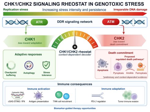
The DNA damage response (DDR) is a central determinant of tumor cell fate and therapeutic response. Checkpoint kinase 1 (CHK1) and checkpoint kinase 2 (CHK2) are canonical mediators of DDR, traditionally viewed as regulators of cell-cycle arrest and genome stability. Emerging evidence reveals broader roles in coordinating cell fate decisions and tumor-immune interactions. However, how checkpoint signaling translates genotoxic stress into divergent cell fate and immune outcomes remains unresolved. Here, we synthesize current evidence to propose a graded rheostat framework in which CHK1 and CHK2 integrate genotoxic stress signals to regulate diverse cell-fate and immune outputs. Although CHK1 and CHK2 share partially overlapping functions, current evidence suggests that CHK1 often contributes to stress-adaptive buffering, whereas CHK2 is more frequently implicated in damage-associated apoptotic commitment; however, these functional tendencies vary with p53 status, tumor lineage, damage characteristics including intensity and duration, treatment context, and cellular state. These distinct yet interconnected roles may be associated with emerging non-canonical programmed cell death pathways beyond canonical apoptosis, although current evidence remains limited and context-dependent. Importantly, CHK1/CHK2 signaling also shapes the tumor immune microenvironment (TIME) by regulating cytosolic DNA accumulation, cGAS-STING activation, antigen presentation, and immune checkpoint regulation. This suggests a mechanistic interface between genomic instability and anti-tumor immunity. Together, this review provides a mechanistic and translational perspective on how DDR signaling governs cell fate and immune outcomes, thereby providing a rationale for biomarker-guided combination strategies.
Keywords: CHK1, CHK2, DNA damage response, programmed cell death, tumor immune microenvironment, cancer therapy
1. Introduction
DNA is often exposed to both exogenous and endogenous sources of damage throughout an individual's life [1, 2]. Exogenous factors mainly include ultraviolet (UV) radiation, ionizing radiation (IR), and harmful chemicals, whereas endogenous sources often arise from replication stress, reactive oxygen species (ROS), and the intrinsic chemical instability of DNA, including processes such as telomere shortening, oncogene activation, and tumor suppressor inactivation [3]. To counteract these threats, cells activate the DNA damage response (DDR), a highly coordinated surveillance network that detects DNA lesions, halts cell-cycle progression, promotes DNA repair, and triggers programmed cell death pathways when damage becomes irreparable. Central to this network are CHK1 and CHK2, which function as key signal transducers downstream of ATR (ataxia telangiectasia and Rad3-related kinase) and ATM (ataxia telangiectasia mutated kinase), respectively [4-6]. Specifically, CHK1 primarily governs replication-stress responses and S/G2 checkpoint buffering, thereby preventing premature mitotic entry [4]. In contrast, CHK2 is more often activated by DNA double-strand breaks (DSBs) and is more closely linked to sustained damage signaling, G1/S and G2/M checkpoint control, and p53-dependent irreversible cell-fate outputs in specific cellular contexts [4, 7].
Although CHK1 and CHK2 have been extensively characterized as central regulators of cell-cycle checkpoints and genome stability, their roles in shaping the outcomes of different forms of cell death remain incompletely defined or controversial [8-11]. Beyond classical apoptosis, checkpoint signaling has increasingly been linked to stress-adaptive autophagy and emerging forms of regulated cell death, including ferroptosis and pyroptosis, although the mechanistic links between CHK1/CHK2 signaling and these non-canonical death programs remain incompletely defined. Concurrently, accumulating evidence indicates that CHK1/CHK2 signaling not only influences the fate of damaged cells but also shapes the TIME and modulates responses to immunotherapy [12-14]. However, the overlapping and distinct roles of CHK1 and CHK2 in regulating diverse PCD pathways and linking genotoxic stress to innate and adaptive immune responses have not yet been synthesized. Here, we propose a graded-evidence rheostat framework, in which evidence support is strongest for apoptosis and replication-stress buffering, moderate for checkpoint-coupled autophagy and DDR-immune crosstalk, and currently preliminary for non-canonical death programs such as ferroptosis and pyroptosis.
1.1 Scope of the review and evidence framework
To support this conceptual synthesis, we performed a focused literature search of PubMed and Web of Science through August 2026 using combinations of terms related to CHK1, CHK2, DNA damage response, cell death, autophagy, ferroptosis, tumor immunity, immune checkpoints, and therapeutic targeting. Because this review was designed as a mechanistic narrative synthesis rather than a systematic review or meta-analysis, the available evidence was qualitatively evaluated according to its consistency, mechanistic directness, and biological validation. Strong evidence was defined as consistent findings supported by multiple lines of evidence, including independent studies, direct mechanistic interrogation, and validation across complementary experimental models or in vivo settings. Moderate evidence referred to reproducible mechanistic observations supported by experimental evidence but influenced by specific cellular contexts, stress conditions, or limited broader validation. Preliminary evidence included findings based on a limited number of studies, indirect associations, or highly context-dependent experimental observations. These categories are intended to reflect the relative maturity and confidence of the available evidence rather than represent a formal quantitative scoring system or evidence-ranking framework.
2. Cell-fate outputs downstream of checkpoint signaling
2.1 Apoptosis
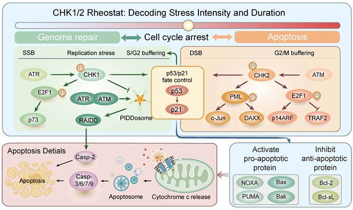
Apoptosis is the best-characterized form of PCD in the context of DDR. At low levels of genotoxic stress, a modest activation of p53 is sufficient to induce p21 transcription, suppress cyclin-dependent kinases (CDKs), and promote cell-cycle arrest [15-19]. However, when DSBs become extensive or persistent, p53 accumulates beyond a critical threshold and triggers the intrinsic apoptotic pathway through transcriptional activation of pro-apoptotic genes, including BAX, BAK, PUMA, and NOXA [20-26]. Moreover, p53 can physically interact with anti-apoptotic proteins such as Bcl-2, Bcl-xL, and Mcl-1, thereby neutralizing their function and indirectly promoting apoptosis [27, 28]. In parallel, E2F1 (E2F transcription factor 1) can be activated downstream of checkpoint signaling and contribute to apoptosis through both p53-dependent and p53-independent mechanisms [29] (Figure 1).
Conceptual model of CHK1/CHK2-associated regulation of the apoptotic threshold. In response to DNA damage, CHK1 and CHK2 orchestrate a graded transition from cell-cycle arrest to irreversible apoptosis. At low genotoxic stress, CHK1 frequently contributes to checkpoint buffering by enforcing p53/p21-dependent S/G2 arrest and by suppressing a distinct caspase-2-dependent apoptotic pathway, thereby preserving a repair-permissive state and restraining premature death. Conversely, under severe or persistent double-strand breaks, CHK2 contributes to pro-apoptotic signaling through multiple effectors: it promotes PML nuclear body formation and facilitates p53 stabilization, activates E2F1-dependent apoptotic pathways, and enhances p53-mediated transcriptional induction of BAX, PUMA, and NOXA while counteracting anti-apoptotic Bcl-2 and Bcl-xL. Thus, rather than operating as binary on/off switches, CHK1 and CHK2 are proposed to function as a dynamic rheostat, integrating the intensity and duration of genotoxic stress to bias the balance between checkpoint maintenance and apoptotic commitment. The left portion of the figure corresponds to Section 2.1.1 describing CHK1-associated checkpoint buffering, whereas the right portion corresponds to Section 2.1.2 illustrating CHK2-associated reinforcement of apoptotic commitment.
2.1.1 CHK1 frequently contributes to stress-adaptive buffering
In several settings, CHK1 acts as a checkpoint buffer that restrains premature apoptotic engagement rather than functioning solely as a pro-apoptotic kinase. This is exemplified by its ability to suppress a caspase-2-dependent apoptotic pathway independently of p53, E2F1, Bcl-2, and caspase-3 [30]. In this setting, CHK1 inhibition promotes ATM/ATR-dependent PIDDosome (p53-induced protein with a death domain-containing complex) assembly and caspase-2 activation, thereby triggering apoptosis outside the conventional mitochondrial or death receptor pathway [31-33]. CHK1 also buffers genotoxic stress through p53/p21-dependent checkpoint enforcement, as shown by ATR-CHK1-mediated cell-cycle arrest [34]. In head and neck squamous cell carcinoma (HNSCC) models, CHK1 suppression causes replication catastrophe and triggers apoptosis in only a fraction of the tumor cell population [35]. Consistent with this buffering role, live-cell imaging showed that increasing replication stress induces stepwise CHK1 activation, delays S-phase progression, and can ultimately trigger p53/p21-dependent arrest in daughter cells after division [36]. In summary, these studies imply that CHK1 often contributes to buffering stress responses in which checkpoint maintenance helps preserve a repair-permissive state and delay premature apoptosis under replication or genotoxic stress.
2.1.2 CHK2 is frequently implicated in apoptotic commitment under sustained genotoxic stress
Compared with CHK1, CHK2 is more frequently linked to apoptotic commitment when DNA damage becomes severe or persistent. Sustained CHK2 activity has been implicated in influencing whether cells undergo p53-dependent arrest or apoptosis, suggesting that the duration and intensity of CHK2 signaling are important factors influencing cell fate decisions. Mechanistically, CHK2 can contribute to apoptotic commitment through several downstream effectors. It facilitates Promyelocytic Leukemia (PML) nuclear body formation and enhances the recruitment of apoptosis-related regulators, including p53, c-Jun, and Daxx, thereby enhancing p53 transcriptional activity and promoting the expression of pro-apoptotic target genes such as BAX and PUMA, which can contribute to caspase-3-mediated apoptosis [37-39]. CHK2 also strengthens p53 signaling by phosphorylating p53 and its regulator Mouse double minute 4 homolog (Mdm4), while RTN3-associated CHK2/p53 activation facilitates p53 nuclear accumulation, collectively leading to p53 stabilization, enhanced nuclear retention, and transcriptional activation of its pro-apoptotic target genes [40-42]. In parallel, CHK2 activates E2F1-dependent death pathways, which may be further amplified through positive feedback to the ATM/CHK2 axis [43, 44]. In addition, CHK2 may contribute to transcriptional control of pro-apoptotic genes, such as FOS and BCL2L11, through the Nuclear Receptor Corepressor (NCoR)/Silencing Mediator of Retinoid and Thyroid hormone receptors (SMRT) complex, further shifting the balance toward cell death [45].
Experimental studies with amentoflavone, cinobufotalin, and nitidine chloride further provide evidence linking CHK2 activation with apoptotic regulation. Amentoflavone activates ATM/CHK2-associated DNA damage signaling and induces caspase-dependent and caspase-independent apoptosis in a p53-independent manner [46]. Cinobufotalin promotes apoptosis through the ATM/CHK2/p53 axis, accompanied by increased expression of FAS, DR4, and DR5 [47], whereas nitidine chloride activates CHK2 and engages p53/Bim-associated mitochondrial apoptotic signaling [48]. Under conditions of persistent genotoxic stress, characterized by sustained unrepaired DSBs, as evidenced by prolonged γH2AX and 53BP1 foci, together with continuous ATM/CHK2 signaling, apoptotic commitment is associated with induction of pro-apoptotic genes, caspase-3-mediated proteolytic cleavage, mitochondrial outer membrane permeabilization (MOMP), and cytochrome c release, contributing to a transition toward cell death when repair capacity becomes insufficient [49-51]. Collectively, these studies support a model in which CHK2 may frequently contribute to apoptotic commitment under conditions of sustained or severe genotoxic stress.
While CHK2 is more frequently implicated in apoptotic commitment, CHK1 can also contribute under conditions of persistent damage. In mouse oocytes, persistent DSBs activate CHK1, which participates in p53/TAp63-mediated apoptotic elimination alongside CHK2, forming a semi-redundant pathway in which both kinases contribute to apoptotic execution when damage exceeds a critical threshold [52]. These complementary functions, together with the buffering role of CHK1 described above, suggest that the balance between CHK1-mediated checkpoint maintenance and CHK2-associated apoptotic signaling influences whether a cell is more likely to maintain survival with DNA repair or progress toward a terminal cell-fate transition.
Overall, these findings support a model in which CHK1 and CHK2 collectively influence the apoptotic threshold downstream of DDR. In this framework, CHK1 more often acts to maintain checkpoint control and preserve a repair-permissive state, whereas CHK2 is more frequently associated with propagation of damage signaling and progression toward apoptotic commitment. Rather than acting as simple on/off switches, CHK1 and CHK2 appear to modulate the timing, intensity, and outcome of apoptotic signaling according to p53 status, tumor lineage, molecular background, damage severity, cellular state, and stress conditions.
2.2 Autophagy as a stress-adaptive extension of checkpoint signaling
Autophagy is a fundamental homeostatic pathway that functions constitutively under basal conditions to maintain cellular integrity through the clearance of damaged organelles and protein aggregates. Importantly, autophagy should not be considered synonymous with programmed cell death. Instead, it primarily represents an adaptive and reversible cellular program that modulates cellular vulnerability and influences subsequent survival, senescence, or death outcomes depending on cellular context. Beyond this housekeeping role, autophagy also contributes to antigen presentation and immune signaling [53]. In this context, accumulating evidence suggests that both CHK1 and CHK2 can participate in the regulation of autophagic programs. However, the role of CHK1 is better mechanistically defined in the context of checkpoint-coupled adaptive autophagy. CHK1 can phosphorylate RhoB, leading to tuberous sclerosis complex (TSC) translocation to lysosomes, inhibition of mTORC1, and induction of autophagy. This effect has been observed under genotoxic stress induced by UV or the alkylating agent methyl methanesulphonate (MMS), suggesting that CHK1 promotes a stress-adaptive, pro-autophagic response in certain DNA damage contexts [54]. In KRAS-mutant pancreatic ductal adenocarcinoma (PDAC), CHK1 inhibition activates AMP-activated protein kinase (AMPK) signaling and induces autophagy, suggesting that autophagy may function as a compensatory survival response when CHK1-biased checkpoint buffering is disrupted. Consistent with this interpretation, concurrent inhibition of CHK1 and autophagy further enhances growth suppression and apoptosis, indicating a close link between CHK1 signaling and stress-adaptive metabolic buffering [55].
In arsenite-exposed cancer cells, IKKα promotes CHK1-dependent DRAM1 activation and autophagy, and its subsequent selective autophagic degradation facilitates apoptosis, highlighting that stress-induced autophagy can shape cell fate rather than merely serving a protective role [56]. In cancer cell models, the G-quadruplex ligand 20A induces ATM-dependent autophagy that sustains CHK1 activation, favoring senescence over apoptosis; conversely, disruption of autophagy impairs CHK1 signaling and sensitizes cancer cells to apoptosis [57]. These cancer-based findings indicate that CHK1-mediated autophagic responses vary among tumor types and stress conditions and may influence therapeutic sensitivity and cell fate decisions.
By contrast, CHK2 appears to regulate autophagy through multiple downstream effectors that connect ATM-dependent DNA damage signaling to the core autophagic machinery. Our group previously reported that the ROS-ATM-CHK2-Beclin 1 axis promotes autophagosome formation by disrupting the Beclin 1-Bcl-2 complex, whereas CHK2-mediated phosphorylation of Unc-51 Like Autophagy Activating Kinase 1 (ULK1) enhances autophagy under conditions of metabolic stress [58, 59]. In addition to post-translational control of autophagy, DNA damage can also activate autophagy at the transcriptional level through an ATM-CHK2-FOXK axis, in which CHK2-mediated phosphorylation of FOXK1/2 promotes their cytoplasmic sequestration, relieves repression of ATG genes, and facilitates autophagic flux [60]. Under oxidative and metabolic stress, ROS-dependent ATM-CHK2 signaling initiates autophagy through CHK2-mediated phosphorylation of tripartite motif containing 32 (TRIM32), which promotes ATG7 ubiquitination and supports a protective stress-adaptive autophagic response [61]. Together, these findings suggest that CHK2 can contribute to stress-associated autophagic reprogramming through multiple downstream mechanisms.
Collectively, rather than uniformly promoting survival or death, CHK1/CHK2-associated autophagic responses function as an adaptive intermediate layer that shapes cellular states and influences subsequent fate transitions, including survival, senescence, or apoptosis, depending on the context of DNA damage and checkpoint signaling status. These observations also highlight substantial functional overlap between CHK1 and CHK2. Although CHK1 is more frequently associated with checkpoint maintenance and stress adaptation, it can also contribute to apoptotic elimination when DNA damage is severe or persistent, as demonstrated by the coordinated involvement of CHK1 and CHK2 in p53/TAp63-mediated apoptosis under persistent DSBs [52]. Conversely, CHK2, despite its frequent association with sustained damage signaling and apoptotic commitment, can also participate in adaptive responses, including autophagy, through mechanisms involving Beclin 1, ULK1, FOXK1/2, and TRIM32 [58-61]. Thus, the relative contributions of CHK1 and CHK2 are better viewed as functional biases rather than a strict division of labor, with their biological outcomes determined by the type, intensity, and duration of DNA damage, as well as p53 status and cellular context [17, 52, 58-61].
3. Immune consequences of checkpoint dysregulation
With the rapid development of immunotherapy, increasing attention has been directed toward the interplay between tumors and the host immune system, giving rise to the concept of the TIME. Within the TIME, diverse immune cell populations, including T cells, dendritic cells, macrophages, and natural killer (NK) cells, dynamically interact with tumor cells and stromal components to shape a balance between anti-tumor immunity and pro-tumor effects [62-64]. In this context, the interplay can result in either immune-mediated tumor elimination or immune evasion and tumor progression [65, 66]. Beyond their canonical checkpoint functions, accumulating evidence suggests that CHK1 and CHK2 contribute to distinct aspects of DDR-immune crosstalk within the TIME. To organize the available evidence, here we propose a three-layer conceptual model.
3.1 Layer I: innate sensing of genomic stress through cytosolic DNA accumulation and cGAS-STING activation
This layer is supported most directly by evidence linking CHK1 dysregulation to replication stress-associated innate immune activation. As a canonical regulator of the replication fork and the S-phase checkpoint, CHK1 normally restrains the accumulation of aberrant DNA species leaking into the cytosol. When this buffering function is compromised, genomic instability increases, leading to cytosolic DNA accumulation, cyclic GMP-AMP synthase (cGAS)-stimulator of interferon genes (STING) activation, type I interferon (IFN) responses, and SASP-associated chemokines such as CXCL10 and CCL5 [12, 67]. In AT-rich interactive domain-containing protein 1A (ARID1A)-deficient colorectal cancer, CHK1 protein stability is markedly increased. Consistent with this, pharmacological CHK1 inhibition induces cytosolic single-stranded DNA (ssDNA), thereby activating the cGAS-STING pathway and downstream IFN signaling, which enhances dendritic cell maturation, increases cytotoxic T-cell infiltration, and boosts antitumor immunity, improving therapeutic responses in ARID1A-deficient colorectal cancer [10]. Moreover, replication stress-induced STING activation within tumor cells, rather than non-tumor host-derived STING, can act as an intrinsic immune adjuvant, highlighting tumor cells themselves as active sensors of genomic stress [68]. Collectively, these findings support a model in which CHK1 dysregulation promotes tumor cell-intrinsic cGAS-STING activation and downstream type I IFN signaling in response to genomic stress.
3.2 Layer II: enhanced tumor immunogenicity through antigen presentation, neoantigen generation, and immune-cell recruitment
The second layer centers on enhanced tumor immunogenicity, whereby DDR disruption increases the recognizability of tumor cells to the adaptive immune system. Downstream of STING activation, DDR disruption enhances antigen presentation and promotes the recruitment of effector lymphocytes such as CD4+ T cells and effector memory CD8+ T cells [10]. Analyses of The Cancer Genome Atlas (TCGA, https://www.cancer.gov/about-nci/organization/ccg/research/structural-genomics/tcga) and The Cancer Immunome Atlas (TCIA, https://tcia.at) databases revealed that mutations in DDR pathway genes, such as ATM and ATR, are positively correlated with increased neoantigen load across multiple cancer types, which expands the repertoire of targets available for T-cell recognition [66]. Consistently, Germano et al. demonstrated that loss of DNA repair function increases frameshift-derived neoantigens and promotes CD8⁺ T-cell-dependent tumor control in vivo [69]. CHK2-deficient tumors (e.g., MC38 and B16 murine models) further exemplify checkpoint-linked immunogenicity and display a more inflamed, “hot” phenotype, characterized by increased mutational burden, enhanced antigen presentation, and greater CD8⁺ T-cell infiltration [70].
DDR-associated stress can also amplify immunogenicity through immunogenic cell death (ICD)-like outputs. STING activation not only induces IFN signaling but may also promote the release or exposure of damage-associated molecular patterns (DAMPs), such as adenosine triphosphate (ATP) and high mobility group box 1 (HMGB1), thereby facilitating dendritic cell maturation and antigen presentation. For instance, in Osi-resistant Lewis lung carcinoma (LLC/OR) cancer model, disruption of the DNA metabolism regulator ribonucleotide reductase subunit M2 (RRM2) activates cGAS-STING signaling and a downstream ROS-driven program that enhances ferroptosis, DAMP release, and STING-dependent ICD, ultimately promoting dendritic cell activation and CD8⁺ T-cell infiltration in vivo [71].
Beyond neoantigen generation and ICD-like amplification, DDR signaling may also directly enhance tumor visibility by upregulating antigen-presentation machinery. Irradiated tumor cells (murine B16-F10 melanoma or MC38 colon carcinoma) release microparticles that induce DSBs and activate the ATM/ATR/CHK1 axis, thereby increasing MHC-I expression via JAK-STAT signaling and enhancing CD8⁺ T-cell-mediated tumor killing [72]. CHK1 inhibition, by either prexasertib or genetic approaches, de-represses interferon regulatory factor 1 (IRF1) in hepatocellular carcinoma, promoting MICA expression and increasing NK and CD8⁺ T-cell tumor infiltration [73, 74]. Collectively, these observations support the view that this second layer reflects a transition from innate stress sensing to adaptive immune engagement. CHK1 and CHK2 may contribute to tumor immunogenicity through overlapping but non-identical mechanisms, with CHK1 more often linked to replication stress-associated immune activation and CHK2 contributing to adaptive TIME remodeling. However, some immunogenic consequences are also associated with broader DDR alterations beyond individual checkpoint kinases.
3.3 Layer III: immune adaptation and evasion through checkpoint signaling
This layer appears to be more frequently associated with CHK2-linked immune adaptation, particularly through immune checkpoint regulation and tumor-intrinsic resistance programs. However, CHK1-dependent checkpoint signaling can also influence immune-evasion pathways.
CHK1 and CHK2 also participate in the regulation of immune checkpoints within the TIME [70]. Accumulating evidence indicates that DDR activation can directly modulate programmed death-ligand 1 (PD-L1) expression in tumor cells. For instance, in vitro studies in cancer cell models have shown that DSBs activate the ATM-CHK2-STAT1-IRF1 axis, thereby upregulating PD-L1 expression and potentially promoting adaptive immune resistance following radiotherapy or chemotherapy [75]. Beyond direct checkpoint induction, CHK2 deficiency has also been associated with improved responsiveness to PD-1 blockade, accompanied by remodeling of the TIME and increased CD8⁺ T-cell infiltration, suggesting that CHK2 may contribute more broadly to immune-adaptive tumor states [70]. By comparison, disruption of the ATR-CHK1 pathway, particularly through ATR inhibition, can suppress PD-L1 expression [76, 77]. ATR-CHK1 inhibition activates the CDK1-SPOP axis, promoting PD-L1 ubiquitination and subsequent degradation, thereby reducing PD-L1 stability and enhancing anti-tumor efficacy in preclinical models. Consistently, preclinical studies and early-phase clinical investigations combining ATR inhibitors with PD-L1 blockade have provided initial evidence supporting enhanced antitumor activity and immune activation [78].
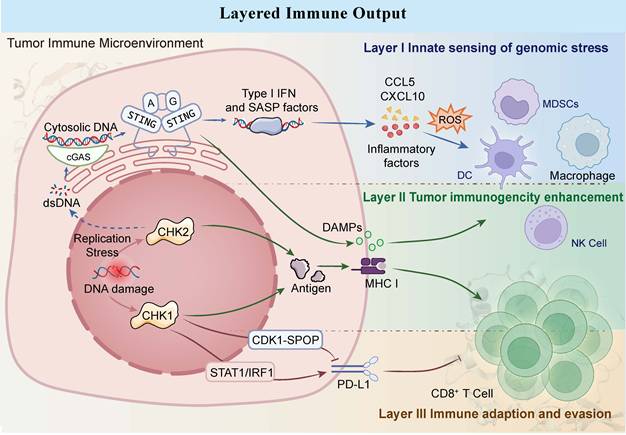
Overall, these observations suggest that immune checkpoint regulation represents a major mode of DDR-driven immune adaptation (Figure 2). Beyond PD-L1 regulation, ATR-CHK1 signaling also shapes the tumor immune microenvironment through innate immune activation and modulation of immune responsiveness, providing a mechanistic basis for combining ATR pathway inhibition with immune checkpoint blockade [78]. These apparently opposing effects reflect distinct regulatory layers rather than a direct antagonistic relationship between CHK1 and CHK2. CHK2 mainly regulates PD-L1 at the transcriptional level through STAT1/IRF1 activation, whereas ATR-CHK1 inhibition promotes PD-L1 destabilization through activation of the CDK1-SPOP pathway. Consistent with this dual-layer regulation, CHK2 is preferentially linked to stress-induced checkpoint activation and tumor-intrinsic resistance programs, whereas ATR-CHK1 inhibition may expose vulnerabilities that enhance responsiveness to PD-1/PD-L1 blockade. In the broader context of DDR-immune crosstalk, this pattern is consistent with a functional tendency in which CHK1 is more frequently associated with early immune activation, whereas CHK2 may contribute more prominently to downstream immune adaptation.
Conceptual three-layer model of CHK1/CHK2-associated immune outputs. DDR-immune crosstalk can be conceptualized as a hierarchical three-layer model, illustrating how CHK1 and CHK2 dysregulation shape the transition from innate stress sensing to adaptive immune evasion. In Layer I, the inhibition or dysregulation of CHK1 leads to the accumulation of dsDNA or ssDNA in the cytosol. This cytosolic DNA activates the cGAS-STING pathway, which induces the production of IFN and SASP factors. These inflammatory mediators create a proinflammatory milieu that initiates the recruitment and activation of innate immune cells such as DCs. Layer II depicts the transition toward adaptive immunity, where tumor cells become increasingly visible to the immune system. DDR disruption, including selected CHK1/CHK2-perturbed contexts, may increase genomic instability and, in some settings, enhance neoantigen generation and antigen presentation. The release of DAMPs including HMGB1 or ATP promotes the maturation of DCs. This enhanced immunogenic context facilitates the priming and proliferation of cytotoxic CD8⁺ T cells. Layer III highlights the compensatory mechanisms tumors employ to survive chronic immune pressure, largely influenced by the ATM-CHK2 axis. Persistent genotoxic stress activates the STAT1-IRF1 signaling pathway, which directly upregulates PD-L1 expression. Surface PD-L1 suppresses T-cell effector function and promotes immune evasion through engagement with the PD-1 receptor. The three-layer immune model represents a conceptual framework for organizing DDR-associated immune consequences rather than a strict CHK1/CHK2-specific pathway map. Layer I reflects innate sensing of genomic stress through cytosolic DNA accumulation and cGAS-STING activation; Layer II represents enhanced tumor immunogenicity involving antigen presentation and immune-cell recruitment; Layer III describes immune adaptation and evasion mediated by checkpoint-related resistance programs.
To clarify the central concept of this review, we propose a simplified framework in which CHK1 and CHK2 function as molecular rheostats that decode the intensity, persistence, and context of genotoxic stress into graded cell-fate and immune outputs. In this model, CHK1 is generally associated with replication stress buffering and adaptive checkpoint maintenance, whereas CHK2 more often reinforces irreversible damage signaling and death-associated programs, although substantial variability remains across cellular states and stress conditions.
4. Therapeutic exploitation of checkpoint vulnerability
4.1 Therapeutic rationale for targeting CHK1/CHK2
From a therapeutic perspective, CHK1 has emerged as the more actionable target. Many tumors experience chronic replication stress caused by oncogene activation, defective G1/S control, or DNA repair deficiency, making them highly dependent on CHK1-mediated S-phase surveillance and replication fork stabilization [79]. As a result, CHK1 inhibition can disrupt this adaptive buffering capacity and create a therapeutically exploitable vulnerability [80-82]. By contrast, CHK2 appears to be more closely associated with sustained DNA damage signaling, apoptotic reinforcement, and stimulus-specific immune regulation in selected biological contexts, suggesting a comparatively narrower and more selective therapeutic scope [7, 70, 83] (Table 1).
Representative clinical studies of CHK1-targeting inhibitors in tumors
| I. Prexasertib (LY2606368): most clinically advanced CHK1 inhibitor | |||||||
| Agent | Trial ID | Phase | Status | Disease | Combination | Enrollment (n) | Reported outcome / note |
| Prexasertib(LY2606368/ ACR-368) | NCT06597565 | II | Recruiting | Recurrent/ metastatic HNSCC | Low-dose gemcitabine | 43 (est.) | No results posted [107]‡ |
| NCT05548296 | II | Recruiting | Endometrial adenocarcinoma | Ultra-low dose gemcitabine | 350 (est.) | Biomarker-guided; no results posted [155]‡ | |
| NCT03414047 | II | Completed | Platinum-resistant/refractory ovarian cancer | Monotherapy | 172 | ORR: 12.1% (platinum-resistant); 6.9% (platinum-refractory) [156] | |
| NCT04032080 | II | Completed | Metastatic TNBC | LY3023414 | 10 | Registry results only | |
| NCT02873975 | II | Completed | Biomarker-selected solid tumors | Monotherapy | 27 | 4-month PFS: 44.4% (CCNE1-amplified; registry) | |
| NCT02735980 | II | Completed | Extensive-stage SCLC | Monotherapy | 133 | ORR: 5.2% (platinum-sensitive); 0% (resistant/refractory) [157] | |
| NCT02203513 | II | Terminated | BRCA-associated cancers and BRCA-WT HGSOC | Monotherapy | 111 | PR: 8/24 (33%), BRCA-WT HGSOC [101] | |
| NCT04095221 | I/II | Completed | DSRCT and rhabdomyosarcoma | Irinotecan ± temozolomide | 21 | Registry results submitted but not yet posted | |
| NCT03057145 | I | Completed | Advanced solid tumors | Olaparib | 29 | RP2D established; PR: 4/18 [105] | |
| NCT03495323 | I | Completed | Advanced solid tumors | PD-L1 inhibitor LY3300054 | 17 | 3 PRs; peripheral CD8+ T-cell activation [120] | |
| NCT02124148 | I | Completed | Advanced solid tumors | Cisplatin, cetuximab, pemetrexed, 5-FU, or LY3023414 | 167, registry target | Feasible; schedule-dependent hematologic toxicity; preliminary activity [158, 159] | |
| NCT02555644 | I | Completed | Locally advanced HNSCC | Cisplatin or cetuximab + radiotherapy | 70, registry target; 25 treated in published report | Cisplatin-RT: no safe dose; cetuximab-RT: feasible [160] | |
| II. Early dual CHK1/CHK2 inhibitors: proof-of-concept but limited by toxicity | |||||||
| AZD7762 | NCT00937664 | I | Terminated | Advanced solid tumors | Gemcitabine | 24 | Safety/PK characterized [88] |
| NCT00473616 | I | Terminated | Advanced solid tumors | Irinotecan | 60 (est.) | No results posted | |
| NCT00413686 | I | Completed | Advanced solid tumors | Gemcitabine | 42 | Cardiac toxicity limited development [87] | |
| PF-00477736 | NCT00437203 | I | Terminated | Advanced solid tumors | Gemcitabine | 43 | Abstract-only clinical data [161]‡ |
| III. Selective CHK1 inhibitors: combination-oriented development | |||||||
| MK-8776 (SCH900776) | NCT00779584 | Ⅰ | Completed | Advanced solid tumors / lymphoma | Gemcitabine | 45 | RP2D defined; 2 PR, 13 SD [94] |
| NCT00907517 | Ⅰ | Terminated | Relapsed/refractory acute leukemias | Cytarabine | 24, treated | CR: 8/24 (33%) [162] | |
| NCT01870596 | Ⅱ | Completed | Relapsed AML | Cytarabine | 32, treated | CR/CRi: 36% vs 44%; no survival benefit [163] | |
| GDC-0575 | NCT01564251 | Ⅰ | Completed | Refractory solid tumors / lymphoma | Gemcitabine | 104 | 4 PRs; frequent hematologic toxicity [92] |
| Rabusertib (LY2603618) | Multiple trials | I/II | Completed | Advanced/metastatic NSCLC | Pemetrexed | 55 | ORR 9.1%; clinical benefit 45.5%; PFS 2.3 months [95] |
| IV. Next-generation CHK1 inhibitors: biomarker-driven and orally bioavailable agents | |||||||
| PEP07 | NCT05983523 | I | Recruiting | Advanced/metastatic solid tumors | Monotherapy | 54 (est.) | No results posted [97]‡ |
| NCT05659732 | Ib | Recruiting | Relapsed/refractory AML and MCL | Monotherapy | 32 (est.) | No results posted | |
| LY2880070 | NCT02632448 | Ib/IIa | Completed | Advanced/metastatic cancers | Monotherapy ± gemcitabine | 229 (est.) | No objective responses in pancreatic cohort [98] |
| NCT05275426 | II | Completed | Ewing/ Ewing-like sarcoma, and DSRCT | Low-dose gemcitabine | 14 | No results posted | |
| BBI-355 | NCT05827614 | I | Terminated | Oncogene-amplified solid tumors | Monotherapy or biomarker-directed combinations | 85 | Preliminary Phase I data reported [96]‡; terminated for non-safety reasons |
| SRA737 (CCT245737) | NCT02797964 | I/II | Completed | Advanced cancer | Monotherapy | 107 | RP2D: 800 mg QD; no CR/PR [164] |
| NCT02797977 | I/II | Completed | Advanced cancer | Low-dose gemcitabine ± cisplatin | 153 | RP2D: 500 mg + 250 mg/m² gemcitabine; ORR 10.8% [115] | |
Table note: Trial status and enrollment were verified using ClinicalTrials.gov as of August 2026. Outcomes are based on peer-reviewed publications when available; otherwise, registry data are reported. Enrollment is registry-reported unless otherwise indicated; est., estimated; treated, number of patients reported in the corresponding publication. ‡ Conference abstract or presentation. RT, radiotherapy
4.1.1 Pharmacologic landscape of CHK1 inhibitors
CHK1 inhibitors are predominantly ATP-competitive small molecules that can be broadly grouped into three developmental categories: early dual inhibitors, selective CHK1 inhibitors, and next-generation orally available CHK1 inhibitors guided by biomarker-based development [84, 85]. The comparative selectivity, pharmacokinetic properties, major toxicities, and clinical development status of representative CHK1-targeted inhibitors, including selective CHK1 inhibitors and dual CHK1/CHK2 inhibitors, are summarized in Table 2. Early dual inhibitors, such as AZD7762 and PF-00477736, provided proof of concept for checkpoint-targeted therapy by sensitizing tumor cells to DNA-damaging agents. For instance, AZD7762 synergizes with gemcitabine to suppress KRAS-driven, LKB1-deficient lung adenocarcinoma by exploiting its high endogenous DNA damage and CHK1 dependence [86]. In Phase I trials, AZD7762 combined with gemcitabine demonstrated preliminary efficacy in advanced solid tumors. However, these early agents revealed limitations including a narrow therapeutic window and severe off-target toxicities; notably, the development of AZD7762 was terminated due to unpredictable cardiotoxicity [87, 88]. Additionally, the orally bioavailable selective CHK1 inhibitor SRA737 showed a comparatively manageable safety profile, with gastrointestinal adverse events being common, although hematologic toxicities, including neutropenia and thrombocytopenia, were also reported [85, 89]. Clinical development has therefore shifted toward rational combinations and molecularly selected tumor contexts, particularly those characterized by high replication stress or defective G1/S checkpoints, including tumors harboring TP53 mutations, MYC amplification, or Cyclin E1 amplification [85, 90].
Selectivity, pharmacokinetics, and toxicity profiles of clinical-stage CHK1/CHK2 inhibitors
| Agent | Selectivity | Biochemical potency | Administration / dosing / key human PK features | Main toxicities / DLTs | Ref |
|---|---|---|---|---|---|
| Prexasertib (LY2606368) | CHK1-dominant CHK1/2 | CHK1: IC₅₀ <1 nM, Ki 0.9 nM; CHK2: IC₅₀ 8 nM | IV; RP2D 105 mg/m² q14d; t₁/₂ ~12 h; minor accumulation | Neutropenia, thrombocytopenia, anemia; febrile neutropenia | [99, 165] |
| AZD7762 | Dual CHK1/CHK2 | CHK1: IC₅₀ 5 nM, Ki 3.6 nM; CHK2: IC₅₀ 5 nM | IV; MTD 30 mg + gemcitabine 1000 mg/m² q21d; t₁/₂ ~8-15.5 h | Cardiac DLTs; neutropenia; nausea/vomiting | [87, 166] |
| MK-8776 (SCH900776) | Selective CHK1 | CHK1: IC₅₀ 3 nM | IV; RP2D 200 mg + gemcitabine 1000 mg/m² q21d; t₁/₂ ~6-10 h | Thrombocytopenia, neutropenia; fatigue, nausea; transient QTc prolongation | [94, 167] |
| GDC-0575 | Selective CHK1 | CHK1: IC₅₀ 1.2 nM | Oral; RP2D 45 mg + gemcitabine 1000 mg/m² or 80 mg + 500 mg/m²; Tmax ≤2 h; t₁/₂ ~23 h | Neutropenia, thrombocytopenia; grade 3-4 febrile neutropenia | [92] |
| Rabusertib (LY2603618) | Selective CHK1 | CHK1: IC₅₀ 7 nM | IV; t₁/₂ ~5-25 h; dose-dependent exposure; high interpatient variability | Fatigue, nausea/diarrhea, thrombocytopenia, neutropenia; infusion-related hypersensitivity | [109, 168] |
| SRA737 (CCT245737) | Selective CHK1 | CHK1: IC₅₀ 1.4 ± 0.3 nM | Oral; RP2D 800 mg QD; Tₘₐₓ ~2-4 h; t₁/₂ ~6-10 h; Cmin ~546 nM | Gastrointestinal events; neutropenia; thrombocytopenia | [85, 89] |
| UCN-01 | Multikinase with CHK1 activity | CHK1: IC₅₀ ~7-11 nM | IV; t₁/₂~250-1660 h due to α1-acid glycoprotein binding | Hyperglycemia, hypotension, nausea/vomiting; pulmonary toxicity reported | [169-171] |
| PF-00477736 | CHK1-dominant | CHK1: Ki 0.49 nM; CHK2: Ki 47 nM | IV; 24-h infusion; MTD 270 mg + gemcitabine 750 mg/m² q21d; t₁/₂ ~8-20 h | Neutropenia, GI toxicity; DLTs: thrombocytopenia, mucositis, elevated lipase, sudden death | [161, 172, 173] |
| LY2880070 | Selective CHK1 | CHK1: IC₅₀ 0.5 nM | Oral; MTD 200 mg BID, days 1-5 q21d; t₁/₂ 5.35 ± 2.3 h | Nausea, vomiting, fatigue; toxicity associated with peak exposure | [98, 174] |
| PEP07 | Selective CHK1 | CHK1: IC₅₀ 1 nM; CHK2: IC₅₀ 1630 nM | Oral; QD for 2 days followed by 5 days off weekly; human PK under evaluation | Not yet established | [175] |
| BBI-355 | Selective CHK1 | CHK1: IC₅₀ 0.6 nM | Oral, Q2D; preliminary MTD 60 mg; t₁/₂ ~40 h; ~2-3-fold accumulation. | Hematologic DLTs, including grade 4 neutrophil- and platelet-count decreases | [176] |
Table note: BID, twice daily; Cmin, minimum plasma concentration; DLT, dose-limiting toxicity; IV, intravenous; MTD, maximum tolerated dose; PK, pharmacokinetics; QD, once daily; q14d, every 14 days; RP2D, recommended phase 2 dose; Tmax, time to maximum plasma concentration; t₁/₂, elimination half-life. Biochemical potency values were derived from preclinical enzyme assays and are assay-dependent; they should not be interpreted as direct cross-compound comparisons. PK values are regimen- and study-dependent and should not be interpreted as direct cross-compound comparisons.
Subsequent efforts shifted toward selective CHK1 inhibition to better exploit tumor dependence on replication stress responses while improving pharmacologic specificity. Representative agents in this class include prexasertib, MK-8776, rabusertib, GDC-0575, and SRA737 [85, 87, 88, 91-95]. In clinical studies, these agents were primarily evaluated in combination with DNA-damaging chemotherapy. For instance, prexasertib showed modest single-agent activity in patients with squamous cell carcinoma in a phase Ib study [93]. MK-8776 was assessed as monotherapy and in combination with gemcitabine in advanced solid tumors, with dose-limiting neutropenia and thrombocytopenia being the main toxicities [94]; it also exhibited enhanced radiosensitizing effects by exacerbating radiation-induced aberrant mitosis in preclinical models [91]. GDC-0575 combined with gemcitabine in refractory solid tumors, but the regimen demonstrated limited clinical efficacy and significant myelosuppression [92]. In a single-arm phase II study in advanced non-small-cell lung cancer, Rabusertib (LY2603618) plus pemetrexed showed no significant clinical activity, with an ORR of 9.1% and a median PFS of 2.3 months [95]. SRA737, an orally administered CHK1 inhibitor, was tested as monotherapy in a phase 1/2 trial in advanced cancer; although it achieved target plasma concentrations, no partial or complete responses were observed, and gastrointestinal toxicity was dose-limiting [85].
More recently, the field has moved further toward orally bioavailable next-generation compounds, such as BBI-355, PEP07, and LY2880070, with increasing emphasis on molecular selection strategies [96-98]. BBI-355 is under investigation in patients with oncogene amplification on extrachromosomal DNA (ecDNA), representing a biomarker-driven approach [96]. PEP07 is being evaluated in a phase I trial for advanced or metastatic solid tumors, focusing on safety and pharmacokinetics [97]. LY2880070 was evaluated in combination with low-dose gemcitabine in a phase I expansion cohort of patients with treatment-refractory metastatic pancreatic adenocarcinoma; although the regimen was feasible, no objective radiologic responses were observed [98]. This evolution reflects a broader conceptual transition in the field: from nonspecific checkpoint blockade toward precision deployment in tumors characterized by high replication stress, ecDNA amplification, or defined DDR-related vulnerabilities. Collectively, these studies indicate that improvements in CHK1 selectivity and pharmacokinetic properties have not translated proportionally into greater clinical efficacy, as dose-limiting toxicity and modest objective response rates remain recurrent challenges across the class.
4.1.2 Prexasertib as the leading clinical prototype
Among clinically investigated CHK1 inhibitors, prexasertib is the most extensively studied and best represents the translational potential of CHK1-directed therapy. Early phase I studies (NCT01115790 and NCT02514603) established an intermittent intravenous dosing schedule and demonstrated pharmacodynamic activity, but also identified hematologic toxicity as the principal dose-limiting liability [99, 100]. Together, these results suggest that the therapeutic value of CHK1 inhibition depends strongly on optimized scheduling and appropriate patient selection [99, 100].
The most convincing clinical signal for prexasertib has been observed in BRCA wild-type recurrent high-grade serous ovarian cancer, especially in platinum-resistant disease. In this setting, phase II studies (NCT02203513 and NCT03414047) support the concept that tumors with high endogenous replication stress may be particularly susceptible to CHK1 inhibition, even in the absence of canonical BRCA deficiency [101, 102]. By contrast, prexasertib has shown limited activity in biomarker-unselected settings, including triple-negative breast cancer (TNBC), extensive-stage small-cell lung cancer, and squamous malignancies, suggesting that its clinical utility as a broadly active monotherapy may be limited [93, 103, 104]. Consequently, current clinical development has shifted toward biomarker-enriched and combination-based strategies, rather than indiscriminate application across tumor types [105-107].
4.1.3 Other CHK1 inhibitors: from proof of concept to biomarker-oriented development
Other selective CHK1 inhibitors have mainly contributed to defining the mechanistic and translational boundaries of this therapeutic class. MK-8776 provided clinical proof of concept that CHK1 inhibition can sensitize tumor cells to DNA-damaging chemotherapy, particularly gemcitabine-based regimens, and supports the feasibility of combination strategies [94]. Rabusertib similarly helped establish the feasibility of combining CHK1 inhibition with cytotoxic chemotherapy, although its clinical efficacy remained limited in later-stage testing [95, 108, 109].
Preclinical evidence further indicates that CHK1 inhibition possesses anti-proliferative activity in soft-tissue sarcoma models, supporting its potential clinical utility in this tumor type [110]. Rather than aiming for broad cytotoxic activity, these agents are increasingly being evaluated in molecular contexts predicted to confer checkpoint dependence, such as replication-stress-high tumors, ecDNA-driven oncogene amplification, and certain sarcoma subtypes [111]. This trend indicates that the future of CHK1-targeted therapy likely lies not in class-wide expansion, but in identifying tumor states in which checkpoint buffering is especially indispensable.
4.1.4 Critical Analysis: Why Has CHK1 Inhibition Shown Limited Clinical Translation?
Despite a strong biological rationale and compelling preclinical efficacy, more than 15 CHK1 inhibitors have entered clinical development, yet none has achieved regulatory approval [84]. This translational gap appears to reflect several interconnected pharmacological and biological constraints.
First, CHK1 inhibition is constrained by a relatively narrow therapeutic window. Clinical studies of prexasertib have consistently identified hematologic toxicities, particularly neutropenia, thrombocytopenia, and febrile neutropenia, as major dose-limiting events, thereby restricting dose intensity and sustained target inhibition [99, 100]. More broadly, across multiple clinical programs, pharmacologically effective CHK1 inhibition has frequently been accompanied by hematologic or other dose-limiting toxicities, while objective antitumor responses have remained modest. Second, tumor cells can activate compensatory checkpoint and DNA-damage response pathways that reduce dependence on CHK1. WEE1 up-regulation has been identified as a mechanism of acquired resistance to CHK1 inhibition in SCLC [112], whereas tumor-intrinsic PD-L1 signaling can stabilize CHK2-dependent DNA-damage responses and reduce sensitivity to CHK1 inhibitors [83, 112]. These observations suggest that resistance may arise through functional redundancy and adaptive rewiring within the DDR network rather than through restoration of CHK1 activity itself.
Third, the absence of prospectively validated predictive biomarkers has limited effective patient selection. Preclinical and translational studies suggest that tumors characterized by high replication stress, CCNE1 amplification, or ARID1A deficiency may exhibit increased CHK1 dependence and sensitivity to CHK1 inhibition [79, 113, 114]. However, these candidate biomarkers have not yet been prospectively validated for routine clinical selection, potentially diluting treatment effects in heterogeneous patient populations.
Fourth, the efficacy of CHK1 inhibition is highly dependent on treatment schedule and combination strategy. Although DNA-damaging agents can increase tumor dependence on CHK1, overlapping myelosuppression constrains the intensity of chemotherapy combinations. The SRA737 trial illustrates this therapeutic trade-off: combining 500 mg SRA737 with 250 mg/m² low-dose gemcitabine improved tolerability but produced an overall objective response rate of only 10.8% [115], indicating that pharmacological optimization alone may be insufficient to overcome intrinsic and adaptive resistance.
Collectively, the limited clinical translation of CHK1 inhibitors likely reflects the combined effects of a restricted therapeutic index, compensatory DDR signaling, insufficient biomarker-based patient selection, and schedule-dependent efficacy rather than a lack of biological relevance of CHK1 itself. Future development should therefore prioritize prospectively validated replication-stress biomarkers, pharmacodynamically informed dosing schedules, and rational combinations targeting complementary or compensatory pathways, including PARP inhibition, immune checkpoint blockade, and WEE1 inhibition [81, 105, 116, 117].
4.2 Combination therapy as the principal therapeutic direction
At present, the strongest rationale for CHK1/CHK2 inhibition lies in combination treatment rather than monotherapy. Checkpoint inhibitors are best viewed as stress amplifiers. DNA-damaging chemotherapies, particularly gemcitabine, have become the most extensively studied partners, because they induce replication fork stalling and create a cellular state of heightened CHK1 dependence [86, 90, 108, 116]. This principle underlies the development of multiple agents across the class, including AZD7762, MK-8776, LY2603618, and prexasertib [86, 108, 116].
Beyond chemotherapy, CHK1 inhibition has shown translational promise in combination with PARP inhibitors, radiotherapy, and targeted therapies. The combination of prexasertib with olaparib is particularly notable in PARP inhibitor-resistant ovarian cancer, where checkpoint blockade may overcome adaptive resistance by further destabilizing replication fork integrity [105]. Likewise, in tumors driven by extreme replication stress, next-generation CHK1 inhibitors may cooperate with other agents that intensify cell-cycle or oncogenic burden, such as FGFR inhibitors or CDK4/6-directed strategies [96]. Taken together, current evidence suggests that CHK1 inhibitors have demonstrated only modest single-agent activity to date, and their greater therapeutic potential lies in combination with treatments that increase checkpoint dependence. Thus, the principal value of CHK1 inhibition may lie less in standalone cytotoxicity than in amplifying pre-existing checkpoint dependence imposed by chemotherapy, PARP inhibition, radiotherapy, or selected targeted therapies.
4.2.1 CHK2-targeted strategies and dual inhibition: mechanistic promise, limited clinical maturity
Compared with CHK1-targeted therapy, CHK2-directed development remains far less advanced. Current evidence suggests that CHK2 is more closely involved in sustained damage signaling, apoptosis reinforcement, and immune adaptation than in the core replication-stress buffering machinery [7, 70, 83]. For this reason, CHK2 inhibition may ultimately prove most useful in selected biologic settings rather than as a universal anti-cancer strategy.
4.2.2 Immunomodulatory potential of CHK1/CHK2 inhibition and combination with immune checkpoint blockade
In addition to directly promoting tumor cell death, CHK1/CHK2 inhibition may reshape the TIME through immunomodulatory mechanisms, although the available evidence is substantially stronger for CHK1 than for CHK2. This emerging dimension is particularly important because it extends the consequences of CHK1/CHK2 targeting beyond cell-intrinsic cytotoxicity. In several preclinical settings, CHK1 inhibition has been shown to increase cytosolic DNA accumulation, activate cGAS-STING signaling, and enhance type I IFN responses, thereby converting replication stress into an immunologically active output [10]. Other studies suggest that CHK1 blockade can increase expression of immune-relevant ligands such as MICA, promote recruitment or activation of NK cells and CD8⁺ T cells, and induce a transiently more inflamed TIME [73]. These observations provide a mechanistic rationale for combining CHK inhibition with immune checkpoint blockade (ICB), particularly in tumors characterized by high replication stress and an immunologically refractory microenvironment.
Preclinical studies have provided growing support for this combination strategy. CHK1 inhibition combined with anti-PD-1/PD-L1 therapy has demonstrated enhanced antitumor activity in several tumor models, accompanied by increased CD8⁺ T-cell infiltration and cGAS-STING-associated immune activation. For example, SRA737 combined with low-dose gemcitabine and anti-PD-L1 therapy induced durable tumor regression in immunocompetent models [14]. CHK2-directed immunotherapy combinations remain considerably less developed. Although CHK2 deficiency has been associated with increased tumor immunogenicity, enhanced antigen presentation, and improved responsiveness to immune checkpoint blockade in selected preclinical settings [70], these genetic observations should not be directly equated with the effects of pharmacological CHK2 inhibition, for which direct evidence in combination with ICB remains limited.
Early clinical observations have also provided preliminary support for this concept. In prexasertib-treated patients, clinical benefit has been associated with favorable immune remodeling, including enhanced Th1- and IFN-γ-related signaling and broader innate and adaptive immune activation, whereas non-responding patients showed expansion of immunosuppressive monocytic myeloid-derived suppressor cells [101, 118, 119]. Furthermore, combination treatment with prexasertib and the anti-PD-L1 antibody LY3300054 showed preliminary clinical activity and peripheral CD8⁺ T-cell activation in CCNE1-amplified high-grade serous ovarian cancer [120]. Within the graded-evidence framework of this review, support for CHK1-mediated immunomodulation is relatively stronger at the preclinical level but remains preliminary clinically, whereas direct evidence for pharmacological CHK2 inhibition in combination with ICB is currently very limited. Accordingly, predictive biomarkers, optimal dosing schedules, and treatment sequences remain to be established.
Collectively, these findings suggest that CHK1 inhibition may create a therapeutically exploitable immunogenic window, during which replication stress-associated immune activation and enhanced tumor visibility could be most effectively leveraged by combination with immune checkpoint blockade [10, 73, 117, 121, 122]. Consistent with the proposed rheostat framework, pharmacological disruption of CHK1-mediated checkpoint buffering may shift tumor cells from relatively immune-silent states toward immunogenically active states, although this transition is highly dependent on the baseline TIME [12, 123]. This transition may involve ICD-like processes characterized by stress-associated DAMP release, enhanced antigen presentation, and subsequent activation of dendritic cells and CD8⁺ T-cell responses [124].
Importantly, the immunomodulatory effects of CHK1/CHK2 inhibition are not uniformly beneficial. Inflammatory responses associated with CHK1 inhibition may arise indirectly from replication stress, DNA damage accumulation, cytosolic DNA sensing, and downstream inflammatory signaling, and may vary according to cellular and metabolic context [125]. However, this response is not universal, as CHK1 inhibition failed to activate STING signaling in a human coculture cancer system, further emphasizing its context dependence [117, 126]. An additional consideration is the potential impact of DDR-targeted therapy on normal immune cells and hematopoietic compartments. CHK1 is required to maintain replication integrity in hematopoietic stem and progenitor cells and in proliferating lymphocyte populations, and excessive CHK1 inhibition may therefore induce replication-associated DNA damage and compromise immune-cell fitness [125]. CHK2 appears to exert distinct and more cell-specific functions, as CHK2 deficiency can reduce DNA damage-induced apoptosis in thymocytes and splenic lymphocytes [127]. These observations indicate that CHK1 and CHK2 targeting may have different consequences for immune-cell homeostasis and should not be considered immunologically equivalent.
These effects are directly relevant to treatment tolerance. Hematologic toxicities, including neutropenia, leukopenia, thrombocytopenia, and anemia, have been frequently observed during CHK1 inhibitor development [101, 115, 120, 128]. Thus, enhanced tumor immunogenicity may coexist with impaired hematopoietic recovery or reduced immune-cell competence. Evidence from SRA737-based regimens further indicates that dose intensity and treatment schedule strongly influence the balance between efficacy and toxicity [85, 115]. Acquired resistance through restoration of replication-stress tolerance, activation of compensatory DDR pathways, or failure to sustain immune activation may further limit treatment durability [83, 129]. Accordingly, successful integration of CHK1/CHK2 targeting with immunotherapy will require a balance between tumor-localized immune activation and preservation of systemic immune and hematopoietic function, together with biomarker-guided patient selection and rational treatment scheduling.
4.3 Beyond CHK1/CHK2: Comparative landscape of DDR targets
CHK1 and CHK2 function within a highly interconnected DDR network rather than as isolated therapeutic targets. ATR and ATM act upstream of CHK1 and CHK2, respectively, whereas WEE1 and PARP regulate complementary checkpoint or DNA-repair processes [129-131]. ATR and CHK1 inhibition therefore share several biological consequences, particularly disruption of replication-stress responses, checkpoint abrogation, and replication catastrophe. However, ATR functions as an upstream coordinator with a broader substrate spectrum, whereas CHK1 represents a major downstream effector that more directly controls replication-fork stability and S/G2 checkpoint activity [131]. By comparison, ATM-CHK2 signaling is more closely associated with double-strand-break responses, highlighting the distinct positioning of the two checkpoint axes within the DDR network [130].
These mechanistic differences also provide a rationale for combination strategies. WEE1 restrains CDK1/2 activity through a checkpoint mechanism complementary to CHK1; therefore, simultaneous disruption of CHK1- and WEE1-dependent control may enhance unscheduled origin firing and premature mitotic entry, thereby intensifying replication stress [132]. PARP inhibition differs mechanistically by impairing single-strand-break repair and replication-fork-associated processes, which can increase tumor dependence on ATR-CHK1 signaling. Accordingly, combined PARP and ATR/CHK1 inhibition may further destabilize replication forks and has been investigated as a strategy to overcome PARP inhibitor resistance [133, 134]. Thus, CHK1/CHK2 inhibition should be viewed as one component of a broader DDR-targeting framework, in which therapeutic benefit may arise either from disabling complementary checkpoint pathways or from exploiting compensatory dependencies induced by inhibition of another DDR node [129]. These mechanistic distinctions and potential cooperative interactions are summarized in Table 3.
Comparison of key DDR targets and their relationship to CHK1/CHK2 inhibition
| Target | Main DDR role | Functional relationship toCHK1/CHK2 | Key difference in inhibition | Potential combination rationale | Refs. |
|---|---|---|---|---|---|
| CHK1 | Replication-stress checkpoint | Core component of the ATR-CHK1 axis | Directly impairs fork protection and checkpoint control | Enhances PARPi- or WEE1i-induced replication stress | [105, 177] |
| CHK2 | DSB checkpoint signaling | Core component of the ATM-CHK2 axis | More closely linked to DSB response and apoptosis | Combination strategies remain less established | [7] |
| ATR | Replication-stress sensor kinase | Upstream activator of CHK1 | Acts more broadly than CHK1 in replication-stress signaling | ATR inhibition can enhance PARPi-induced replication stress | [178, 179] |
| WEE1 | CDK1/2 checkpoint control | Functionally complementary to CHK1 | Promotes premature cell-cycle progression when inhibited | CHK1/WEE1 co-inhibition induces checkpoint failure and mitotic entry | [81, 177] |
| PARP | SSB and replication-associated repair | Functionally coupled to ATR-CHK1 signaling | Generates DNA lesions rather than directly blocking checkpoints | PARPi increases dependence on ATR-CHK1 signaling | [178-180] |
Table note: DDR, DNA damage response; DSB, double-strand break; SSB, single-strand break; ATR, ataxia telangiectasia and Rad3-related kinase; ATM, ataxia telangiectasia mutated kinase; PARP, poly(ADP-ribose) polymerase; PARPi, PARP inhibitor; WEE1i, WEE1 inhibitor.
5. Current challenges and future directions
Despite a strong mechanistic rationale, the clinical development of CHK1/CHK2 inhibitors remains constrained by several persistent challenges, including toxicity, especially hematologic toxicity, schedule dependence, and the lack of robust predictive biomarkers [87, 88, 99, 100]. Future progress will likely require a shift from empirical development toward biomarker-guided precision deployment. Features such as high replication stress signatures, ecDNA amplification, ARID1A deficiency, and related DDR-associated vulnerabilities may help identify tumors most likely to benefit, particularly if these biomarkers are prospectively incorporated into trial design [10, 82, 96]. Overall, the next phase of clinical translation will depend less on expanding the number of checkpoint inhibitors and more on improving patient selection, optimizing treatment scheduling, and refining biomarker-guided combination strategies.
5.1 Future perspectives: Emerging and context-dependent cell-death outputs associated with CHK1/CHK2 signaling
In keeping with the graded-evidence framework introduced above, support for these non-canonical outputs remains preliminary and is supported mainly by stimulus-specific observations. Importantly, current evidence is largely derived from indirect signaling intermediates or condition-specific observations, and direct kinase-substrate phosphorylation events linking CHK1/CHK2 with core execution machinery remain unestablished.
5.1.1 Ferroptosis
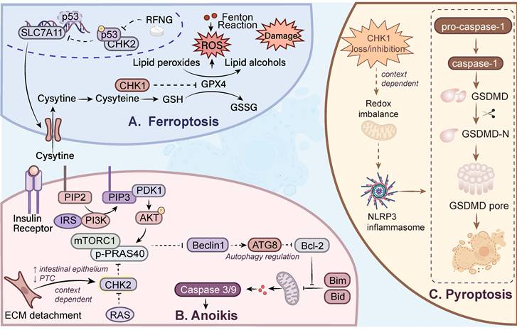
Ferroptosis is an iron-dependent form of PCD characterized by iron overload, uncontrolled lipid peroxidation, accumulation of lipid ROS, and plasma membrane rupture [135-137]. Both glutathione peroxidase 4 (GPX4) and solute carrier family 7 member 11 (SLC7A11, also known as xCT) are negative regulators of ferroptosis [138-141]. Crosstalk between DDR and ferroptosis is increasingly recognized as a largely noncanonical regulatory interface. ATM/ATR, p53, and MDM2/MDMX have all been implicated in ferroptosis control through mechanisms involving iron handling, cystine transport, lipid peroxidation, and antioxidant defense, rather than through their classical roles in cell-cycle arrest and DNA repair alone [142]. In colorectal cancer, oxaliplatin-induced apoptosis and ferroptosis can be suppressed by radical fringe (RFNG)-mediated disruption of the CHK2-p53 axis, suggesting that CHK2-dependent p53 signaling may favor both apoptotic and ferroptotic outputs in a wild-type p53 context [143]. In non-cancer murine models, a role for CHK1 in ferroptosis has been suggested in spinal cord injury, where p-CHK1 and γ-H2AX were increased, whereas GPX4 and SLC7A11 were reduced after injury. Functional studies showed that CHK1 knockdown restored GPX4/SLC7A11 expression and improved motor outcomes, whereas CHK1 overexpression aggravated injury-associated dysfunction. Although this work was performed in a neurotraumatic rather than tumor context, it supports the possibility that CHK1 may also contribute to ferroptotic output under severe stress conditions [144]. Importantly, no direct phosphorylation of ferroptosis execution regulators, such as GPX4, by CHK1 or CHK2 has been demonstrated. Current evidence for a direct CHK1/CHK2-ferroptosis axis in cancer remains limited, with stronger support for indirect regulation through p53/SLC7A11-associated pathways than for a conserved CHK1/CHK2-centered ferroptosis module (Figure 3a). Collectively, these findings support predominantly indirect and stress-dependent links between checkpoint signaling and ferroptosis. Available evidence suggests that ferroptotic susceptibility may be influenced by checkpoint-associated stress responses, with some observations involving CHK2-related signaling; however, its placement within the CHK1/CHK2 rheostat framework remains speculative.
Proposed links between CHK1/CHK2 signaling and emerging or context-dependent cell-death outputs. A. Ferroptosis: In certain contexts, chemotherapy-induced DNA damage may activate CHK2-associated signaling and promote p53 phosphorylation. As a transcription factor, p53 binds to the promoter of SLC7A11 (xCT) and suppresses its transcription. The system Xc⁻ transporter mediates cystine uptake and, together with GPX4, reduces cytotoxic lipid peroxides, thereby inhibiting ferroptosis. B. Anoikis: ECM detachment was associated with CHK2 downregulation in PTC cells. CHK2 has been proposed to promote cell death in a p53-independent manner through regulation of PRAS40 activation. Upon activation of the insulin receptor, the downstream PI3K-Akt pathway is initiated, leading to phosphorylation and inactivation of mTORC1 inhibitors and thereby promoting mTORC1 activity. PRAS40 forms a complex with mTORC1 and inhibits both substrate-recruitment sites. C. Pyroptosis: Proposed context-dependent intersection between CHK1-linked redox stress and pyroptosis-associated injury. These pathways represent proposed associations based on current evidence and do not indicate established direct kinase-substrate regulatory mechanisms.
5.1.2 Anoikis
Anoikis refers to apoptosis induced by detachment from the extracellular matrix (ECM), and loss of anoikis sensitivity is a key mechanism underlying tumor metastasis [145]. Foundational evidence for a CHK2-dependent role in anoikis came from intestinal epithelial models, in which ECM detachment induced CHK2 upregulation and activation, whereas CHK2 depletion protected cells from anoikis in a p53-independent manner. Importantly, oncogenic Ras activation blocked both detachment-induced CHK2 upregulation and the anoikis-promoting effect of enforced CHK2 expression, supporting the idea that CHK2-driven anoikis is strongly constrained by oncogenic context [146]. This concept was later extended to papillary thyroid cancer (PTC), where CHK2 knockdown reduced detachment-induced apoptosis, while CHK2 overexpression enhanced apoptosis only under suspension conditions. Mechanistically, although ECM detachment was associated with CHK2 downregulation, functional analyses indicated that CHK2 promotes anoikis through regulation of Proline-rich AKT1 substrate (PRAS40) activation in a p53-independent manner. Notably, the direction of CHK2 expression changes following ECM detachment differs across experimental models, further underscoring the dependence of the CHK2-anoikis relationship on cellular background. Notably, CHK2 and p-CHK2 levels were reduced in metastatic lymph nodes relative to matched primary tumors, supporting the idea that loss of CHK2-dependent anoikis may facilitate survival during metastatic dissemination [147] (Figure 3b). These findings suggest that CHK2 may promote anoikis under anchorage-loss conditions; however, this relationship appears to depend on tumor type and extracellular signaling context, and its general relevance across tumor models remains unclear. Therefore, CHK2-associated anoikis regulation should currently be viewed as a model-specific hypothesis rather than a conserved downstream pathway. However, direct mechanistic evidence demonstrating how CHK2 signaling controls anoikis execution remains limited.
5.1.3 Pyroptosis
Pyroptosis is an inflammatory form of PCD characterized by inflammasome activation, gasdermin D (GSDMD) cleavage, and release of pro-inflammatory cytokines [148-152]. To date, direct phosphorylation of GSDMD or other core pyroptotic execution components by CHK1/CHK2 has not been demonstrated. Current studies more often suggest an indirect association that varies among experimental systems, in which checkpoint-linked stress responses may bias cells toward pyroptotic outcomes rather than directly triggering a defined CHK1/CHK2-pyroptosis pathway. For example, in pancreatic ductal adenocarcinoma (PDAC) models, lipid raft disruption induced oxidative and proteotoxic stress, activated ATR-CHK1-associated DDR signaling, and was accompanied by caspase-1-dependent pyroptotic features in selected cell lines, such as BxPC-3 and MIA PaCa-2, whereas others, such as L3.6pl, preferentially underwent apoptosis [153]. In addition, evidence from non-tumor murine cardiomyocytes suggests that CHK1 loss can intersect with mitochondrial redox imbalance, NLRP3 activation, and pyroptotic injury, although these findings mainly inform tissue-specific toxicity rather than tumor cell fate [154]. Collectively, these studies suggest that pyroptosis may be a possible inflammatory endpoint of checkpoint-associated stress or checkpoint dysregulation (Figure 3c). However, whether pyroptosis represents a true downstream program or merely a stress-overflow phenomenon remains unresolved and requires more direct mechanistic interrogation.
Taken together, current studies suggest that ferroptosis, anoikis, and pyroptosis may represent emerging extensions of checkpoint-associated cell-fate regulation. However, the available evidence remains insufficient to firmly position these non-canonical PCD modalities within the CHK1/CHK2 rheostat framework. Although some observations implicate CHK1 or CHK2 in these emerging pathways, the available evidence does not yet support a consistent kinase-specific bias. Establishing direct kinase-substrate relationships between CHK1/CHK2 and core components of these death programs will therefore be essential to determine whether these associations represent bona fide downstream regulatory pathways or indirect consequences of checkpoint-associated cellular stress.
6. Conclusions
We propose that CHK1 and CHK2 function not merely as linear transducers of DNA damage signals, but as components of a dynamic decision module that governs cell-fate bifurcation under genotoxic stress. In this framework, checkpoint signaling acts as a molecular rheostat that integrates damage intensity, replication stress, and signal duration into graded biological outputs. Under lower levels of genotoxic or replication stress, CHK1-associated signaling more often sustains replication and enforces repair-permissive checkpoints. As stress intensifies, this buffering capacity may destabilize, giving rise to heterogeneous intermediate states and divergent cell-fate outcomes. As damage becomes more severe or persistent, CHK2-biased signaling becomes increasingly associated with the amplification of irreversible damage cues and commitment to cell death. This transition reflects a bifurcation tendency rather than a binary switch, where modest variations in checkpoint activity or cellular context produce qualitatively distinct outcomes. Thus, CHK1 and CHK2 can be conceptualized as an overlapping, signal-strength-responsive module that biases cells toward survival adaptation or irreversible death. Importantly, this framework extends to tumor immunity, as distinct checkpoint states and death modalities encode differential immunogenic outputs, ranging from immune-silent clearance to cGAS-STING-driven activation. In practical terms, the future of checkpoint-targeted therapy may depend on identifying tumor states in which checkpoint buffering, stress adaptation or immune evasion have become selectively indispensable (Table 4).
Conceptual framework of CHK1/CHK2 as molecular rheostats that decode genotoxic stress into graded cell-fate and immune outputs
| Evidence strength | Representative stress context | CHK bias | Cell-fate tendency | Layered immune output |
|---|---|---|---|---|
| Strong evidence | Mild to intermediate genotoxic/ replication stress | CHK1-biased | Checkpoint maintenance, survival adaptation [4] | Layer I bias: restrained innate immune activation [10, 67] |
| Moderate evidence | Persistent replication stress | Predominantly CHK1-biased | Autophagy/adaptive intermediate state [54-57, 181, 182] | Layer I-II transition: cGAS-STING activation and enhanced tumor immunogenicity [72, 73] |
| Strong evidence | Persistent or irreparable DNA damage | CHK2-biased | Irreversible death commitment [43-45, 47, 48] | Variable immune consequence; may remain immune-silent unless accompanied by DAMP release [71] |
| Preliminary evidence | Severe or persistent stress; anchorage loss | No consistent CHK1/ CHK2 bias | Ferroptosis [143], anoikis [146, 147] and pyroptosis [153, 154]. | Potential immune effects remain context-dependent |
Table note: Evidence strength primarily refers to the CHK1/CHK2-cell-fate association; evidence supporting the corresponding immune outputs may differ in maturity.
6.1 Model boundaries and exceptions
It is important to emphasize that the CHK1-adaptation versus CHK2-death framework should not be interpreted as a binary model with mutually exclusive functional assignments, but rather as a continuum of functional biases. CHK1 more frequently favors stress buffering and adaptive checkpoint maintenance, whereas CHK2 is more often associated with reinforcement of irreversible damage responses. However, the position along this continuum can be shifted by factors including damage intensity and duration, p53 status, cellular lineage, metabolic state, and other signaling contexts. Importantly, several exceptions define the boundaries of this framework. CHK1 can contribute to apoptosis under persistent or severe DNA damage, whereas CHK2 can, in specific contexts, participate in adaptive or pro-survival responses, including autophagy under metabolic or other stress conditions [17, 58]. These observations indicate that CHK1 and CHK2 do not determine cell fate through fixed kinase-specific assignments. Instead, the final outcome emerges from the integration of checkpoint signaling strength and duration with the broader cellular context. Accordingly, the proposed rheostat should be viewed as a conceptual framework describing predominant functional tendencies and organizing current evidence, rather than as a deterministic rule applicable to all biological contexts.
6.2 Future directions
To advance this framework further, future work may need to converge along three major axes: quantitative DDR signaling, temporal dynamics, and immunotherapy integration. A more quantitative view may be necessary to determine how different amplitudes and durations of CHK1/CHK2 signaling generate graded cell-fate outputs. Meanwhile, resolving the temporal dynamics of checkpoint signaling may clarify whether damaged cells remain repair-permissive, undergo irreversible death, or transiently adopt immunogenic states. Finally, integrating these insights with immunotherapy may be essential, particularly for defining how checkpoint inhibition can be aligned with immune checkpoint blockade or radiotherapy in a schedule-optimized manner.
Abbreviations
cGAS: cyclic GMP-AMP synthase; CHK1: checkpoint kinase 1; CHK2: checkpoint kinase 2; DAMPs: danger-associated molecular patterns; DDR: DNA damage response; ecDNA: extrachromosomal DNA; ECM: extracellular matrix; HNSCC: head and neck squamous cell carcinoma; ICD: immunogenic cell death; IR: ionizing radiation; PCD: programmed cell death; PDAC: pancreatic ductal adenocarcinoma; PD-L1: programmed death-ligand 1; STING: stimulator of interferon genes; TIME: tumor immune microenvironment; TNBC: triple-negative breast cancer.
Acknowledgements
Funding
This work was supported by the Department of Science and Technology of Liaoning Province (2026JH2/101300140) and the 2024 Special Science and Technology Project for High-Quality Development of China Medical University (24-219-3-02) awarded to X. Li, the National Natural Science Foundation of China (32570923) awarded to L. Cao, the Department of Science and Technology of Liaoning Province (2024-MS-058, 2024-MS-021) awarded to L. Wang and N. Bai, respectively, National Natural Science Foundation of China (82570464), the Natural Science Foundation Program Projects of Liaoning Province, China (2025-YQ-12) to Y. Zhang and Liaoning Provincial Department of Education Natural Science Basic Research Project for Higher Education Institutions (LJ212510162011) to X. Zhao.
Author contributions
X. Li, L. Cao, L. Wang, X. Zhao and Y. Zhang conceived and supervised the review. J. Yang, Z. Liu, and Y. Z. Chen drafted the manuscript. Y. Feng and Y. Mu prepared the figures and tables. N. Bai, L. Yuan, M. Yang, Y. Liu, H. Zheng, Y. Zhao, Y.T. Chen, and H. Fang contributed to literature analysis and manuscript revision. X. Zhao performed language and reference editing. All authors reviewed and approved the final manuscript.
AI statement
During the preparation of this manuscript, the authors used ChatGPT (OpenAI) solely for language editing. All scientific content, analysis, interpretation, and conclusions were developed by the authors. The authors reviewed and revised the AI-assisted text and take full responsibility for the final content.
Competing Interests
The authors have declared that no competing interest exists.
References
1. Milano L, Gautam A, Caldecott KW. DNA damage and transcription stress. Mol Cell. 2024;84:70-9
2. Chakarov S, Petkova R, Russev GC, Zhelev N. DNA damage and mutation. Types of DNA damage. BioDiscovery. 2014;11:1
3. Lindahl T. Instability and decay of the primary structure of DNA. Nature. 1993;362:709-15
4. Smith J, Tho LM, Xu N, Gillespie DA. The ATM-Chk2 and ATR-Chk1 pathways in DNA damage signaling and cancer. Adv Cancer Res. 2010;108:73-112
5. Zhao P, Chen L, Li LH, Wei ZF, Tong B, Jia YG. et al. SC-III3, a novel scopoletin derivative, induces cytotoxicity in hepatocellular cancer cells through oxidative DNA damage and ataxia telangiectasia-mutated nuclear protein kinase activation. BMC Cancer. 2014;14:987
6. Sim MY, Go ML, Yuen JSP. The mechanistic effects of the dioxonaphthoimidazolium analog YM155 in renal cell carcinoma cell cycling and apoptosis. Life Sci. 2018;203:282-90
7. Zannini L, Delia D, Buscemi G. CHK2 kinase in the DNA damage response and beyond. J Mol Cell Biol. 2014;6:442-57
8. Zhou X, Zhao Y, Huang S, Shu H, Zhang Y, Yang H. et al. TRIM32 promotes neuronal ferroptosis by enhancing K63-linked ubiquitination and subsequent p62-selective autophagic degradation of GPX4. Int J Biol Sci. 2025;21:1259-74
9. Liu S, Liu J, Wang Y, Deng F, Deng Z. Oxidative Stress: Signaling Pathways, Biological Functions, and Disease. MedComm (2020). 2025;6:e70268
10. Chen JY, Ke TW, Chiang SF, Hong WZ, Chang HY, Liang JA. et al. CHK1 inhibition increases the therapeutic response to radiotherapy via antitumor immunity in ARID1A-deficient colorectal cancer. Cell Death Dis. 2025;16:584
11. Zhang S, Guan M, Ren T, Li N, Ding Q, Sun D. et al. Prexasertib exerts a synergistic effect on the antitumor activity of Lenvatinib through ALOX15-mediated ferroptosis in hepatocellular carcinoma. Int Immunopharmacol. 2025;150:114278
12. Sen T, Rodriguez BL, Chen L, Della Corte CM, Morikawa N, Fujimoto J. et al. Targeting DNA Damage Response Promotes Antitumor Immunity through STING-Mediated T-cell Activation in Small Cell Lung Cancer. Cancer Discov. 2019;9:646-61
13. Wang L, Yang L, Wang C, Zhao W, Ju Z, Zhang W. et al. Inhibition of the ATM/Chk2 axis promotes cGAS/STING signaling in ARID1A-deficient tumors. J Clin Invest. 2020;130:5951-66
14. Sen T, Della Corte CM, Milutinovic S, Cardnell RJ, Diao L, Ramkumar K. et al. Combination Treatment of the Oral CHK1 Inhibitor, SRA737, and Low-Dose Gemcitabine Enhances the Effect of Programmed Death Ligand 1 Blockade by Modulating the Immune Microenvironment in SCLC. J Thorac Oncol. 2019;14:2152-63
15. Williams AB, Schumacher B. p53 in the DNA-Damage-Repair Process. Cold Spring Harb Perspect Med. 2016;6:a026070
16. Tibbetts RS, Brumbaugh KM, Williams JM, Sarkaria JN, Cliby WA, Shieh SY. et al. A role for ATR in the DNA damage-induced phosphorylation of p53. Genes Dev. 1999;13:152-7
17. van Jaarsveld MTM, Deng D, Wiemer EAC, Zi Z. Tissue-Specific Chk1 Activation Determines Apoptosis by Regulating the Balance of p53 and p21. iScience. 2019;12:27-40
18. Abbas T, Dutta A. p21 in cancer: intricate networks and multiple activities. Nat Rev Cancer. 2009;9:400-14
19. Myers K, Gagou ME, Zuazua-Villar P, Rodriguez R, Meuth M. ATR and Chk1 suppress a caspase-3-dependent apoptotic response following DNA replication stress. PLoS genetics. 2009;5:e1000324
20. Carlsson MJ, Vollmer AS, Demuth P, Heylmann D, Reich D, Quarz C. et al. p53 triggers mitochondrial apoptosis following DNA damage-dependent replication stress by the hepatotoxin methyleugenol. Cell Death & Disease. 2022;13:1009
21. Oda E, Ohki R, Murasawa H, Nemoto J, Shibue T, Yamashita T. et al. Noxa, a BH3-only member of the Bcl-2 family and candidate mediator of p53-induced apoptosis. Science. 2000;288:1053-8
22. Nakano K, Vousden KH. PUMA, a novel proapoptotic gene, is induced by p53. Mol Cell. 2001;7:683-94
23. Chen Y, Zhang X, Dantas Machado AC, Ding Y, Chen Z, Qin PZ. et al. Structure of p53 binding to the BAX response element reveals DNA unwinding and compression to accommodate base-pair insertion. Nucleic Acids Res. 2013;41:8368-76
24. Kracikova M, Akiri G, George A, Sachidanandam R, Aaronson SA. A threshold mechanism mediates p53 cell fate decision between growth arrest and apoptosis. Cell Death Differ. 2013;20:576-88
25. Miyashita T, Reed JC. Tumor suppressor p53 is a direct transcriptional activator of the human bax gene. Cell. 1995;80:293-9
26. Graupner V, Alexander E, Overkamp T, Rothfuss O, De Laurenzi V, Gillissen BF. et al. Differential regulation of the proapoptotic multidomain protein Bak by p53 and p73 at the promoter level. Cell Death Differ. 2011;18:1130-9
27. Wei H, Qu L, Dai S, Li Y, Wang H, Feng Y. et al. Structural insight into the molecular mechanism of p53-mediated mitochondrial apoptosis. Nat Commun. 2021;12:2280
28. Yao H, Mi S, Gong W, Lin J, Xu N, Perrett S. et al. Anti-apoptosis proteins Mcl-1 and Bcl-xL have different p53-binding profiles. Biochemistry. 2013;52:6324-34
29. Urist M, Tanaka T, Poyurovsky MV, Prives C. p73 induction after DNA damage is regulated by checkpoint kinases Chk1 and Chk2. Genes Dev. 2004;18:3041-54
30. Sidi S, Sanda T, Kennedy RD, Hagen AT, Jette CA, Hoffmans R. et al. Chk1 suppresses a caspase-2 apoptotic response to DNA damage that bypasses p53, Bcl-2, and caspase-3. Cell. 2008;133:864-77
31. Hubackova S, Davidova E, Boukalova S, Kovarova J, Bajzikova M, Coelho A. et al. Replication and ribosomal stress induced by targeting pyrimidine synthesis and cellular checkpoints suppress p53-deficient tumors. Cell Death & Disease. 2020;11:110
32. Manzl C, Fava LL, Krumschnabel G, Peintner L, Tanzer MC, Soratroi C. et al. Death of p53-defective cells triggered by forced mitotic entry in the presence of DNA damage is not uniquely dependent on Caspase-2 or the PIDDosome. Cell Death Dis. 2013;4:e942
33. Ando K, Kernan JL, Liu PH, Sanda T, Logette E, Tschopp J. et al. PIDD death-domain phosphorylation by ATM controls prodeath versus prosurvival PIDDosome signaling. Mol Cell. 2012;47:681-93
34. Rong JJ, Hu R, Song XM, Ha J, Lu N, Qi Q. et al. Gambogic acid triggers DNA damage signaling that induces p53/p21(Waf1/CIP1) activation through the ATR-Chk1 pathway. Cancer Lett. 2010;296:55-64
35. van Harten AM, Buijze M, van der Mast R, Rooimans MA, Martens-de Kemp SR, Bachas C. et al. Targeting the cell cycle in head and neck cancer by Chk1 inhibition: a novel concept of bimodal cell death. Oncogenesis. 2019;8:38
36. Lebrec V, Poteau M, Morretton JP, Gavet O. Chk1 dynamics in G2 phase upon replication stress predict daughter cell outcome. Dev Cell. 2022;57:638-53.e5
37. Hofmann TG, Will H. Body language: the function of PML nuclear bodies in apoptosis regulation. Cell Death Differ. 2003;10:1290-9
38. Zhong S, Muller S, Ronchetti S, Freemont PS, Dejean A, Pandolfi PP. Role of SUMO-1-modified PML in nuclear body formation. Blood. 2000;95:2748-52
39. Guo A, Salomoni P, Luo J, Shih A, Zhong S, Gu W. et al. The function of PML in p53-dependent apoptosis. Nat Cell Biol. 2000;2:730-6
40. Huang RX, Zhou PK. DNA damage response signaling pathways and targets for radiotherapy sensitization in cancer. Signal Transduct Target Ther. 2020;5:60
41. Song S, Shi Y, Wu W, Wu H, Chang L, Peng P. et al. Reticulon 3-mediated Chk2/p53 activation suppresses hepatocellular carcinogenesis and is blocked by hepatitis B virus. Gut. 2021;70:2159-71
42. Chen L, Gilkes DM, Pan Y, Lane WS, Chen J. ATM and Chk2-dependent phosphorylation of MDMX contribute to p53 activation after DNA damage. Embo j. 2005;24:3411-22
43. Powers JT, Hong S, Mayhew CN, Rogers PM, Knudsen ES, Johnson DG. E2F1 uses the ATM signaling pathway to induce p53 and Chk2 phosphorylation and apoptosis. Mol Cancer Res. 2004;2:203-14
44. Rogoff HA, Pickering MT, Frame FM, Debatis ME, Sanchez Y, Jones S. et al. Apoptosis associated with deregulated E2F activity is dependent on E2F1 and Atm/Nbs1/Chk2. Mol Cell Biol. 2004;24:2968-77
45. Scafoglio C, Smolka M, Zhou H, Perissi V, Rosenfeld MG. The co-repressor SMRT delays DNA damage-induced caspase activation by repressing pro-apoptotic genes and modulating the dynamics of checkpoint kinase 2 activation. PLoS One. 2013;8:e59986
46. Lin CH, Lin KH, Ku HJ, Lee KC, Lin SS, Hsu FT. Amentoflavone induces caspase-dependent/-independent apoptosis and dysregulates cyclin-dependent kinase-mediated cell cycle in colorectal cancer in vitro and in vivo. Environ Toxicol. 2023;38:1078-89
47. Xia Z, Li M, Hu M, Lin Y, Atteh LL, Fu W. et al. Phosphoproteomics reveals that cinobufotalin promotes intrahepatic cholangiocarcinoma cell apoptosis by activating the ATM/CHK2/p53 signaling pathway. Front Oncol. 2022;12:982961
48. Kwon HJ, Kim LH, Ahn CH, Yang IH, Hong KO, Hong SD. et al. A new insight into the apoptotic effect of nitidine chloride targeting Checkpoint kinase 2 in human cervical cancer in vitro. J Clin Biochem Nutr. 2019;65:193-202
49. Paek AL, Liu JC, Loewer A, Forrester WC, Lahav G. Cell-to-Cell Variation in p53 Dynamics Leads to Fractional Killing. Cell. 2016;165:631-42
50. Groelly FJ, Fawkes M, Dagg RA, Blackford AN, Tarsounas M. Targeting DNA damage response pathways in cancer. Nat Rev Cancer. 2023;23:78-94
51. Shah RB, Li Y, Yu H, Kini E, Sidi S. Stepwise phosphorylation and SUMOylation of PIDD1 drive PIDDosome assembly in response to DNA repair failure. Nat Commun. 2024;15:9195
52. Rinaldi VD, Bloom JC, Schimenti JC. Oocyte Elimination Through DNA Damage Signaling from CHK1/CHK2 to p53 and p63. Genetics. 2020;215:373-8
53. Deretic V. Autophagy in inflammation, infection, and immunometabolism. Immunity. 2021;54:437-53
54. Liu M, Zeng T, Zhang X, Liu C, Wu Z, Yao L. et al. ATR/Chk1 signaling induces autophagy through sumoylated RhoB-mediated lysosomal translocation of TSC2 after DNA damage. Nat Commun. 2018;9:4139
55. Klomp JE, Lee YS, Goodwin CM, Papke B, Klomp JA, Waters AM. et al. CHK1 protects oncogenic KRAS-expressing cells from DNA damage and is a target for pancreatic cancer treatment. Cell Rep. 2021;37:110060
56. Tan Q, Zou S, Jin R, Hu Y, Xu H, Wang H. et al. Selective degradation of IKKalpha by autophagy is essential for arsenite-induced cancer cell apoptosis. Cell Death Dis. 2020;11:222
57. Beauvarlet J, Bensadoun P, Darbo E, Labrunie G, Rousseau B, Richard E. et al. Modulation of the ATM/autophagy pathway by a G-quadruplex ligand tips the balance between senescence and apoptosis in cancer cells. Nucleic Acids Res. 2019;47:2739-56
58. Guo QQ, Wang SS, Zhang SS, Xu HD, Li XM, Guan Y. et al. ATM-CHK2-Beclin 1 axis promotes autophagy to maintain ROS homeostasis under oxidative stress. EMBO J. 2020;39:e103111
59. Guo R, Wang SS, Jiang XY, Zhang Y, Guo Y, Cui HY. et al. CHK2 Promotes Metabolic Stress-Induced Autophagy through ULK1 Phosphorylation. Antioxidants (Basel). 2022;11:1166
60. Chen Y, Wu J, Liang G, Geng G, Zhao F, Yin P. et al. CHK2-FOXK axis promotes transcriptional control of autophagy programs. Sci Adv. 2020;6:eaax5819
61. Liu J, Lu S, Zheng L, Guo Q, Cao L, Xiao Y. et al. ATM-CHK2-TRIM32 axis regulates ATG7 ubiquitination to initiate autophagy under oxidative stress. Cell Rep. 2023;42:113402
62. Lei X, Lei Y, Li JK, Du WX, Li RG, Yang J. et al. Immune cells within the tumor microenvironment: Biological functions and roles in cancer immunotherapy. Cancer Lett. 2020;470:126-33
63. Zhou Y, Cheng L, Liu L, Li X. NK cells are never alone: crosstalk and communication in tumour microenvironments. Mol Cancer. 2023;22:34
64. Ran GH, Lin YQ, Tian L, Zhang T, Yan DM, Yu JH. et al. Natural killer cell homing and trafficking in tissues and tumors: from biology to application. Signal Transduct Target Ther. 2022;7:205
65. Li Q, Lei X, Zhu J, Zhong Y, Yang J, Wang J. et al. Radiotherapy/Chemotherapy-Immunotherapy for Cancer Management: From Mechanisms to Clinical Implications. Oxid Med Cell Longev. 2023;2023:7530794
66. Kakoti S, Sato H, Laskar S, Yasuhara T, Shibata A. DNA Repair and Signaling in Immune-Related Cancer Therapy. Front Mol Biosci. 2020;7:205
67. Lowery CD, Dowless M, Renschler M, Blosser W, VanWye AB, Stephens JR. et al. Broad Spectrum Activity of the Checkpoint Kinase 1 Inhibitor Prexasertib as a Single Agent or Chemopotentiator Across a Range of Preclinical Pediatric Tumor Models. Clin Cancer Res. 2019;25:2278-89
68. Alshebremi M, Tomchuck SL, Myers JT, Kingsley DT, Eid S, Abiff M. et al. Functional tumor cell-intrinsic STING, not host STING, drives local and systemic antitumor immunity and therapy efficacy following cryoablation. J Immunother Cancer. 2023 11
69. Germano G, Lamba S, Rospo G, Barault L, Magri A, Maione F. et al. Inactivation of DNA repair triggers neoantigen generation and impairs tumour growth. Nature. 2017;552:116-20
70. Xu P, Gao Y, Jiang S, Cui Y, Xie Y, Kang Z. et al. CHEK2 deficiency increase the response to PD-1 inhibitors by affecting the tumor immune microenvironment. Cancer Lett. 2024;588:216595
71. Zhu J, Cui K, Zhou W, Zhou X, Yao Y, Zhao C. et al. Targeting RRM2 with dual-modal theranostic smart nanoresponder overcomes osimertinib resistance and triggers immune remodeling in NSCLC. Sci Adv. 2025;11:eady7930
72. Deng S, Wang J, Hu Y, Sun Y, Yang X, Zhang B. et al. Irradiated tumour cell-derived microparticles upregulate MHC-I expression in cancer cells via DNA double-strand break repair pathway. Cancer Lett. 2024;592:216898
73. Li X, Huang J, Wu Q, Du Q, Wang Y, Huang Y. et al. Inhibition of Checkpoint Kinase 1 (CHK1) Upregulates Interferon Regulatory Factor 1 (IRF1) to Promote Apoptosis and Activate Anti-Tumor Immunity via MICA in Hepatocellular Carcinoma (HCC). Cancers (Basel). 2023;15:850
74. Yan Y, Zheng L, Du Q, Cui X, Dong K, Guo Y. et al. Interferon regulatory factor 1 (IRF-1) downregulates Checkpoint kinase 1 (CHK1) through miR-195 to upregulate apoptosis and PD-L1 expression in Hepatocellular carcinoma (HCC) cells. Br J Cancer. 2021;125:101-11
75. Sato H, Niimi A, Yasuhara T, Permata TBM, Hagiwara Y, Isono M. et al. DNA double-strand break repair pathway regulates PD-L1 expression in cancer cells. Nat Commun. 2017;8:1751
76. Tang Z, Pilie PG, Geng C, Manyam GC, Yang G, Park S. et al. ATR Inhibition Induces CDK1-SPOP Signaling and Enhances Anti-PD-L1 Cytotoxicity in Prostate Cancer. Clin Cancer Res. 2021;27:4898-909
77. Mouw KW, Konstantinopoulos PA. From checkpoint to checkpoint: DNA damage ATR/Chk1 checkpoint signalling elicits PD-L1 immune checkpoint activation. Br J Cancer. 2018;118:933-5
78. Ngoi NYL, Peng G, Yap TA. A Tale of Two Checkpoints: ATR Inhibition and PD-(L)1 Blockade. Annu Rev Med. 2022;73:231-50
79. da Costa A, Chowdhury D, Shapiro GI, D'Andrea AD, Konstantinopoulos PA. Targeting replication stress in cancer therapy. Nat Rev Drug Discov. 2023;22:38-58
80. Neizer-Ashun F, Bhattacharya R. Reality CHEK: Understanding the biology and clinical potential of CHK1. Cancer Letters. 2021;497:202-11
81. Kumar NA, Manasa P, Chandrasekaran J. Revisiting CHK1 and WEE1 kinases as therapeutic targets in cancer. Biochim Biophys Acta Rev Cancer. 2025;1880:189482
82. Kciuk M, Wanke K, Marciniak B, Kołat D, Aleksandrowicz M, Mujwar S. et al. Targeting ATR-CHK1 and ATM-CHK2 Axes in Pancreatic Cancer-A Comprehensive Review of Literature. Int J Mol Sci. 2026;27:1152
83. Murray CE, Kornepati AVR, Ontiveros C, Liao Y, de la Pena Avalos B, Rogers CM. et al. Tumour-intrinsic PDL1 signals regulate the Chk2 DNA damage response in cancer cells and mediate resistance to Chk1 inhibitors. Mol Cancer. 2024;23:242
84. Liu T, Su M, Wang Y, Gao A, Jiang K, Xu L. et al. A patent review of CHK1 inhibitors (2019 - present). Expert Opin Ther Pat. 2025;35:915-31
85. Kristeleit R, Plummer R, Jones R, Carter L, Blagden S, Sarker D. et al. A Phase 1/2 trial of SRA737 (a Chk1 inhibitor) administered orally in patients with advanced cancer. Br J Cancer. 2023;129:38-45
86. Liu Y, Li Y, Wang X, Liu F, Gao P, Quinn MM. et al. Gemcitabine and Chk1 Inhibitor AZD7762 Synergistically Suppress the Growth of Lkb1-Deficient Lung Adenocarcinoma. Cancer Res. 2017;77:5068-76
87. Sausville E, Lorusso P, Carducci M, Carter J, Quinn MF, Malburg L. et al. Phase I dose-escalation study of AZD7762, a checkpoint kinase inhibitor, in combination with gemcitabine in US patients with advanced solid tumors. Cancer Chemother Pharmacol. 2014;73:539-49
88. Seto T, Esaki T, Hirai F, Arita S, Nosaki K, Makiyama A. et al. Phase I, dose-escalation study of AZD7762 alone and in combination with gemcitabine in Japanese patients with advanced solid tumours. Cancer Chemother Pharmacol. 2013;72:619-27
89. Walton MI, Eve PD, Hayes A, Henley AT, Valenti MR, De Haven Brandon AK. et al. The clinical development candidate CCT245737 is an orally active CHK1 inhibitor with preclinical activity in RAS mutant NSCLC and Eµ-MYC driven B-cell lymphoma. Oncotarget. 2016;7:2329-42
90. Derenzini E, Agostinelli C, Imbrogno E, Iacobucci I, Casadei B, Brighenti E. et al. Constitutive activation of the DNA damage response pathway as a novel therapeutic target in diffuse large B-cell lymphoma. Oncotarget. 2015;6:6553-69
91. Suzuki M, Yamamori T, Bo T, Sakai Y, Inanami O. MK-8776, a novel Chk1 inhibitor, exhibits an improved radiosensitizing effect compared to UCN-01 by exacerbating radiation-induced aberrant mitosis. Transl Oncol. 2017;10:491-500
92. Italiano A, Infante JR, Shapiro GI, Moore KN, LoRusso PM, Hamilton E. et al. Phase I study of the checkpoint kinase 1 inhibitor GDC-0575 in combination with gemcitabine in patients with refractory solid tumors. Ann Oncol. 2018;29:1304-11
93. Hong DS, Moore K, Patel M, Grant SC, Burris HA 3rd, William WN Jr. et al. Evaluation of Prexasertib, a Checkpoint Kinase 1 Inhibitor, in a Phase Ib Study of Patients with Squamous Cell Carcinoma. Clin Cancer Res. 2018;24:3263-72
94. Daud AI, Ashworth MT, Strosberg J, Goldman JW, Mendelson D, Springett G. et al. Phase I dose-escalation trial of checkpoint kinase 1 inhibitor MK-8776 as monotherapy and in combination with gemcitabine in patients with advanced solid tumors. J Clin Oncol. 2015;33:1060-6
95. Scagliotti G, Kang JH, Smith D, Rosenberg R, Park K, Kim SW. et al. Phase II evaluation of LY2603618, a first-generation CHK1 inhibitor, in combination with pemetrexed in patients with advanced or metastatic non-small cell lung cancer. Invest New Drugs. 2016;34:625-35
96. Drilon A, Rodriguez Rivera II, Spira AI, Sharma MR, Hansen R, Chowdhry S. et al. 682P Preclinical and ongoing phase I study data of the CHK1 inhibitor BBI-355 in development for patients with oncogene amplification on extrachromosomal DNA (ecDNA). Annals of Oncology. 2023;34:S477
97. Bai LY, Chiang NJ, Wu YS, Lin CC. 1027eTiP A multicenter phase I study of PEP07 (Chk1i) to evaluate safety, tolerability, and pharmacokinetics in patients with advanced or metastatic solid tumors. Annals of Oncology. 2025;36:S626-S7
98. Huffman BM, Feng H, Parmar K, Wang J, Kapner KS, Kochupurakkal B. et al. A Phase I Expansion Cohort Study Evaluating the Safety and Efficacy of the CHK1 Inhibitor LY2880070 with Low-dose Gemcitabine in Patients with Metastatic Pancreatic Adenocarcinoma. Clin Cancer Res. 2023;29:5047-56
99. Hong D, Infante J, Janku F, Jones S, Nguyen LM, Burris H. et al. Phase I Study of LY2606368, a Checkpoint Kinase 1 Inhibitor, in Patients With Advanced Cancer. J Clin Oncol. 2016;34:1764-71
100. Iwasa S, Yamamoto N, Shitara K, Tamura K, Matsubara N, Tajimi M. et al. Dose-finding study of the checkpoint kinase 1 inhibitor, prexasertib, in Japanese patients with advanced solid tumors. Cancer Sci. 2018;109:3216-23
101. Lee JM, Nair J, Zimmer A, Lipkowitz S, Annunziata CM, Merino MJ. et al. Prexasertib, a cell cycle checkpoint kinase 1 and 2 inhibitor, in BRCA wild-type recurrent high-grade serous ovarian cancer: a first-in-class proof-of-concept phase 2 study. Lancet Oncol. 2018;19:207-15
102. Giudice E, Huang TT, Nair JR, Zurcher G, McCoy A, Nousome D. et al. The CHK1 inhibitor prexasertib in BRCA wild-type platinum-resistant recurrent high-grade serous ovarian carcinoma: a phase 2 trial. Nat Commun. 2024;15:2805
103. Byers LA, Navarro A, Schaefer E, Johnson M, Özgüroğlu M, Han J-Y. et al. A Phase II Trial of Prexasertib (LY2606368) in Patients With Extensive-Stage Small-Cell Lung Cancer. Clinical Lung Cancer. 2021;22:531-40
104. Gatti-Mays ME, Karzai FH, Soltani SN, Zimmer A, Green JE, Lee MJ. et al. A Phase II Single Arm Pilot Study of the CHK1 Inhibitor Prexasertib (LY2606368) in BRCA Wild-Type, Advanced Triple-Negative Breast Cancer. Oncologist. 2020;25:1013-e824
105. Do KT, Kochupurakkal B, Kelland S, de Jonge A, Hedglin J, Powers A. et al. Phase 1 Combination Study of the CHK1 Inhibitor Prexasertib and the PARP Inhibitor Olaparib in High-grade Serous Ovarian Cancer and Other Solid Tumors. Clin Cancer Res. 2021;27:4710-6
106. Lee J, MacLaughlan S, Matei D, Song M, Brubaker L, Rimmel B. et al. 744P A phase II study of ACR-368 in patients with ovarian (OvCa) or endometrial carcinoma (EnCa) and prospective validation of OncoSignature patient selection (NCT05548296). Annals of Oncology. 2024;35:S564-S5
107. Park R, Zandberg DP, Cuillerot J-M, Gamelin E, Maharaj SS, Kirtane K. et al. A phase II study of ACR-368 and low dose gemcitabine combination therapy in patients with recurrent and/or metastatic head and neck squamous cell carcinoma. Journal of Clinical Oncology. 2025;43:TPS6110-TPS
108. Doi T, Yoshino T, Shitara K, Matsubara N, Fuse N, Naito Y. et al. Phase I study of LY2603618, a CHK1 inhibitor, in combination with gemcitabine in Japanese patients with solid tumors. Anticancer Drugs. 2015;26:1043-53
109. Weiss GJ, Donehower RC, Iyengar T, Ramanathan RK, Lewandowski K, Westin E. et al. Phase I dose-escalation study to examine the safety and tolerability of LY2603618, a checkpoint 1 kinase inhibitor, administered 1 day after pemetrexed 500 mg/m(2) every 21 days in patients with cancer. Invest New Drugs. 2013;31:136-44
110. Laroche-Clary A, Lucchesi C, Rey C, Verbeke S, Bourdon A, Chaire V. et al. CHK1 inhibition in soft-tissue sarcomas: biological and clinical implications. Ann Oncol. 2018;29:1023-9
111. Tang J, Weiser NE, Wang G, Chowdhry S, Curtis EJ, Zhao Y. et al. Enhancing transcription-replication conflict targets ecDNA-positive cancers. Nature. 2024;635:210-8
112. Zhao X, Kim IK, Kallakury B, Chahine JJ, Iwama E, Pierobon M. et al. Acquired small cell lung cancer resistance to Chk1 inhibitors involves Wee1 up-regulation. Mol Oncol. 2021;15:1130-45
113. Xu H, Gitto SB, Ho GY, Medvedev S, Shield-Artin K, Kim H. et al. CHK1 inhibitor SRA737 is active in PARP inhibitor resistant and CCNE1 amplified ovarian cancer. iScience. 2024;27:109978
114. Scholtes MP, Akbarzadeh M, Galaras A, Nakauma-Gonzáles JA, Bazrafshan A, Solanki V. et al. Integrative analysis of patient-derived tumoroids and ex vivo organoid modelling of ARID1A loss in bladder cancer reveals therapeutic molecular targets. Cancer Lett. 2025;614:217506
115. Jones R, Plummer R, Moreno V, Carter L, Roda D, Garralda E. et al. A Phase I/II Trial of Oral SRA737 (a Chk1 Inhibitor) Given in Combination with Low-Dose Gemcitabine in Patients with Advanced Cancer. Clin Cancer Res. 2023;29:331-40
116. Calvo E, Braiteh F, Von Hoff D, McWilliams R, Becerra C, Galsky MD. et al. Phase I Study of CHK1 Inhibitor LY2603618 in Combination with Gemcitabine in Patients with Solid Tumors. Oncology. 2016;91:251-60
117. Proctor M, Gonzalez Cruz JL, Daignault-Mill SM, Veitch M, Zeng B, Ehmann A. et al. Targeting Replication Stress Using CHK1 Inhibitor Promotes Innate and NKT Cell Immune Responses and Tumour Regression. Cancers (Basel). 2021;13:3733
118. Clark CA, della Manna D, Nabell L, Willey CD, Bonner JA, Dobelbower MC. et al. Immune and Tumor Genome Signatures Associated with Response to Radiotherapy and Chk-1 Inhibition in a Phase 1b Clinical Trial for Patients with Head and Neck Squamous Cell Carcinoma. International Journal of Radiation Oncology*Biology*Physics. 2022;112:e45
119. Giudice E, Zurcher G, Nair J, Huang T-T, Lee M-J, Trepel JB. et al. Abstract 1562: Correlative biomarker analysis of the phase II study of prexasertib, a cell cycle checkpoint kinase 1 (CHK1) inhibitor, in BRCA wild-type (BRCAwt), platinum-resistant recurrent, high-grade serous ovarian cancer (PR-HGSOC). Cancer Research. 2023;83:1562 -
120. Do KT, Manuszak C, Thrash E, Giobbie-Hurder A, Hu J, Kelland S. et al. Immune modulating activity of the CHK1 inhibitor prexasertib and anti-PD-L1 antibody LY3300054 in patients with high-grade serous ovarian cancer and other solid tumors. Cancer Immunol Immunother. 2021;70:2991-3000
121. Dmello C, Zhao J, Chen L, Gould A, Castro B, Arrieta VA. et al. Checkpoint kinase 1/2 inhibition potentiates anti-tumoral immune response and sensitizes gliomas to immune checkpoint blockade. Nat Commun. 2023;14:1566
122. Fu Y, Kang X, Li W, Wang Z, Luo W, Yang B. et al. The low-dose CHK1 inhibitor prexasertib triggers VDAC1 dephosphorylation to activate mtDNA-STING signaling and synergize immunotherapy. Cell Rep. 2025;44:115605
123. Kwon J, Bakhoum SF. The Cytosolic DNA-Sensing cGAS-STING Pathway in Cancer. Cancer Discovery. 2020;10:26-39
124. Gabrielli BG, Proctor M, Cruz JG, Zeng B, Dolcetti R, Keane C. et al. Abstract 1443: CHKing melanoma: CHK1 inhibitor +low dose hydroxyurea triggers immunogenic cell death and immunostimulatory cytokine expression to drive an anti-tumor immune response. Cancer Research. 2021;81:1443 -
125. Schuler F, Afreen S, Manzl C, Häcker G, Erlacher M, Villunger A. Checkpoint kinase 1 is essential for fetal and adult hematopoiesis. EMBO Rep. 2019;20:e47026
126. Brooks T, Wayne J, Massey AJ. Checkpoint Kinase 1 (Chk1) inhibition fails to activate the Stimulator of Interferon Genes (STING) innate immune signalling in a human coculture cancer system. Mol Biomed. 2021;2:19
127. Takai H, Naka K, Okada Y, Watanabe M, Harada N, Saito S. et al. Chk2-deficient mice exhibit radioresistance and defective p53-mediated transcription. Embo j. 2002;21:5195-205
128. Choi W, Lee ES. Therapeutic Targeting of DNA Damage Response in Cancer. Int J Mol Sci. 2022;23:1701
129. Drew Y, Zenke FT, Curtin NJ. DNA damage response inhibitors in cancer therapy: lessons from the past, current status and future implications. Nature Reviews Drug Discovery. 2025;24:19-39
130. Ampolini EA, Jimenez-Sainz J, Long DT. The Development of ATM Inhibitors in Cancer Therapy. Target Oncol. 2025;20:281-97
131. Hou S, Sun Y, Zhou L, Tang L, Tang R, Li F. et al. Recent progress in developing ATR inhibitors as anticancer agents. Biochem Pharmacol. 2026;243:117521
132. Wang C, Lu X. WEE-family kinases in cancer: synthetic lethal interactions and drug discovery. Trends Pharmacol Sci. 2026;47:184-97
133. Lin X, Qiu Y, Soni A, Stuschke M, Iliakis G. Reversing regulatory safeguards: Targeting the ATR pathway to overcome PARP inhibitor resistance. Mol Ther Oncol. 2025;33:200934
134. König P, Bade L, Eichhorn JM, Skalski H, Cinatl J, Michaelis M. et al. Increased efficacy of PARP inhibitors against cisplatin-sensitive and -resistant ovarian cancer cells mediated via ATR and ATM inhibition. Cell Death Discov. 2025;11:438
135. Dixon SJ, Olzmann JA. The cell biology of ferroptosis. Nat Rev Mol Cell Biol. 2024;25:424-42
136. Dixon SJ, Lemberg KM, Lamprecht MR, Skouta R, Zaitsev EM, Gleason CE. et al. Ferroptosis: an iron-dependent form of nonapoptotic cell death. Cell. 2012;149:1060-72
137. Cai K, Jiang H, Zou Y, Song C, Cao K, Chen S. et al. Programmed death of cardiomyocytes in cardiovascular disease and new therapeutic approaches. Pharmacol Res. 2024;206:107281
138. Yan Y, Teng H, Hang Q, Kondiparthi L, Lei G, Horbath A. et al. SLC7A11 expression level dictates differential responses to oxidative stress in cancer cells. Nat Commun. 2023;14:3673
139. Koppula P, Zhuang L, Gan B. Cystine transporter SLC7A11/xCT in cancer: ferroptosis, nutrient dependency, and cancer therapy. Protein Cell. 2021;12:599-620
140. He F, Zhang P, Liu J, Wang R, Kaufman RJ, Yaden BC. et al. ATF4 suppresses hepatocarcinogenesis by inducing SLC7A11 (xCT) to block stress-related ferroptosis. J Hepatol. 2023;79:362-77
141. Chen F, Kang R, Tang D, Liu J. Ferroptosis: principles and significance in health and disease. J Hematol Oncol. 2024;17:41
142. Chen PH, Tseng WH, Chi JT. The Intersection of DNA Damage Response and Ferroptosis-A Rationale for Combination Therapeutics. Biology (Basel). 2020;9:187
143. Di Y, Zhang X, Wen X, Qin J, Ye L, Wang Y. et al. MAPK Signaling-Mediated RFNG Phosphorylation and Nuclear Translocation Restrain Oxaliplatin-Induced Apoptosis and Ferroptosis. Adv Sci (Weinh). 2024;11:e2402795
144. Fan J, Du X, Chen M, Xu Y, Xu J, Lu L. et al. Critical role of checkpoint kinase 1 in spinal cord injury-induced motor dysfunction in mice. Int Immunopharmacol. 2024;138:112521
145. D'Amore T, Bravoco D, Di Paola G, Albano F, Brancaccio M, Sabato C. et al. Anoikis resistance in gastric cancer: a comprehensive review. Cell Death Dis. 2025;16:528
146. Yoo BH, Berezkin A, Wang Y, Zagryazhskaya A, Rosen KV. Tumor suppressor protein kinase Chk2 is a mediator of anoikis of intestinal epithelial cells. International journal of cancer. 2012;131:357-66
147. Zhao W, Chen S, Hou X, Chen G, Zhao Y. CHK2 Promotes Anoikis and is Associated with the Progression of Papillary Thyroid Cancer. Cell Physiol Biochem. 2018;45:1590-602
148. Rao Z, Zhu Y, Yang P, Chen Z, Xia Y, Qiao C. et al. Pyroptosis in inflammatory diseases and cancer. Theranostics. 2022;12:4310-29
149. Fu J, Wu H. Structural Mechanisms of NLRP3 Inflammasome Assembly and Activation. Annu Rev Immunol. 2023;41:301-16
150. Shi X, Sun Q, Hou Y, Zeng H, Cao Y, Dong M. et al. Recognition and maturation of IL-18 by caspase-4 noncanonical inflammasome. Nature. 2023;624:442-50
151. Coll RC, Schroder K, Pelegrin P. NLRP3 and pyroptosis blockers for treating inflammatory diseases. Trends Pharmacol Sci. 2022;43:653-68
152. Kumari P, Vasudevan SO, Russo AJ, Wright SS, Fraile-Agreda V, Krajewski D. et al. Host extracellular vesicles confer cytosolic access to systemic LPS licensing non-canonical inflammasome sensing and pyroptosis. Nat Cell Biol. 2023;25:1860-72
153. Hamilton JS, Garg RR, Thomas RM. Dynasore as a dual modulator of lipid rafts and cell death in pancreatic cancer. Neoplasia. 2026;71:101254
154. Chen JW, Shan TK, Wei TW, Jiang QQ, Du C, Gu LF. et al. SIRT3-dependent mitochondrial redox homeostasis mitigates CHK1 inhibition combined with gemcitabine treatment induced cardiotoxicity in hiPSC-CMs and mice. Arch Toxicol. 2023;97:3209-26
155. Konstantinopoulos P, Song M, Eskander RN, Matei D, Winer I, Bradley WH. et al. Clinical Activity of Acr-368 in Patients with Endometrial Carcinoma Prospectivly Selected by Oncosignature - a Phase 2 Study - Acr-368-201/Gog3082 (Nct05548296). International Journal of Gynecological Cancer. 2026;36:104573
156. Konstantinopoulos PA, Lee J-m, Gao B, Miller R, Lee J-Y, Colombo N. et al. A Phase 2 study of prexasertib (LY2606368) in platinum resistant or refractory recurrent ovarian cancer. Gynecologic Oncology. 2022;167:213-25
157. Camps-Fajol C, Minguillón J, Surrallés J. Synthetic lethality in cancer: mechanisms, therapeutic exploitation and clinical translation. Signal Transduct Target Ther. 2026;11:403
158. Moore KN, Hong DS, Patel MR, Pant S, Ulahannan SV, Jones S. et al. A Phase 1b Trial of Prexasertib in Combination with Standard-of-Care Agents in Advanced or Metastatic Cancer. Target Oncol. 2021;16:569-89
159. Hong DS, Moore KN, Bendell JC, Karp DD, Wang JS, Ulahannan SV. et al. Preclinical Evaluation and Phase Ib Study of Prexasertib, a CHK1 Inhibitor, and Samotolisib (LY3023414), a Dual PI3K/mTOR Inhibitor. Clin Cancer Res. 2021;27:1864-74
160. Yang ES, Deutsch E, Mehmet A, Fayette J, Tao Y, Nabell L. et al. A Phase 1b trial of prexasertib in combination with chemoradiation in patients with locally advanced head and neck squamous cell carcinoma. Radiother Oncol. 2021;157:203-9
161. Brega N, McArthur GA, Britten C, Wong SG, Wang E, Wilner KD. et al. Phase I clinical trial of gemcitabine (GEM) in combination with PF-00477736 (PF-736), a selective inhibitor of CHK1 kinase. Journal of Clinical Oncology. 2010;28:3062 -
162. Karp JE, Thomas BM, Greer JM, Sorge C, Gore SD, Pratz KW. et al. Phase I and pharmacologic trial of cytosine arabinoside with the selective checkpoint 1 inhibitor Sch 900776 in refractory acute leukemias. Clin Cancer Res. 2012;18:6723-31
163. Webster JA, Tibes R, Morris L, Blackford AL, Litzow M, Patnaik M. et al. Randomized phase II trial of cytosine arabinoside with and without the CHK1 inhibitor MK-8776 in relapsed and refractory acute myeloid leukemia. Leuk Res. 2017;61:108-16
164. Kristeleit R, Plummer R, Jones R, Carter L, Blagden S, Sarker D. et al. A Phase 1/2 trial of SRA737 (a Chk1 inhibitor) administered orally in patients with advanced cancer. British Journal of Cancer. 2023;129:38-45
165. King C, Diaz HB, McNeely S, Barnard D, Dempsey J, Blosser W. et al. LY2606368 Causes Replication Catastrophe and Antitumor Effects through CHK1-Dependent Mechanisms. Mol Cancer Ther. 2015;14:2004-13
166. Zabludoff SD, Deng C, Grondine MR, Sheehy AM, Ashwell S, Caleb BL. et al. AZD7762, a novel checkpoint kinase inhibitor, drives checkpoint abrogation and potentiates DNA-targeted therapies. Mol Cancer Ther. 2008;7:2955-66
167. Guzi TJ, Paruch K, Dwyer MP, Labroli M, Shanahan F, Davis N. et al. Targeting the replication checkpoint using SCH 900776, a potent and functionally selective CHK1 inhibitor identified via high content screening. Mol Cancer Ther. 2011;10:591-602
168. King C, Diaz H, Barnard D, Barda D, Clawson D, Blosser W. et al. Characterization and preclinical development of LY2603618: a selective and potent Chk1 inhibitor. Invest New Drugs. 2014;32:213-26
169. Kawabe T. G2 checkpoint abrogators as anticancer drugs. Mol Cancer Ther. 2004;3:513-9
170. Fuse E, Tanii H, Kurata N, Kobayashi H, Shimada Y, Tamura T. et al. Unpredicted clinical pharmacology of UCN-01 caused by specific binding to human alpha1-acid glycoprotein. Cancer Res. 1998;58:3248-53
171. Dees EC, Baker SD, O'Reilly S, Rudek MA, Davidson SB, Aylesworth C. et al. A phase I and pharmacokinetic study of short infusions of UCN-01 in patients with refractory solid tumors. Clin Cancer Res. 2005;11:664-71
172. Blasina A, Hallin J, Chen E, Arango ME, Kraynov E, Register J. et al. Breaching the DNA damage checkpoint via PF-00477736, a novel small-molecule inhibitor of checkpoint kinase 1. Mol Cancer Ther. 2008;7:2394-404
173. Warren NJH, Eastman A. Comparison of the different mechanisms of cytotoxicity induced by checkpoint kinase I inhibitors when used as single agents or in combination with DNA damage. Oncogene. 2020;39:1389-401
174. Miller WH, Chu QS, Bouganim N, Jonker DJ, Batist G, Buhlaiga NJ. et al. A phase Ib study of oral Chk1 inhibitor LY2880070 as monotherapy in patients with advanced or metastatic cancer. Journal of Clinical Oncology. 2020;38:3579 -
175. Cheng C-F, Chen H-L, Lee FY, Liu C-H, Li C-F, Wang H-R. Abstract 5638: Preclinical and clinical studies of PEP07, a novel brain penetrating Chk1 inhibitor, on solid tumor treatments. Cancer Research. 2025;85:5638 -
176. Meyer ST, Chowdhry S, Elsdon RJ, Mauger J, Truong YPH, Hansen R. et al. Discovery of BBI-355, a Potent, Selective, and Orally Available Checkpoint Kinase 1 Inhibitor for the Treatment of Extrachromosomal DNA Oncogene-Amplified Cancers. Journal of Medicinal Chemistry. 2026;69:17814-24
177. Chao Y, Chen Y, Zheng W, Demanelis K, Liu Y, Connelly JA. et al. Synthetic lethal combination of CHK1 and WEE1 inhibition for treatment of castration-resistant prostate cancer. Oncogene. 2024;43:789-803
178. Biegała Ł, Statkiewicz M, Gajek A, Szymczak-Pajor I, Rusetska N, Śliwińska A. et al. Molecular mechanisms restoring olaparib efficacy through ATR/CHK1 pathway inhibition in olaparib-resistant BRCA1/2(MUT) ovarian cancer models. Biochim Biophys Acta Mol Basis Dis. 2025;1871:167574
179. Smith HL, Willmore E, Prendergast L, Curtin NJ. ATR, CHK1 and WEE1 inhibitors cause homologous recombination repair deficiency to induce synthetic lethality with PARP inhibitors. Br J Cancer. 2024;131:905-17
180. Ray Chaudhuri A, Nussenzweig A. The multifaceted roles of PARP1 in DNA repair and chromatin remodelling. Nat Rev Mol Cell Biol. 2017;18:610-21
181. Xu X, Wang H, Liu S, Xing C, Liu Y, Aodengqimuge. et al. TP53-dependent autophagy links the ATR-CHEK1 axis activation to proinflammatory VEGFA production in human bronchial epithelial cells exposed to fine particulate matter (PM2.5). Autophagy. 2016;12:1832-48
182. Xu H, Xu X, Wang H, Qimuge A, Liu S, Chen Y. et al. LKB1/p53/TIGAR/autophagy-dependent VEGF expression contributes to PM2.5-induced pulmonary inflammatory responses. Sci Rep. 2019;9:16600
Author contact
Corresponding authors: Xiaoman Li, xmli56edu.cn; Liu Cao, lcaoedu.cn; Xiangxuan Zhao, xiangxuanzhaocom; Liang Wang, lwangedu.cn; Ying Zhang, yzhang02edu.cn; Ning Bai, nbaiedu.cn.