Impact Factor ISSN: 1449-2288




Int J Biol Sci 2026; 22(15):8689-8706. doi:10.7150/ijbs.139760 This issue Cite
Review
Orchestrating Physiological Homeostasis: The Spatial and Cellular Heterogeneity of Tryptophan Transport and Metabolism
Xiaofeng Zhu1#, Yuling Zhang1#, Songlin Zhang1, Sitong Chen1, Yulong Yin2,3  , Xiongzhuo Tang1,2
1. Animal Nutritional Genome and Germplasm Innovation Research Center, College of Animal Science and Technology, Hunan Agricultural University, Changsha, Hunan 410128, China.
2. Yuelushan Laboratory, Changsha, Hunan, 410128, China.
3. Laboratory of Animal Nutritional Physiology and Metabolic Process, Key Laboratory of Agro-Ecological Processes in Subtropical Region, Institute of Subtropical Agriculture, Chinese Academy of Sciences, Changsha, Hunan, 410125, China.
# These authors contributed equally.
Received 2026-6-23; Accepted 2026-9-7; Published 2026-10-2
Abstract
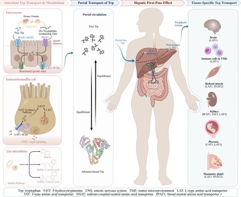
In addition to being incorporated into proteins, tryptophan (Trp) is increasingly recognized as a critical metabolic hub orchestrating systemic physiological homeostasis through interactions among the digestive, nervous and immune systems. This review systematically delineates the latest advancements in the molecular mechanisms governing the spatial and cellular heterogeneity of Trp transport. We summarize the sophisticated transmembrane machinery of Trp transport in intestinal epithelial cells, including angiotensin-converting enzyme 2-dependent broad neutral amino acid transporter 1 complex in absorptive enterocytes and L-type amino acid transporter 1/alanine, serine, cysteine transporter 2-mediated uptake in secretory enterochromaffin cells. Additionally, the albumin-binding dynamics of Trp in tryptophan 2,3-dioxygenase-mediated hepatic first-pass gating and its metabolic shunting to peripheral tissues are also discussed in this review. By bridging micro-level transport mechanisms with macro-level physiological regulation, this review provides a comprehensive framework for understanding the pleiotropic functions of Trp and identifies transporter-enzyme coupling as a potential target for therapeutic intervention in metabolic disorders, neurodegenerative diseases, and immune dysfunction.
Keywords: amino acid, enterochromaffin cell, tryptophan transport, hepatic first-pass
Introduction
Amino acids (AAs) are the building blocks of proteins and are essential for normal cellular growth and function [1]. Tryptophan (Trp) is an essential amino acid (EAA) for maintaining nitrogen balance and facilitating protein synthesis [2]. However, the versatile biological roles of Trp have been substantially revealed by recent metabolomic, immunological and neuroendocrinological research [3]. The metabolic networks of Trp serve as a central hub that connects the neural, immune, and microbial systems in the host. Like in the gut, Trp-derived metabolites act together with other microbial generated bioactive compounds or particles including short chain fatty acids, secondary bile acids and extracellular vesicles that are essential for maintaining gut homeostasis and modulating immune functions [4-6].
These diverse functions are governed by specific membrane transporters and distinct metabolic networks. Intestinal Trp modulation exhibits substantial spatial and cellular heterogeneity due to the presence of specialized transporter systems within different intestinal epithelial compartments [7, 8]. Absorptive enterocytes mediate Trp transport through dedicated membrane transporters, whereas secretory cells (such as enterochromaffin (EC) cells) convert intracellular Trp into 5-hydroxytryptamine (5-HT, serotonin) through tryptophan hydroxylase 1 (TPH1)-dependent metabolism [9, 10].
Within tissue and cellular microenvironments, Trp generates several vital bioactive compounds, predominantly through three core metabolic pathways: the kynurenine pathway (KP), 5-HT pathway, and gut microbiota-mediated indole pathway [11]. These metabolites are central nodes for chemical signaling within the microbiota-gut-brain axis and play important roles in mediating energy metabolism, modulating inflammation and oxidative stress, inducing immune tolerance, and maintaining intestinal barrier homeostasis [9, 12].
Elucidating the transport and metabolic networks of Trp is essential for understanding metabolic diseases, neurodegenerative disorders, and immune dysregulation. Most current studies have focused on the host and microbiota mediated Trp metabolism. The mechanisms governing its transmembrane transport and tissue distribution remain incompletely defined. In this review, we first outline the major metabolic pathways of Trp and then focus on its transport processes in the host. As the first-pass effect describes how the liver extracts and metabolizes the absorbed Trp before it enters the systemic circulation [8, 10, 13], the route of Trp from intestinal absorption to hepatic portal circulation is discussed in this review. Finally, the distribution of Trp and uptake by peripheral tissues has also been included.
Tryptophan Metabolic Pathways
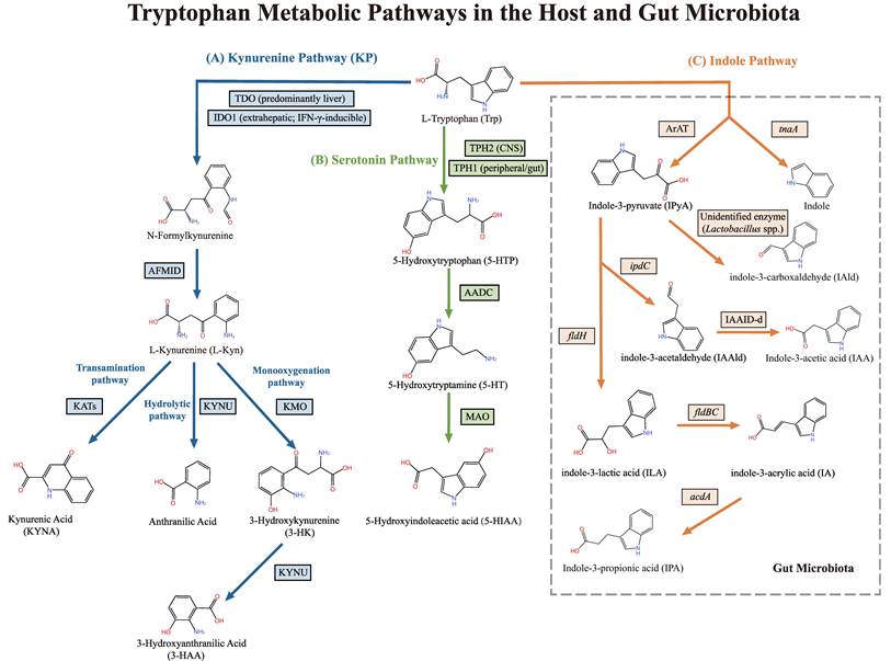
Trp metabolism primarily involves two major systems: host cellular and gut microbiota metabolism (Figure 1). In host cells, Trp is mainly catabolized via two distinct pathways: the kynurenine and 5-HT pathways [4]. Similar to other AAs, Trp metabolism is highly organ-specific and relies on distinct local enzymes [8]. The liver is the central site of Trp metabolism, where the KP accounts for over 90% of total Trp degradation [14]. The gastrointestinal tract and central nervous system (CNS) are secondary sites for Trp metabolism, in which the 5-HT pathway plays a pivotal role. More than 90% of the systemic 5-HT is synthesized in the gut, mainly by EC cells [15]. The CNS contains less than 5% of total body 5-HT [16]. In addition to host cellular metabolism, the gut microbiota can metabolize Trp. Therefore, unabsorbed Trp in the gut is primarily converted by the microbiota via the indole pathway [17].
The Metabolic Pathways of Tryptophan in the Host and Gut Microbiota. Host cells and gut microbes metabolize Trp through three major pathways. (A) The kynurenine pathway is the principal route of host Trp catabolism. TDO acts mainly in the liver, whereas IDO1 is widely expressed in extrahepatic tissues and can be induced by inflammatory cytokines such as IFN-γ. Both enzymes catalyze the first step from Trp to N-formylkynurenine. AFMID then converts N-formylkynurenine to L-Kyn. KATs convert L-Kyn to KYNA, KYNU converts L-Kyn to anthranilic acid, and KMO converts L-Kyn to 3-HK. KYNU further converts 3-HK to 3-HAA. (B) Approximately 1-2% of Trp enters the serotonin pathway. TPH1 acts mainly in peripheral tissues, especially gut enterochromaffin cells, whereas TPH2 functions in the CNS. Both enzymes convert Trp to 5-HTP. AADC converts 5-HTP to 5-HT, and MAO metabolizes 5-HT to 5-HIAA. (C) About 4-6% of dietary Trp reaches the colon. Gut microbes convert this fraction to indole and related metabolites. TnaA converts Trp to indole, whereas ArAT converts Trp to IPyA. IpdC converts IPyA to IAAld, and IAAID-d converts IAAld to IAA. A separate branch forms IAld, mainly in Lactobacillus spp. The responsible enzyme remains unidentified. FldH converts IPyA to ILA, FldBC converts ILA to IA, and AcdA converts IA to IPA. Abbreviations: Trp, tryptophan; IFN-γ, interferon-γ; TDO, tryptophan 2,3-dioxygenase; IDO1, indoleamine 2,3-dioxygenase 1; AFMID, arylformamidase; L-Kyn, L-kynurenine; KATs, kynurenine aminotransferases; KYNA, kynurenic acid; KMO, kynurenine 3-monooxygenase; 3-HK, 3-hydroxykynurenine; KYNU, kynureninase; 3-HAA, 3-hydroxyanthranilic acid; TPH1, tryptophan hydroxylase 1; TPH2, tryptophan hydroxylase 2; CNS, central nervous system; 5-HTP, 5-hydroxytryptophan; AADC, aromatic L-amino acid decarboxylase; 5-HT, 5-hydroxytryptamine; MAO, monoamine oxidase; 5-HIAA, 5-hydroxyindoleacetic acid; tnaA, gene encoding tryptophanase; ArAT, aromatic amino acid aminotransferase; IPyA, indole-3-pyruvate; ipdC, gene encoding indolepyruvate decarboxylase; IAAld, indole-3-acetaldehyde; IAAID-d, indole-3-acetaldehyde dehydrogenase; IAA, indole-3-acetic acid; IAld, indole-3-carboxaldehyde; fldH, gene encoding phenyllactate dehydrogenase; ILA, indole-3-lactic acid; fldBC, gene encoding phenyllactate dehydratase; IA, indole-3-acrylic acid; acdA, gene encoding acyl-CoA dehydrogenase; IPA, indole-3-propionic acid.
The Kynurenine Pathway
The KP is the principal pathway for Trp metabolism. The cascade of this pathway is initiated by the oxidative cleavage of the indole ring of Trp [18]. This rate-limiting step is catalyzed either by tryptophan 2,3-dioxygenase (TDO, highly expressed in the liver, encoded by TDO2) or by indoleamine 2,3-dioxygenase 1 (IDO1, widely distributed in extrahepatic tissues and induced by inflammatory cytokines, such as interferon-γ [IFN-γ]) [18, 19]. This reaction generates the unstable intermediate N-formylkynurenine, which is subsequently and rapidly hydrolyzed by arylformamidase (kynurenine formamidase) to yield L-kynurenine (L-Kyn), the central metabolite of the pathway [19]. The downstream metabolism of L-Kyn primarily diverges into transamination, monooxygenation, and hydrolysis pathways [20]. In the transamination pathway, L-Kyn is catalyzed by kynurenine aminotransferases to form kynurenic acid [20]. In the monooxygenation branch, L-Kyn is first oxidized by kynurenine-3-monooxygenase to form 3-hydroxykynurenine, which is subsequently cleaved into 3-hydroxyanthranilic acid by kynureninase (KYNU) [12]. In the direct hydrolytic pathway, L-Kyn is cleaved by KYNU to generate anthranilic acid [21].
The Serotonin Pathway
Although the majority of Trp in the body is degraded via the KP, approximately 1-2% enters the 5-HT pathway [22]. During the biosynthesis of 5-HT, Trp is hydroxylated to produce 5-hydroxytryptophan (5-HTP), a reaction catalyzed by the rate-limiting enzyme tryptophan hydroxylase (TPH) [23]. Subsequently, 5-HTP is decarboxylated by aromatic L-amino acid decarboxylase (AADC) to generate the bioactive molecule, 5-HT [4]. Furthermore, TPH exists as two distinct isoforms, TPH1 and tryptophan hydroxylase 2 (TPH2), which mediate 5-HT synthesis in peripheral tissues (primarily ECs) and the CNS, respectively [24-26]. In addition to exerting direct receptor-mediated physiological functions, 5-HT is metabolized by monoamine oxidase (MAO) into 5-hydroxyindoleacetic acid, which is then excreted in urine [27].
The Indole Pathway
Approximately 4-6% of unabsorbed dietary Trp reaches the colon, where it is metabolized into indole and its derivatives by the commensal gut microbiota [28]. This process predominantly relies on the bacteria-expressed enzyme, tryptophanase, which catalyzes the cleavage of Trp into indole, pyruvate, and ammonia [29]. In addition to indole, certain gut microbes use specialized enzymes to convert Trp into other active metabolites [30, 31]. In one major branch, aromatic amino acid aminotransferase (ArAT) transforms Trp into the central intermediate indole-3-pyruvate (IPyA) [8, 32]. From IPyA, several metabolic routes diverge. Decarboxylation by indolepyruvate decarboxylase (IpdC) yields indole-3-acetaldehyde, which is oxidized by indole-3-acetaldehyde dehydrogenase (IAAID-d) to produce indole-3-acetic acid (IAA) [4, 32]. In another branch, IPyA is converted into indole-3-carboxaldehyde (IAld), primarily by Lactobacillus species, although the precise enzyme is unidentified [8, 30]. The third pathway involves phenyllactate dehydrogenase (FldH), which reduces IPyA to indole-3-lactic acid (ILA). For many strains, metabolism ends at ILA. However, Clostridium sporogenes uses phenyllactate dehydratase (FldBC) to convert it into indole-3-acrylic acid (IA). Subsequently, acyl-CoA dehydrogenase (AcdA) reduces this IA into indole-3-propionic acid (IPA) [30, 32].
These three metabolic pathways determine the chemical fate of Trp in the host and their initiation and efficiency are dependent on the spatial availability of the substrate. This availability is governed by a highly orchestrated network of transmembrane transporters located in the intestinal epithelium, a primary site for nutrient absorption.
Mechanisms of Intestinal Absorption, Transmembrane Transport, and Metabolism of Tryptophan
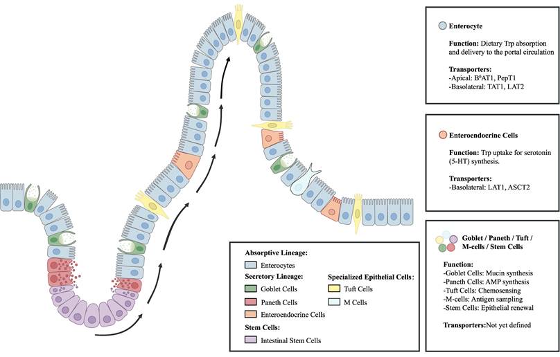
Most dietary Trp absorption occurs in the small intestine, especially in the jejunum and ileum [30]. Intestinal epithelial cells (IECs) form the first cellular barrier between luminal Trp and the host circulation [33, 34]. IECs comprise several distinct populations: the absorptive lineage, primarily consisting of enterocytes for nutrient uptake; the secretory lineage, which includes mucin-secreting goblet cells, antimicrobial peptide (AMP)-producing Paneth, and enteroendocrine cells (EECs); specialized functional or immune-related cells, such as chemosensory tuft and antigen-sampling microfold cells; and intestinal stem cells for epithelial renewal [35-37] (Figure 2). The fate of Trp differs between intestinal absorptive and secretory cells. To satisfy the systemic nutritional demands of the body, the absorptive lineage uses dedicated transporters to actively shuttle Trp across the membrane [38]. In contrast, the secretory lineage, particularly EC cells, converts Trp into bioactive metabolites such as 5-HT, which contributes to intestinal motility and local immune and inflammatory regulation [39].
Spatial Distribution, Cellular Heterogeneity, and Transporter Annotation of Intestinal Epithelial Cells in the Jejunum and Ileum. The intestinal epithelium has distinct lineages. Enterocytes, the primary absorptive site, mediate the directional transcellular shuttling of luminal Trp. This directional transport is powered by specific apical uptake (B0AT1 and PepT1) and is functionally coupled with basolateral efflux (TAT1 and LAT2). The secretory lineages include goblet, Paneth, and enteroendocrine cells (EECs). Among EECs, enterochromaffin cells (EC) rely on basolateral transporters (LAT1 and ASCT2) for Trp uptake to synthesize bioactive signaling molecules, such as serotonin. Cells with special functions (tuft and microfold cells) coordinate localized mucosal immunity. Concurrently, the intestinal stem cells at the crypt base drive epithelial renewal. However, the Trp transporters in the goblet, Paneth, tuft, microfold, and stem cells remain unclear. Abbreviations: AMP, antimicrobial peptide; ASCT2, alanine, serine, cysteine transporter 2; B0AT1, broad neutral amino acid transporter 1; EC, enterochromaffin; EECs, enteroendocrine cells; LAT1/2, L-type amino acid transporter 1/2; M cells, microfold cells; PepT1, peptide transporter 1; TAT1, T-type amino acid transporter 1; Trp, tryptophan; 5-HT, 5-hydroxytryptamine.
Tryptophan Transport Pathways and Molecular Mechanisms in Intestinal Absorptive Cells
After ingestion by monogastric hosts, dietary proteins undergo rapid enzymatic cleavage by pepsin and pancreatic proteases. Subsequently, these massive complexes are stripped down into a readily absorbable pool consisting mostly of free amino acids (FAAs) and short oligopeptides, such as di- and tripeptides [40]. Enterocytes, the dominant absorptive cell types among IECs, are critical sites for Trp uptake and its subsequent translocation into the systemic circulation. Efficient intestinal Trp uptake is important for growth and development [32]. Notably, the efficiency of intestinal Trp transport not only determines its bioavailability, but also modulates its subsequent metabolic fate. It is the main physiological requirement for sustaining nitrogen balance and host homeostasis [41].
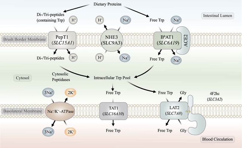
Under normal physiological conditions, Trp absorption is highly efficient; the majority is taken up by the small intestine, with only 4-6% reaching the colon for microbial degradation [28, 32]. Trp absorption is a highly orchestrated energy-dependent physiological process regulated by the synergistic action of multiple carrier proteins, and involves both facilitated diffusion and active transport mechanisms. This process involves uptake at the brush border membrane (BBM) and efflux across the basolateral membrane (BLM), primarily relying on the coordinated expression and functional coupling of the solute carrier (SLC) family members [10] (Figure 3).
Molecular Mechanisms of Tryptophan Transmembrane Transport in Absorptive Enterocytes. At the brush border membrane, free luminal Trp is primarily cotransported with Na+ by the B0AT1 transporter (encoded by SLC6A19). B0AT1 depends on ACE2 for proper membrane localization and transport function. Di- and tripeptides (Trp-containing) are internalized via the H+-coupled peptide transporter, PepT1 (encoded by SLC15A1), and hydrolyzed by cytosolic peptidases into free Trp. NHE3 exports H+ in exchange for luminal Na+ and helps maintain the proton gradient required for PepT1 activity. The basolateral Na+/K+-ATPase maintains the Na+ gradient that supports apical transport. At the basolateral membrane, the efflux of Trp into portal circulation is primarily facilitated by the uniporter, TAT1 (encoded by SLC16A10). The antiporter, LAT2 (encoded by SLC7A8), which is covalently linked to the heavy chain, 4F2hc (encoded by SLC3A2), primarily functions as an exchanger to maintain the intracellular AA pool. Abbreviations: B0AT1, broad neutral amino acid transporter 1; ACE2, angiotensin-converting enzyme 2; PepT1, peptide transporter 1; NHE3, Na+/H+ exchanger 3; AA, amino acid; TAT1, T-type amino acid transporter 1; LAT2, L-type amino acid transporter 2; 4F2hc, 4F2 cell-surface antigen heavy chain; Gly, glycine; Trp, tryptophan.
Mechanism of Tryptophan Uptake via the Enterocyte Brush Border Membrane
The uptake of Trp across the enterocyte BBM is the main absorption step. Luminal free Trp is mainly transported across the BBM via the Na+-dependent transport, system B0 [42]. The key transporter responsible for apical Trp uptake is broad neutral amino acid transporter 1 (B0AT1), encoded by the SLC6A19 gene [43, 44].
B0AT1 is a broad-spectrum neutral AA symporter. It harnesses the Na+ electrochemical gradient established by the basolateral Na+/K+-ATPase to co-transport one Trp molecule and one Na+ ion into the cell [42, 45]. The functional expression and localization of B0AT1 on the BBM do not occur independently; they are highly dependent on angiotensin-converting enzyme 2 (ACE2), which is a chaperone and accessory subunit to maintain its transport activity and membrane stability [23, 44]. Because ACE2 is a key regulator of the renin-angiotensin system (RAS) [46], the RAS may participate in regulating Trp absorption via a non-classical pathway. The dependence of B0AT1 on ACE2 also makes this transport system sensitive to changes in ACE2 abundance. This becomes relevant during SARS-CoV-2 infection because ACE2 also serves as the viral entry receptor [23, 47]. Viral binding and subsequent internalization are associated with reduced surface ACE2 expression. This disruption compromises B0AT1-ACE2 transport complex surface expression, potentially leading to impaired intestinal Trp absorption. Mechanistic studies indicate that localized Trp deficiency reduces epithelial antimicrobial peptide expression, promotes gut dysbiosis, and increases susceptibility to mucosal inflammation [48]. This cascade pathology is a potential contributing explanation for the gastrointestinal symptoms frequently observed in patients with COVID-19 [49, 50]. Under normal dietary conditions, Trp also competes with other neutral amino acids for B0AT1-mediated uptake. These include other large neutral amino acids (LNAAs), particularly branched-chain amino acids (BCAAs, including leucine (Leu), isoleucine (Ile), and valine (Val)) and phenylalanine [38, 51, 52].
In addition to FAA transport, a portion of Trp is absorbed as Trp-containing di- or tri-peptides via peptide transporter 1 (PepT1) [53, 54]. Encoded by SLC15A1, PepT1 is a high-capacity low-affinity transporter predominantly located in the BBM that mediates the transport of small peptides [55]. PepT1 functions as a proton (H+)-coupled symporter. It co-transports one neutral or cationic di-peptide with one H+, whereas one anionic di-peptide is co-transported with two H+ ions [56, 57]. The co-transported H+ is subsequently extruded by Na+/H+ exchanger 3 (NHE3, encoded by SLC9A3) on the BBM [58]. Intracellular peptides are rapidly hydrolyzed by cytosolic peptidases into FAAs, which enter the intracellular AA pool [59]. Furthermore, PepT1 expression is highly adaptable to dietary changes. Both high-protein diets and short-term fasting can upregulate PepT1 expression for peptide absorption [58]. The absorption of peptide-bound Trp is a compensatory mechanism. If primary transporters, such as B0AT1, become saturated or pathologically impaired, the peptide-driven bypass provides an alternative absorptive route that can partially compensate for reduced amino acid uptake [40]. This compensatory pathway helps preserve intestinal Trp uptake, thereby contributing to the maintenance of systemic Trp homeostasis and providing a mechanistic basis for improving precision nutrition. Therefore, future studies are needed to determine whether diets rich in Trp-containing dipeptides could exploit the PepT1 bypass to circumvent the ACE2/B0AT1 blockade and rescue acute AA starvation during viral enteritis.
The luminal substrate environment also affects apical Trp uptake. Dietary protein source and high-fat feeding can change the intestinal amino acid profile and the timing of substrate availability [60-63]. These changes may modify the degree of substrate competition among LNAAs for B0AT1-mediated transport [38, 52], thereby influencing Trp absorption.
Efflux and Uptake of Tryptophan at the Basolateral Membrane of Enterocytes
Following uptake by enterocytes via transporters on the BBM, free Trp is rarely involved in local synthesis or metabolism. Instead, it normally undergoes simple transcellular transit. It is directly exported across the BLM into the lamina propria, subsequently entering the capillaries and joining portal circulation [41]. This efflux process primarily involves facilitated diffusion by T-type amino acid transporter 1 (TAT1, encoded by SLC16A10) and exchange transport by L-type amino acid transporter 2 (LAT2, encoded by SLC7A8) [40].
TAT1 is a specific passive uniporter for aromatic amino acids (AAAs) [64, 65]. TAT1 channels intracellular Trp outward using only the transmembrane concentration gradient. This substrate-independent facilitated diffusion allows TAT1 to directly mediate net basolateral Trp efflux toward the portal circulation [64].
LAT2 typically forms a heterodimeric complex with the heavy chain, 4F2 cell-surface antigen heavy chain (4F2hc, alias for cluster of differentiation 98 (CD98), encoded by SLC3A2), via a disulfide bond to localize to the BLM and function properly [66]. Unlike TAT1, LAT2 mainly functions as an amino acid exchanger rather than a simple efflux route. LAT2 exchanges intracellular and extracellular AAs in a 1:1 stoichiometric ratio [67]. LAT2 mediates cellular uptake of neutral AAs, including glutamine (Gln), Trp, and Ile. This exchange directly couples the inward transport of these neutral AAs to the efflux of smaller, more polar AAs, such as glycine. Despite a minor capacity for unidirectional transport by LAT2, the actual efflux of AAAs, such as Trp and phenylalanine, is largely supported by TAT1-mediated facilitated diffusion [40].
Transport Pathways and Molecular Mechanisms of Tryptophan in EECs
Although EECs account for only approximately 1% of IECs [68], EC cells play a pivotal role in Trp metabolism [69]. Unlike enterocytes, which primarily mediate the transmembrane transport of Trp, EC cells, the largest endocrine cell population in the gut [70], specifically convert Trp into 5-HT, serving as a core node in the chemical signaling of the gut-brain axis [71].
Uptake and Metabolism of Tryptophan in EC Cells
Trp required for 5-HT synthesis in EC cells is thought to be supplied largely from the basolateral circulation [72]. In EC cells, Trp uptake is mediated primarily by L-type amino acid transporter 1 (LAT1, encoded by SLC7A5) [73], with alanine, serine, and cysteine transporter 2 (ASCT2, encoded by SLC1A5) indirectly supporting LAT1-dependent transport [74]. Similar to LAT2, LAT1 associates with 4F2hc via a conserved disulfide linkage to form a heterodimeric complex that supports proper plasma membrane localization and transport function [75-77]. LAT1 can utilize intracellular Gln as an exchange substrate, coupling Gln efflux to extracellular Trp uptake [75, 78]. ASCT2 mediates Na+-dependent exchange of neutral AAs, including Gln, and helps maintain the intracellular exchange-substrate pool available to LAT1 [79, 80]. In EC cells, this arrangement may support LAT1-mediated Trp uptake and provide Trp for 5-HT synthesis (Figure 4).
Metabolic Coupling of Tryptophan Uptake and Serotonin Biosynthesis in Enterochromaffin Cells. Trp for serotonin synthesis in ECs is supplied largely from the basolateral circulation. LAT1 (encoded by SLC7A5) forms a complex with 4F2hc (encoded by SLC3A2) and exchanges intracellular glutamine (Gln) for extracellular Trp. ASCT2 (encoded by SLC1A5) supports this process by maintaining the intracellular Gln pool available for LAT1 exchange. TPH1 converts intracellular Trp to 5-hydroxytryptophan (5-HTP), and AADC converts 5-HTP to serotonin (5-HT). Free cytosolic 5-HT is susceptible to MAO-mediated degradation. VMAT1 (encoded by SLC18A1) transports newly synthesized 5-HT into secretory granules for storage and subsequent release. Abbreviations: AADC, aromatic L-amino acid decarboxylase; ASCT2, alanine, serine, cysteine transporter 2; 4F2hc, 4F2 cell-surface antigen heavy chain; Gln, glutamine; LAT1, L-type amino acid transporter 1; MAO, monoamine oxidase; TPH1, tryptophan hydroxylase 1; Trp, tryptophan; 5-HT, 5-hydroxytryptamine; 5-HTP, 5-hydroxytryptophan; AA, amino acid; ECs, enterochromaffin cells; VMAT1, vesicular monoamine transporter 1.
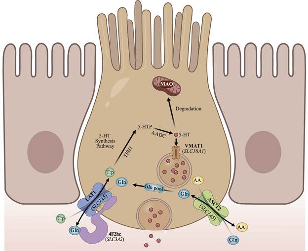
This LAT1-ASCT2 coupling may stabilize Trp uptake when nutrient availability changes. ASCT2 maintains the intracellular Gln pool that LAT1 uses for amino acid exchange. This mechanism may help EC cells maintain 5-HT synthesis during fasting [81, 82].
Once Trp enters an EC cell, it can enter the 5-HT pathway. TPH1 first converts Trp to 5-HTP in the presence of tetrahydrobiopterin. AADC then converts 5-HTP to 5-HT [24, 82]. Newly synthesized 5-HT is prone to degradation by MAO if left free in the cytoplasm. Therefore, it is rapidly sequestered into basolateral secretory granules by vesicular monoamine transporter 1 (encoded by SLC18A1) to form a high-concentration reserve that prevents intracellular oxidative degradation [69]. The stored 5-HT can then be released when EC cells detect mechanical or chemical stimuli.
Release of 5-HT from EC Cells
The release of 5-HT is primarily mediated by mechanical and chemical pathways [83, 84] (Figure 5). Mechanical forces associated with intestinal motility and luminal contents can stimulate mechanosensitive EC cells. Physical stretching directly induces the opening of Piezo2, a mechanosensitive ion channel in the EC cell membrane. This influx of nonselective cations (primarily Na+ and Ca2+) down their electrochemical gradients initiates cell membrane depolarization [84]. When the membrane potential reaches the threshold for excitation, voltage-gated Na+ channels are activated, leading to Na+ influx and action potential generation [85]. As action potentials propagate along the membrane, they rapidly activate voltage-gated calcium channels (VGCCs). This activation promotes extracellular Ca2+ influx and produces localized elevations in intracellular Ca2+ near secretory sites. Ultimately, this activates the soluble N-ethylmaleimide-sensitive factor attachment protein receptor (SNARE) complex, driving the exocytosis of 5-HT-containing dense-core vesicles and the release of 5-HT into the lamina propria [86, 87].
Mechanosensory and Chemosensory Control of Serotonin Release from Enterochromaffin Cells and Its Downstream Actions. Mechanical and chemical signals trigger 5-HT release from ECs. Mechanical triggering: Intestinal stretch activates Piezo2 and allows Na+ and Ca2+ to enter the cell. The resulting membrane depolarization activates voltage-gated sodium channels (VGSCs) and generates action potentials. Voltage-gated calcium channels (VGCCs) then open and increase Ca2+ influx. The rise in intracellular Ca2+ triggers SNARE-mediated exocytosis of 5-HT-containing vesicles. Chemical triggering: Luminal nutrients and microbial metabolites provide chemical stimuli to EC cells. Olfr78 couples to Gs. Gs activates adenylyl cyclase (AC) and increases cAMP. cAMP activates PKA, which reduces K+ conductance, and also enhances inward depolarizing currents through HCN channels. FFAR2 couples to Gq and activates PLC. PLC generates IP3, which releases Ca2+ from the endoplasmic reticulum and promotes membrane depolarization. Released 5-HT activates 5-HT3 receptors on vagal afferents and 5-HT4 and other serotonergic receptors in enteric neural circuits. These local signals support gut-brain communication and intestinal motility and secretion. Intestinal epithelial cells take up extracellular 5-HT through SERT, followed by intracellular degradation by MAO. 5-HT that escapes local clearance can enter the circulation, where platelets take it up through SERT and store it. Abbreviations: AC, adenylyl cyclase; cAMP, cyclic adenosine monophosphate; ECs, enterochromaffin cells; ER, endoplasmic reticulum; FFAR2, free fatty acid receptor 2; Gs/Gq, G-protein subtypes; HCN, hyperpolarization-activated cyclic nucleotide-gated; IP3, inositol 1,4,5-trisphosphate; MAO, monoamine oxidase; Olfr78, olfactory receptor 78; PKA, protein kinase A; PLC, phospholipase C; SERT, serotonin transporter; SNARE, soluble N-ethylmaleimide-sensitive factor attachment protein receptor; VGCCs, voltage-gated calcium channels; VGSCs, voltage-gated sodium channels; ENS, enteric nervous system; 5-HT, 5-hydroxytryptamine.
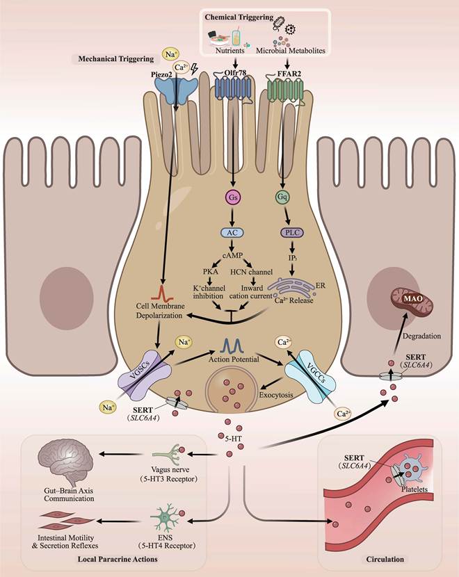
In the chemical-triggering pathway, the chemical microenvironment within the intestinal lumen, particularly nutrients and gut microbiota metabolites, is a crucial chemical stimulus for EC cells. Microbial metabolites, such as short-chain fatty acids (e.g., acetate, propionate, and butyrate), are sensed by G protein-coupled receptors in EC cells that possess an apical domain exposed to the intestinal lumen. These include olfactory receptor 78 (Olfr78, human homolog OR51E2) and the free fatty acid receptor 2, FFAR2 (also known as GPR43) [88]. Olfr78 couples to Gs signaling. Activated Gs stimulates adenylyl cyclase and increases intracellular cyclic adenosine monophosphate (cAMP). Elevated cAMP activates protein kinase A (PKA), which can inhibit K+ channel activity and reduce outward K+ conductance. cAMP can also directly modulate hyperpolarization-activated cyclic nucleotide-gated (HCN) channels and enhance inward depolarizing cation currents. These two cAMP-dependent mechanisms favor membrane depolarization [89-91]. In parallel, FFAR2 can couple to Gq and activate phospholipase C (PLC). PLC generates the second messenger inositol 1,4,5-trisphosphate (IP3), which mobilizes Ca2+ from the endoplasmic reticulum. The increase in cytosolic Ca2+ can activate depolarizing cation conductances and shift the membrane potential toward the threshold for action potential generation [92-94].
Locally, 5-HT released by EC cells activates 5-HT3 receptors on vagal and other primary afferent nerve fibers and thereby conveys sensory information along the gut-brain axis. 5-HT also acts on 5-HT4 and other serotonergic receptors within enteric neural circuits and contributes to peristaltic and secretory reflexes [85, 95]. After completing local signal transmission, the released 5-HT is taken up by the serotonin transporter (SERT, encoded by SLC6A4), which is widely expressed in intestinal epithelial cells and subsequently intracellularly degraded by MAO [96]. Systemically, 5-HT that is not cleared locally can enter the portal circulation [97]. Circulating 5-HT is taken up by platelets via SERT and stored in platelets, whereas a much smaller free fraction remains in plasma [98]. Platelets can release stored 5-HT upon activation [99]. Free circulating gut-derived 5-HT can act as an endocrine signal in peripheral tissues. Gut-derived 5-HT has been implicated in bone formation and contributes to the regulation of glucose and lipid metabolism [81, 97, 100]. These actions illustrate how Trp-derived 5-HT extends the physiological role of Trp beyond nutrient provision to systemic metabolic signaling. This is the fate of the Trp fraction used for intestinal 5-HT synthesis. Most absorbed Trp follows a different route and leaves the intestinal epithelium in unmetabolized form.
Albumin Binding and Circulatory Transport of Tryptophan
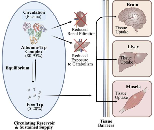
After intestinal absorption, Trp enters the portal circulation. In portal blood, Trp already exists in free and albumin-bound forms. A large fraction binds reversibly to serum albumin, while a smaller fraction remains free in plasma [101]. This equilibrium affects how much free Trp is available when portal blood reaches the liver (Figure 6). The same free-bound equilibrium continues after hepatic passage in the systemic circulation.
Circulatory Transport Forms and Albumin-Binding Dynamics of Tryptophan. Plasma Trp exists as albumin-bound and free fractions that remain in reversible equilibrium. About 80-95% is bound to serum albumin, mainly at Sudlow site II, whereas 5-20% remains free. Albumin-bound Trp forms a circulating reservoir for the free pool. Albumin binding reduces renal Trp loss and limits exposure to catabolic pathways. Free Trp is taken up by peripheral tissues such as the liver and skeletal muscle and can enter the brain after transport across the BBB. Abbreviations: BBB, blood-brain barrier; Trp, tryptophan.
Molecular Mechanism and Specificity of Tryptophan Binding to Albumin
Under physiological conditions, approximately 80-95% of Trp in plasma is non-covalently bound to serum albumin, leaving only 5-20% in the free form [102]. Trp primarily binds to Sudlow site II (the indole-benzodiazepine site) on the albumin molecule, which is located within the hydrophobic pocket of subdomain IIIA [103]. This binding is saturable and governed by the law of mass action, maintaining a dynamic dissociation-association equilibrium. The albumin-bound pool acts as a circulating reservoir and replenishes free Trp as tissues remove it from blood [104, 105].
Albumin binding also limits Trp loss from the circulation. The albumin-Trp complex crosses the glomerular filtration barrier poorly because of its large molecular size. Albumin binding therefore reduces renal Trp loss and limits the amount of Trp that is immediately available to enter catabolic pathways in the liver and other tissues. These effects help maintain a relatively stable circulating Trp pool [101, 106]. The binding is reversible [104]. As tissues take up free Trp, albumin releases part of its bound Trp and replenishes the free pool. Free Trp is the main form available for tissue uptake. It can be taken up by peripheral tissues such as the liver and skeletal muscle through amino acid transporters. Similarly, free Trp must cross the blood-brain barrier (BBB) through specific amino acid transport systems to enter the CNS [107, 108]. As free Trp is taken up by tissues, albumin-bound Trp can contribute to replenishment of the free pool through reversible dissociation and thereby help maintain circulating Trp availability.
Factors That Alter Albumin-Tryptophan Binding
The distribution of Trp between free and albumin-bound pools is influenced by plasma non-esterified fatty acid (NEFA) concentrations. Trp binds mainly to Sudlow site II on albumin. The FA3-FA4 region overlaps with this site and binds fatty acids. Albumin also has several high-affinity sites for long-chain fatty acids [101, 109, 110]. NEFAs can compete with Trp for sites on albumin [111]. Fasting, stress, and prolonged exercise can increase plasma NEFA levels. High-fat feeding can also raise plasma NEFA levels under some conditions [111-113]. Higher NEFA levels can displace Trp from albumin and increase the free Trp fraction. Disease-associated glycation and oxidation can alter human serum albumin (HSA) structure and ligand binding at Sudlow site II. For instance, glycation changes the conformation of this site and alters its affinity for ligands in patients with diabetes [114, 115]. Oxidative modification also changes albumin structure and impair Site II binding for some ligands [116]. The Sudlow site II acts as the major binding site for Trp and structural changes at this site can alter the equilibrium between albumin-bound Trp and free Trp, thus changing the circulating free Trp pool [101]. Subsequently, the changed free Trp pool in portal blood reaches the liver, where part of the absorbed Trp is removed during the first pass.
Hepatic First-Pass Effect and Cell-Specific Metabolic Shunting
Portal blood reaches the liver before the systemic circulation. During this first pass, the liver extracts and metabolizes part of the absorbed Trp. The remainder enters the systemic circulation [8, 10, 13] (Figure 7). Direct quantitative data on hepatic first-pass extraction of Trp in humans remain limited. In sheep, the liver extracts approximately 78% of Trp from the portal-drained viscera, leaving only 22% for the systemic circulation [117].
Hepatic First-Pass Effect and Post-Hepatic Distribution of Tryptophan. After intestinal absorption, Trp enters the portal circulation and reaches the liver before the systemic circulation. The liver takes up and metabolizes part of the absorbed Trp during this first pass. The remaining Trp leaves the liver and enters the systemic circulation for delivery to peripheral tissues. The extent of first-pass Trp extraction in humans remains poorly defined. The liver also accounts for a major share of whole-body Trp catabolism, but this cumulative contribution is distinct from first-pass extraction. Abbreviation: Trp, tryptophan.
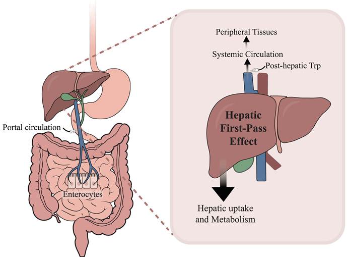
This first-pass extraction differs from the liver's cumulative contribution to whole-body Trp catabolism. The liver is the primary organ for Trp catabolism (accounting for approximately 90% of the total degradation) and serves as a metabolic hub. Through hepatic uptake and TDO-mediated catabolism, the liver regulates how much Trp is degraded locally and how much remains available to the systemic circulation [10, 102].
Hepatic Trp metabolism exhibits substantial cellular heterogeneity [118]. Under homeostatic conditions, Trp crosses the hepatic sinusoidal endothelium into the Space of Disse [119] and is subsequently taken up via the TAT1 transporter located at the sinusoidal membrane of hepatocytes [120]. The intracellular metabolic flux of Trp through the kynurenine pathway is largely regulated by the rate-limiting enzyme TDO [121]. At baseline physiological Trp concentrations, TDO-mediated catabolism is restrained, which helps preserve Trp availability in the systemic circulation. Conversely, Trp availability rises after ingestion of Trp-rich protein meals [122]. When Trp availability rises, increased substrate supply can enhance TDO-mediated Trp catabolism in hepatocytes.
Beyond this substrate-dependent hepatocyte response, an additional IDO1-mediated route of hepatic Trp catabolism can be induced during inflammation. The portal circulation continuously exposes the liver to gut-derived pathogen-associated molecular patterns (PAMPs), including lipopolysaccharide (LPS), and periportal Kupffer cells help restrain excessive responses to these signals [123]. During inflammation, IFN-γ can induce IDO1 in Kupffer cells and dendritic cells, where IDO1 promotes local Trp catabolism, suppresses T-cell responses, and may contribute to hepatic immune tolerance [124, 125]. Thus, IDO1 in hepatic immune cells provides an inflammation-sensitive complement to TDO-mediated control of hepatic Trp metabolism. These findings suggest that inflammatory signaling can shift hepatic Trp metabolism toward local IDO1-dependent catabolism, potentially reducing the amount of Trp that reaches the systemic circulation. This may constitute a gut-liver regulatory link through which inflammatory stress favors local immunoregulatory Trp catabolism over peripheral Trp availability.
Tissue-Targeted Transport and Metabolism of Tryptophan Following the Hepatic First-Pass Effect
Although the liver is the primary site of Trp catabolism and TDO is predominantly expressed in the liver, a fraction of absorbed Trp remains unmetabolized after hepatic passage and enters the systemic circulation [10, 126]. This fraction, here referred to as “post-hepatic Trp,” undergoes further metabolism in peripheral tissues. Although this post-hepatic fraction is smaller than the amount of Trp metabolized by the liver, it remains physiologically important because it supports protein synthesis and serves as a substrate for tissue-specific pathways involved in neuroendocrine and immune regulation [8].
Nervous System
Within the nervous system, the CNS is a critical target organ for post-hepatic Trp metabolism. Trp cannot freely diffuse across the BBB. Instead, its entry into the brain parenchyma is mediated mainly by LAT1 expression in brain capillary endothelial cells [108, 127]. At the BBB, Trp competes with other LNAAs for LAT1-mediated transport [128, 129]. Brain 5-HT synthesis depends not only on dietary Trp intake but also on the plasma Trp/LNAA ratio, which influences Trp availability for LAT1-mediated transport across the BBB [130].
High-protein diets typically provide substantially more competing LNAAs, particularly BCAAs, relative to Trp. This can lower the plasma Trp/LNAA ratio and increase competition for LAT1 at the BBB. As a result, less Trp may become available for brain uptake [122, 130]. Lower brain Trp availability can reduce brain 5-HT synthesis [131]. Conversely, carbohydrate-rich, low-protein meals can stimulate insulin secretion and promote the uptake of competing LNAAs, particularly BCAAs, by peripheral tissues. This can increase the plasma Trp/LNAA ratio and facilitate Trp uptake into the brain, increasing its availability for 5-HT synthesis [132, 133]. The same competitive transport mechanism also becomes evident in liver disease. In patients with liver cirrhosis and hepatic encephalopathy, plasma BCAA levels are decreased relative to levels of aromatic amino acids (AAAs), which further alters the profile of circulating LNAAs [134, 135]. As BCAAs and AAAs belong to the group of LNAAs that compete for LAT1 at the BBB, this imbalance reduces competition from BCAAs and favors brain uptake of AAAs, including Trp [135, 136]. Additionally, BCAA-enriched infusion also reduces Trp transport and 5-HT levels in cerebrospinal fluid [136]. Recent targeted metabolomics further demonstrate the systemic dysregulation of Trp and serotonin metabolism in patients with cirrhosis and hepatic encephalopathy [137]. These findings indicate that pathological changes in the plasma LNAA profile can alter Trp delivery to the brain and consequently modify central 5-HT metabolism. Thus, the Trp/LNAA-LAT1 mechanism described above for dietary regulation also has clinical relevance in liver dysfunction and hepatic encephalopathy. At the BBB, competition for LAT1 controls how much circulating Trp enters the brain. In tumors and immune tissues, local Trp consumption becomes another major determinant of Trp availability.
Immune System and Tumor Microenvironment
In the tumor microenvironment (TME), malignant cells can increase Trp uptake through upregulated amino acid transporters [138]. Tumor and immune cells that express IDO1 or TDO can also enhance local Trp catabolism, reduce Trp availability, and increase the production of kynurenine-pathway metabolites [139]. IFN-γ can further induce IDO1 in antigen-presenting and tumor cells [140]. Reduced Trp availability can limit effector T-cell proliferation. Trp depletion also activates the general control nonderepressible 2 (GCN2)-eukaryotic initiation factor 2α (eIF2α)-activating transcription factor 4 (ATF4) amino-acid stress response and induces GCN2-dependent upregulation of LAT1 [74, 141]. In the kynurenine-rich TME, increased LAT1 expression enhances kynurenine uptake, which promotes aryl hydrocarbon receptor (AHR) signaling and favors regulatory T-cell (Treg) differentiation [141]. Strikingly, despite a strong mechanistic rationale, clinical development of IDO1 inhibitors has yielded disappointing results, a failure that may be partially attributable to compensatory upregulation of upstream transporters such as LAT1, which sustains kynurenine delivery to the tumor microenvironment. For instance, in the Phase 3 ECHO-301/KEYNOTE-252 trial, the addition of epacadostat to pembrolizumab failed to improve progression-free or overall survival compared with pembrolizumab monotherapy [142]. This outcome indicates that selective IDO1 inhibition alone may be insufficient to dismantle the broader Trp-Kyn-AHR immunoregulatory network. Clinical epacadostat treatment induces adaptive metabolic changes within the TME [143]. These findings support therapeutic strategies that target multiple nodes of the Trp-Kyn pathway. Dual IDO1/TDO inhibition can counter compensatory TDO activity, whereas pharmacological blockade of LAT1 can limit kynurenine uptake and restore antitumor T-cell function in preclinical tumor models [144, 145].
The expression of LAT1 also changes during non-neoplastic intestinal inflammation, but its role is different from that in tumors. In active ulcerative colitis, LAT1 expression is increased in regenerating colonic mucosa, particularly in the lower crypt region [146]. Recent experimental evidence further shows that LAT1 supports epithelial recovery from intestinal inflammation, as LAT1 deficiency delays tissue repair and impairs colonic crypt regeneration [147]. Therefore, increased LAT1 expression may help intestinal epithelial cells meet their amino acid demand.
Peripheral Organ Utilization: Muscle Anabolic Metabolism, Renal Reabsorption, and Placental Transfer
Peripheral organs use Trp in different ways. Skeletal muscle contributes to amino acid utilization, the kidney limits urinary Trp loss, and reproductive tissues transfer amino acids to the fetus or milk.
Skeletal Muscle Anabolism
Skeletal muscle is the largest peripheral amino-acid reservoir and contributes potentially to systemic Trp modulation through protein turnover and tissue Trp/Kyn metabolism [10, 148]. Insulin can modulate skeletal-muscle amino-acid handling by increasing the inward transport of selected amino acids and promoting muscle protein anabolism [149]. At the sarcolemma, LAT1 function is closely coupled to sodium-coupled neutral amino acid transporter 2 (SNAT2, encoded by SLC38A2) in skeletal-muscle amino-acid transport [150]. In skeletal muscle, insulin can also increase LAT1 expression through a mechanistic target of rapamycin complex 1 (mTORC1)-dependent mechanism in skeletal muscle cells [151]. SNAT2 complements this regulation by mediating Na+-coupled Gln uptake and increasing intracellular Gln availability. Gln can then serve as an exchange substrate for LAT1-mediated uptake of extracellular LNAAs, including Trp [152]. This mechanism may therefore contribute to intracellular Trp availability in skeletal muscle and to the amino-acid pool available for protein synthesis. In the condition of metabolic dysfunction, however, the regulation of the LAT1/SNAT2 transport network may be altered. Insulin-resistant myotubes accumulate extracellular BCAAs without changing the abundance of LAT1, indicating that amino acid disorders cannot be explained by reduced LAT1 level alone [153]. Additionally, aging also blunts the increased expression of SNAT2 after resistance exercise and EAA ingestion [154]. These findings imply that insulin resistance and aging are capable of altering the LAT1/SNAT2-dependent amino acid transport, thus further affecting Trp delivery to skeletal muscle and contributing to anabolic resistance.
Renal Reabsorption
Although the intestinal tract is the initial site of absorption, renal reabsorption also contributes to plasma Trp homeostasis by limiting urinary Trp loss [10]. At the renal proximal tubule BBM, B0AT1 plays a major role in Trp reabsorption [155]. Furthermore, TAT1 and LAT2 on the BLM are responsible for the efflux of reabsorbed Trp back into systemic circulation [156].
Reproductive System Remodeling in Placental Barrier and Mammary Tissue
During gestation and lactation, tissue-specific AA transport systems adapt to changing nutrient demands, including the increased demand for Trp. In the placenta, System L transporters within the syncytiotrophoblast form a major route for maternal-fetal Trp transfer. In human term placenta, LAT1 is predominantly localized to the maternal-facing microvillous membrane (MVM), whereas LAT2 is detected at both the MVM and the fetal-facing basal membrane (BM) [157]. At the MVM, System L transporters, particularly LAT1, mediate Trp uptake from maternal blood through amino-acid exchange. Placental Trp transport is a critical rate-limiting step that governs placental Trp availability and metabolism, and fetal Trp supply [158]. After entry into the syncytiotrophoblast, Trp must cross the BM to reach the fetal circulation [158].
During lactation, mammary AA transport also adapts to support milk protein synthesis. Prolactin increases SNAT2 expression, and SNAT2 protein abundance can rise by more than 10-fold during early lactation in rat mammary tissue [159]. SNAT2 transports neutral AAs such as Gln, which can expand the intracellular pool of exchange substrates available to System L. Prolactin also increases LAT1 expression and plasma-membrane localization in mammary epithelial cells and enhances LAT1-dependent AA uptake [160]. Coordinated SNAT2 and LAT1 activity may also support mammary Trp uptake during lactation and contribute to the amino-acid supply required for milk protein synthesis.
Conclusions and Perspectives
The spatial and cellular heterogeneity of tryptophan transport and metabolism account for numerous physiological and pathological functions in the host. Trp modulation within host cells or organs involves multiple sequential steps, including intestinal absorption, portal circulation, reversible albumin binding, hepatic first-pass effect, and ultimate distribution to peripheral tissues. These processes collectively determine the Trp availability for protein synthesis or its downstream metabolic processes.
The allocation of Trp levels and the influence of transporter on its metabolic fate within individual tissues have gained increasing attention. Circulating Trp levels alone only offer a preliminary estimate of its tissue-specific bioavailability. Rather, local Trp levels are determined by the integrated actions of transporter activity, substrate competition, albumin binding, and intracellular metabolism. Recently, genetically encoded Trp sensors such as green ratiometric indicator have been shown to permit quantitative measurement of Trp dynamics in living biological systems [2, 161]. Further development of these tools may enable direct comparisons of extracellular, intracellular, and tissue-specific Trp pools, thereby discriminating whether altered Trp levels are determined by transport kinetics or by metabolic consumption.
Trp transport in absorptive enterocytes is relatively well defined, particularly the roles of B0AT1 at the brush-border membrane and TAT1/LAT2 at the basolateral membrane. In contrast, the transport machinery utilized by EC cells is less completely established. The proposed LAT1-ASCT2 interaction provides a plausible mechanism for Trp uptake, while much of the evidence is inferred from transporter biochemistry and lacks experimental validation in EC cells. Moreover, far less is known regarding Trp acquisition by other intestinal epithelial populations, including Paneth cells, goblet cells, tuft cells and intestinal stem cells. Defining the transporter repertoire of these cell types in combination with measurement of cell-specific Trp levels will be essential to elucidate how Trp is distributed and functions among the heterogeneous intestinal epithelial compartment.
The interaction between intestinal transport and microbial metabolism of Trp also requires further investigation. Human studies indicate the association between dietary Trp intake and circulating microbial metabolites such as indole-3-propionic acid [17]. Experimental studies further reveal a close relationship between intestinal amino-acid transport and the gut microbiota. In Chd8+/-mice, changes in microbial composition are associated with altered expression of intestinal amino-acid transporter and circulating amino-acid levels. Likewise, microbiota manipulations also modify intestinal amino-acid transport [40, 162]. However, the molecular signals responsible for these effects remain unclear. Whether specific microbial species or their metabolites directly regulate the expression, membrane localization, or transport activity of Trp transporters such as the B0AT1-ACE2 complex remains unexplored. Dissecting these mechanisms would establish the crosstalk between microbial metabolism of Trp and its transport process in intestinal epithelial cells.
Notably, the mechanism of how Trp is extracted in the process of hepatic first-pass in humans remains incompletely characterized. Current understanding is mainly based on studies focused on hepatocyte uptake and TDO-dependent Trp metabolism under normal condition as well as inducible IDO1-dependent Trp catabolism upon inflammatory responses in the liver. Additionally, the mechanisms of Trp-specific transport in peripheral tissues also lack investigation. For instance, the proposed models of Trp modulation in skeletal muscle and mammary tissue are established by neutral amino acid transport rather than direct transport of Trp. Future studies should quantify the proportion of Trp extracted during the first hepatic passage in humans and investigate how tissue-specific Trp distribution is determined by dietary Trp availability, inflammatory stimulation, and diseased conditions.
Finally, Trp transporters have been considered as potential therapeutic targets in recent years. For instance, Trp depletion increases GCN2-dependent LAT1 expression and Kyn uptake and enhances AHR-dependent Treg cell differentiation [141]. Pharmacological inhibition of LAT1 by compound nanvuranlat (JPH203) has been shown to restrict nutrient acquisition by tumor cells in a clinical trial [163]. Whether LAT1 inhibition modifies the Kyn-AHR immunoregulatory axis in patients remains to be established. Because LAT1 requires its association with 4F2hc for stable plasma-membrane localization and transport [75], disrupting the LAT1-4F2hc interaction may provide an alternative clinical strategy for limiting LAT1 activity. In addition, the gut microbiota-derived metabolites are also capable of modulating the activity of Trp transporters for disease treatment. For instance, indole-3-propionic acid has been shown to inhibit the activity of SLC36 to suppress tumor growth [164]. Engineered Clostridium sporogenes enhances indole-3-propionic acid production to treat postmenopausal osteoporosis [165]. Thus, targeting specific microbial metabolites may provide a new therapeutic strategy for disease treatment or control.
Abbreviations
3-HAA: 3-hydroxyanthranilic acid;
3-HK: 3-hydroxykynurenine;
4F2hc: 4F2 cell-surface antigen heavy chain;
5-HIAA: 5-hydroxyindoleacetic acid;
5-HT: 5-hydroxytryptamine;
5-HTP: 5-hydroxytryptophan;
AAAs: aromatic amino acids;
AADC: aromatic L-amino acid decarboxylase;
AAs: amino acids;
acdA: gene encoding acyl-CoA dehydrogenase;
ACE2: angiotensin-converting enzyme 2;
AFMID: arylformamidase;
AHR: aryl hydrocarbon receptor;
AMP: antimicrobial peptide;
ArAT: aromatic amino acid aminotransferase;
ASCT2: alanine, serine, cysteine transporter 2;
ATF4: activating transcription factor 4;
B0AT1: broad neutral amino acid transporter 1;
BBB: blood-brain barrier;
BBM: brush border membrane;
BCAAs: branched-chain amino acids;
BLM: basolateral membrane;
BM: basal membrane;
cAMP: cyclic adenosine monophosphate;
CD98: cluster of differentiation 98 (alias for 4F2hc);
CNS: central nervous system;
CSF: cerebrospinal fluid;
EAA: essential amino acid;
EC: enterochromaffin;
EECs: enteroendocrine cells;
eIF2α: eukaryotic initiation factor 2α;
FAAs: free amino acids;
FFAR2: free fatty acid receptor 2;
fldBC: gene encoding phenyllactate dehydratase;
fldH: gene encoding phenyllactate dehydrogenase;
GCN2: general control nonderepressible 2;
Gln: glutamine;
HCN: hyperpolarization-activated cyclic nucleotide-gated;
HSA: human serum albumin;
ipdC: gene encoding indolepyruvate decarboxylase;
IA: indole-3-acrylic acid;
IAA: indole-3-acetic acid;
IAAld: indole-3-acetaldehyde;
IAAID-d: indole-3-acetaldehyde dehydrogenase;
IAld: indole-3-carboxaldehyde;
IDO1: indoleamine 2,3-dioxygenase 1;
IECs: intestinal epithelial cells;
IFN-γ: interferon-γ;
ILA: indole-3-lactic acid;
Ile: isoleucine;
IP3: inositol 1,4,5-trisphosphate;
IPA: indole-3-propionic acid;
ipdC: indolepyruvate decarboxylase;
IPyA: indole-3-pyruvate;
KATs: kynurenine aminotransferases;
KMO: kynurenine 3-monooxygenase;
KP: kynurenine pathway;
KYNA: kynurenic acid;
KYNU: kynureninase;
LAT1: L-type amino acid transporter 1;
LAT2: L-type amino acid transporter 2;
Leu: leucine;
L-Kyn: L-kynurenine;
LNAAs: large neutral amino acids;
LPS: lipopolysaccharide;
mTORC1: mechanistic target of rapamycin complex 1;
MAO: monoamine oxidase;
MVM: microvillous membrane;
NEFAs: non-esterified fatty acids;
NHE3: Na+/H+ exchanger 3;
Olfr78: olfactory receptor 78;
PAMPs: pathogen-associated molecular patterns
PepT1: peptide transporter 1;
PLC: phospholipase C;
PKA: protein kinase A;
RAS: renin-angiotensin system;
SERT: serotonin transporter;
SLC: solute carrier;
SNARE: soluble N-ethylmaleimide-sensitive factor attachment protein receptor;
SNAT2: sodium-coupled neutral amino acid transporter 2;
tnaA: gene encoding tryptophanase;
TAT1: T-type amino acid transporter 1;
TDO: tryptophan 2,3-dioxygenase;
TME: tumor microenvironment;
TPH1: tryptophan hydroxylase 1;
TPH2: tryptophan hydroxylase 2;
TPH: tryptophan hydroxylase;
Tregs: regulatory T cells;
Trp: tryptophan;
Val: valine;
VGCCs: voltage-gated calcium channels.
Acknowledgements
This work was supported by Yuelushan Laboratory Talent Program (2026RC4011), Hunan Key Project of Basic Research Program (2024JC0007), Hunan Provincial Natural Science Foundation Basic Research Project for Young Students (2026JJ90262). We would like to thank Zhengguo Hu, Yuezhou Yao and Feiyue Chen for their valuable comments.
Author contributions
All authors revised and approved the final manuscript.
AI usage statement
The authors used ChatGPT-5.6 Sol only for language polishing to improve the readability of the manuscript. The authors reviewed and verified all AI-assisted content and took full responsibility for the accuracy, integrity and scientific content of the manuscript.
Competing Interests
The authors have declared that no competing interest exists.
References
1. Ling ZN, Jiang YF, Ru JN. et al. Amino acid metabolism in health and disease. Signal Transduct Target Ther. 2023;8:345
2. Wang K, Chen TL, Zhang XX. et al. Unveiling tryptophan dynamics and functions across model organisms via quantitative imaging. BMC Biol. 2024;22:258
3. Miao H, Zhang SJ, Wu X. et al. Tryptophan metabolism as a target in gut microbiota, ageing and kidney disease. Int J Biol Sci. 2025;21:4374-87
4. Tang X, Wu J, Hu Z. et al. Tryptophan metabolism in inter-organ communication and its potential applications for disease prevention and control. The Innovation Life. 2025;3:100177
5. Tao S, Song M, Fan J. et al. Lactobacillus johnsonii-derived extracellular vesicles carrying GAPDH protect against ulcerative colitis through modulating macrophage polarization. J Adv Res. 2026;81:409-24
6. Song M, Zhou W, Fan J. et al. Diarrheal microbiota-derived extracellular vesicles drive intestinal homeostasis dysfunction via miR-125b/NF-kappaB-mediated macrophage polarization. Gut Microbes. 2025;17:2541036
7. Zhang Y, Zhao X, Wang H. et al. Integrating host-microbiota metabolic networks: how aromatic amino acids shape immune homeostasis and affect disease progression. Cell Mol Immunol. 2026;23:773-86
8. Xue C, Li G, Zheng Q. et al. Tryptophan metabolism in health and disease. Cell Metab. 2023;35:1304-26
9. Zhang J, Zhou Y, Mei X. et al. Molecular mechanisms and therapeutic potential of tryptophan metabolism in gut-brain signaling transduction: a narrative review. J Neuroinflammation. 2026;23:131
10. Klaessens S, Stroobant V, De Plaen E. et al. Systemic tryptophan homeostasis. Front Mol Biosci. 2022;9:897929
11. Nunzi E, Pariano M, Costantini C. et al. Host-microbe serotonin metabolism. Trends Endocrinol Metab. 2025;36:83-95
12. Qin N, Xie X, Deng R. et al. The role of the tryptophan metabolites in gut microbiota-brain axis and potential treatments: a focus on ischemic stroke. Front Pharmacol. 2025;16:1578018
13. Badawy AA. Tryptophan metabolism and disposition in cancer biology and immunotherapy. Biosci Rep. 2022;42:BSR20221682
14. Gao K, Mu CL, Farzi A. et al. Tryptophan metabolism: a link between the gut microbiota and brain. Adv Nutr. 2020;11:709-23
15. Agus A, Planchais J, Sokol H. Gut microbiota regulation of tryptophan metabolism in health and disease. Cell Host Microbe. 2018;23:716-24
16. Olivier B. Serotonin: a never-ending story. Eur J Pharmacol. 2015;753:2-18
17. Hu Y, Li J, Wang B. et al. Interplay between diet, circulating indolepropionate concentrations and cardiometabolic health in US populations. Gut. 2023;72:2260-71
18. Lin S, Zheng G, Yan Y. et al. Targeting the kynurenine pathway: a novel approach in tumor therapy. Expert Rev Mol Med. 2025;27:1-33
19. Badawy AA. Kynurenine pathway of tryptophan metabolism: regulatory and functional aspects. Int J Tryptophan Res. 2017;10:1178646917691938
20. Lu Z, Zhang C, Zhang J. et al. The kynurenine pathway and indole pathway in tryptophan metabolism influence tumor progression. Cancer Med. 2025;14:e70703
21. Adams S, Braidy N, Bessede A. et al. The kynurenine pathway in brain tumor pathogenesis. Cancer Res. 2012;72:5649-57
22. Haq S, Grondin JA, Khan WI. Tryptophan-derived serotonin-kynurenine balance in immune activation and intestinal inflammation. FASEB J. 2021;35:e21888
23. Yan R, Zhang Y, Li Y. et al. Structural basis for the recognition of SARS-CoV-2 by full-length human ACE2. Science. 2020;367:1444-8
24. Zhu K, Liu C, Gao Y. et al. Cryo-EM structure and activator screening of human tryptophan hydroxylase 2. Front Pharmacol. 2022;13:907437
25. Wesolowski R, Schutz A, Lisurek M. et al. Selectivity and safety characterization of a xanthine-imidazothiazole lead structure: a novel tryptophan hydroxylase inhibitor of peripheral serotonin synthesis. ACS Pharmacol Transl Sci. 2025;8:1678-93
26. Vedel IM, Prestel A, Zhang Z. et al. Structural characterization of human tryptophan hydroxylase 2 reveals that L-Phe is superior to L-Trp as the regulatory domain ligand. Structure. 2023;31:689-99 e6
27. Chen L, Huang S, Wu X. et al. Serotonin signalling in cancer: emerging mechanisms and therapeutic opportunities. Clin Transl Med. 2024;14:e1750
28. Taleb S. Tryptophan dietary impacts gut barrier and metabolic diseases. Front Immunol. 2019;10:2113
29. Davies J, Mayer MJ, Juge N. et al. Bacteroides thetaiotaomicron enhances H(2)S production in Bilophila wadsworthia. Gut Microbes. 2024;16:2431644
30. Roager HM, Licht TR. Microbial tryptophan catabolites in health and disease. Nat Commun. 2018;9:3294
31. Krautkramer KA, Fan J, Backhed F. Gut microbial metabolites as multi-kingdom intermediates. Nat Rev Microbiol. 2021;19:77-94
32. Hu Z, Feng L, Jiang Q. et al. Intestinal tryptophan metabolism in disease prevention and swine production. Anim Nutr. 2023;15:364-74
33. Xie B, Wang M, Xiao Y. et al. Tryptophan metabolic gatekeeping in epithelial repair: GPR35-KLF5 circuitry decodes mucosal damage signals for repair programming. Cell Death Dis. 2026;17:25
34. Katsura K, Hira T. Amino acid transport dynamics in the jejunum and ileum in rats: a regional and time-course analysis. Br J Nutr. 2024;132:1278-88
35. Elmentaite R, Kumasaka N, Roberts K. et al. Cells of the human intestinal tract mapped across space and time. Nature. 2021;597:250-5
36. Allaire JM, Crowley SM, Law HT. et al. The intestinal epithelium: central coordinator of mucosal immunity. Trends Immunol. 2018;39:677-96
37. Barker N. Adult intestinal stem cells: critical drivers of epithelial homeostasis and regeneration. Nat Rev Mol Cell Biol. 2014;15:19-33
38. Xu J, Hu Z, Dai L. et al. Molecular basis of inhibition of the amino acid transporter B(0)AT1 (SLC6A19). Nat Commun. 2024;15:7224
39. Najjar SA, Hung LY, Margolis KG. Serotonergic control of gastrointestinal development, motility, and inflammation. Compr Physiol. 2023;13:4851-68
40. Broer S. Intestinal amino acid transport and metabolic health. Annu Rev Nutr. 2023;43:73-99
41. Wu G, Knabe DA, Flynn NE. Amino acid metabolism in the small intestine: biochemical bases and nutritional significance. In: Burrin DG, Mersmann HJ, editors. Biology of Metabolism in Growing Animals. Edinburgh: Elsevier. 2005:107-26
42. Broer S. Apical transporters for neutral amino acids: physiology and pathophysiology. Physiology (Bethesda). 2008;23:95-103
43. Broer S. Amino acid transporters as disease modifiers and drug targets. SLAS Discov. 2018;23:303-20
44. Kowalczuk S, Broer A, Tietze N. et al. A protein complex in the brush-border membrane explains a Hartnup disorder allele. FASEB J. 2008;22:2880-7
45. Chen Z, Duan R, Chen Z. et al. SLC6A19-mediated tryptophan uptake suppresses renal cell carcinoma metastasis via activating NAD(+)-dependent deacetylase SIRT1. Oncogenesis. 2025;14:38
46. Gheblawi M, Wang K, Viveiros A. et al. Angiotensin-converting enzyme 2: SARS-CoV-2 receptor and regulator of the renin-angiotensin system: celebrating the 20th anniversary of the discovery of ACE2. Circ Res. 2020;126:1456-74
47. Kuba K, Imai Y, Rao S. et al. A crucial role of angiotensin converting enzyme 2 (ACE2) in SARS coronavirus-induced lung injury. Nat Med. 2005;11:875-9
48. Hashimoto T, Perlot T, Rehman A. et al. ACE2 links amino acid malnutrition to microbial ecology and intestinal inflammation. Nature. 2012;487:477-81
49. Mao R, Qiu Y, He JS. et al. Manifestations and prognosis of gastrointestinal and liver involvement in patients with COVID-19: a systematic review and meta-analysis. Lancet Gastroenterol Hepatol. 2020;5:667-78
50. Trottein F, Sokol H. Potential causes and consequences of gastrointestinal disorders during a SARS-CoV-2 infection. Cell Rep. 2020;32:107915
51. Bohmer C, Broer A, Munzinger M. et al. Characterization of mouse amino acid transporter B0AT1 (slc6a19). Biochem J. 2005;389:745-51
52. Li Y, Chen Y, Zhang Y. et al. Structural insight into the substrate recognition and transport mechanism of amino acid transporter complex ACE2-B(0)AT1 and ACE2-SIT1. Cell Discov. 2023;9:93
53. Rohm F, Skurk T, Daniel H. et al. Appearance of di- and tripeptides in human plasma after a protein meal does not correlate with PEPT1 substrate selectivity. Mol Nutr Food Res. 2019;63:e1801094
54. Nassl AM, Rubio-Aliaga I, Fenselau H. et al. Amino acid absorption and homeostasis in mice lacking the intestinal peptide transporter PEPT1. Am J Physiol Gastrointest Liver Physiol. 2011;301:G128-37
55. Stauffer M, Jeckelmann JM, Ilgu H. et al. Peptide transporter structure reveals binding and action mechanism of a potent PEPT1 and PEPT2 inhibitor. Commun Chem. 2022;5:23
56. Lichtinger SM, Parker JL, Newstead S. et al. The mechanism of mammalian proton-coupled peptide transporters. Elife. 2024;13:RP96507
57. Xu G, Zhao W, Yu Z. Intestinal epithelial transport of bioactive di/tripeptides through PepT1: molecular mechanism and influencing factors. Food Chem. 2025;496:146851
58. Spanier B. Transcriptional and functional regulation of the intestinal peptide transporter PEPT1. J Physiol. 2014;592:871-9
59. Miguens-Gomez A, Casanova-Marti A, Blay MT. et al. Glucagon-like peptide-1 regulation by food proteins and protein hydrolysates. Nutr Res Rev. 2021;34:259-75
60. Sakata Y, Yago T, Mori S. et al. Time courses of gastric volume and content after different types of casein ingestion in healthy men: a randomized crossover study. J Nutr. 2022;152:2367-75
61. Vivanco-Maroto SM, Santos-Hernandez M, Sanchon J. et al. In vitro digestion of milk proteins including intestinal brush border membrane peptidases. Transepithelial transport of resistant casein domains. Food Res Int. 2022;157:111238
62. Lee YR, Lee HB, Kim HJ. et al. Integrated serum and fecal metabolomics identifies compartment-specific metabolic remodeling in mice fed high-fat and Western diets. Nutr Metab (Lond). 2026 [Epub ahead of print]
63. Chajadine M, Laurans L, Radecke T. et al. Harnessing intestinal tryptophan catabolism to relieve atherosclerosis in mice. Nat Commun. 2024;15:6390
64. Mariotta L, Ramadan T, Singer D. et al. T-type amino acid transporter TAT1 (Slc16a10) is essential for extracellular aromatic amino acid homeostasis control. J Physiol. 2012;590:6413-24
65. Felmlee MA, Jones RS, Rodriguez-Cruz V. et al. Monocarboxylate transporters (SLC16): function, regulation, and role in health and disease. Pharmacol Rev. 2020;72:466-85
66. Rosell A, Meury M, Alvarez-Marimon E. et al. Structural bases for the interaction and stabilization of the human amino acid transporter LAT2 with its ancillary protein 4F2hc. Proc Natl Acad Sci U S A. 2014;111:2966-71
67. Jiang Q, Shi L. Coordination of the uptake and metabolism of amino acids in Mycobacterium tuberculosis-infected macrophages. Front Immunol. 2021;12:711462
68. Nery Neto JAO, Yariwake VY, Camara NOS. et al. Enteroendocrine cells and gut hormones as potential targets in the crossroad of the gut-kidney axis communication. Front Pharmacol. 2023;14:1248757
69. Li G, Dong S, Liu C. et al. Serotonin signaling to regulate energy metabolism: a gut microbiota perspective. Life Metab. 2025;4:loae039
70. Alcaino C, Knutson KR, Treichel AJ. et al. A population of gut epithelial enterochromaffin cells is mechanosensitive and requires Piezo2 to convert force into serotonin release. Proc Natl Acad Sci U S A. 2018;115:E7632-E41
71. Cryan JF, O'Riordan KJ, Cowan CSM. et al. The microbiota-gut-brain axis. Physiol Rev. 2019;99:1877-2013
72. Rezzani R, Franco C, Franceschetti L. et al. A focus on enterochromaffin cells among the enteroendocrine cells: localization, morphology, and role. Int J Mol Sci. 2022;23:3758
73. Ye D, Xu H, Xia H. et al. Targeting SERT promotes tryptophan metabolism: mechanisms and implications in colon cancer treatment. J Exp Clin Cancer Res. 2021;40:173
74. Timosenko E, Ghadbane H, Silk JD. et al. Nutritional stress induced by tryptophan-degrading enzymes results in ATF4-dependent reprogramming of the amino acid transporter profile in tumor cells. Cancer Res. 2016;76:6193-204
75. Kahlhofer J, Teis D. The human LAT1-4F2hc (SLC7A5-SLC3A2) transporter complex: physiological and pathophysiological implications. Basic Clin Pharmacol Toxicol. 2023;133:459-72
76. Kantipudi S, Jeckelmann JM, Ucurum Z. et al. The heavy chain 4F2hc modulates the substrate affinity and specificity of the light chains LAT1 and LAT2. Int J Mol Sci. 2020;21:7573
77. Yan R, Zhao X, Lei J. et al. Structure of the human LAT1-4F2hc heteromeric amino acid transporter complex. Nature. 2019;568:127-30
78. El Ansari R, Craze ML, Miligy I. et al. The amino acid transporter SLC7A5 confers a poor prognosis in the highly proliferative breast cancer subtypes and is a key therapeutic target in luminal B tumours. Breast Cancer Res. 2018;20:21
79. Liu Y, Zong X, Altea-Manzano P. et al. Amino acid metabolism in breast cancer: pathogenic drivers and therapeutic opportunities. Protein Cell. 2025;16:506-31
80. Borowska AM, Chiariello MG, Garaeva AA. et al. Structural basis of the obligatory exchange mode of human neutral amino acid transporter ASCT2. Nat Commun. 2024;15:6570
81. Sumara G, Sumara O, Kim JK. et al. Gut-derived serotonin is a multifunctional determinant to fasting adaptation. Cell Metab. 2012;16:588-600
82. Grondin JA, Khan WI. Emerging roles of gut serotonin in regulation of immune response, microbiota composition and intestinal inflammation. J Can Assoc Gastroenterol. 2024;7:88-96
83. Szczot M, Nickolls AR, Lam RM. et al. The form and function of PIEZO2. Annu Rev Biochem. 2021;90:507-34
84. Knutson KR, Whiteman ST, Alcaino C. et al. Intestinal enteroendocrine cells rely on ryanodine and IP(3) calcium store receptors for mechanotransduction. J Physiol. 2023;601:287-305
85. Bellono NW, Bayrer JR, Leitch DB. et al. Enterochromaffin cells are gut chemosensors that couple to sensory neural pathways. Cell. 2017;170:185-98 e16
86. Raghupathi R, Duffield MD, Zelkas L. et al. Identification of unique release kinetics of serotonin from guinea-pig and human enterochromaffin cells. J Physiol. 2013;591:5959-75
87. Bayrer JR, Castro J, Venkataraman A. et al. Gut enterochromaffin cells drive visceral pain and anxiety. Nature. 2023;616:137-42
88. Dinsart G, Leprovots M, Lefort A. et al. The olfactory receptor Olfr78 promotes differentiation of enterochromaffin cells in the mouse colon. EMBO Rep. 2024;25:304-33
89. Billesbolle CB, de March CA, van der Velden WJC. et al. Structural basis of odorant recognition by a human odorant receptor. Nature. 2023;615:742-9
90. Shuster MJ, Camardo JS, Siegelbaum SA. et al. Cyclic AMP-dependent protein kinase closes the serotonin-sensitive K+ channels of Aplysia sensory neurones in cell-free membrane patches. Nature. 1985;313:392-5
91. Saponaro A, Bauer D, Giese MH. et al. Gating movements and ion permeation in HCN4 pacemaker channels. Mol Cell. 2021;81:2929-43 e6
92. Le Poul E, Loison C, Struyf S. et al. Functional characterization of human receptors for short chain fatty acids and their role in polymorphonuclear cell activation. J Biol Chem. 2003;278:25481-9
93. Shah BP, Liu P, Yu T. et al. TRPM5 is critical for linoleic acid-induced CCK secretion from the enteroendocrine cell line, STC-1. Am J Physiol Cell Physiol. 2012;302:C210-9
94. Alcaino C, Guccio N, Miedzybrodzka EL. et al. Mechanisms of activation and serotonin release from human enterochromaffin cells. Cell Mol Gastroenterol Hepatol. 2025;19:101610
95. Zhang S, Song X, Wen Y. et al. Chronic constipation and the brain-gut-microbiome axis: the role of 5-HT signaling and Traditional Chinese Medicine in pathophysiology and treatment. Front Med (Lausanne). 2025;12:1706411
96. Mawe GM, Hoffman JM. Serotonin signalling in the gut-functions, dysfunctions and therapeutic targets. Nat Rev Gastroenterol Hepatol. 2013;10:473-86
97. Choi W, Namkung J, Hwang I. et al. Serotonin signals through a gut-liver axis to regulate hepatic steatosis. Nat Commun. 2018;9:4824
98. Suchacki KJ, Ramage LE, Kwok TC. et al. The serotonin transporter sustains human brown adipose tissue thermogenesis. Nat Metab. 2023;5:1319-36
99. Rieder M, Gauchel N, Bode C. et al. Serotonin: a platelet hormone modulating cardiovascular disease. J Thromb Thrombolysis. 2021;52:42-7
100. Yadav VK, Ryu JH, Suda N. et al. Lrp5 controls bone formation by inhibiting serotonin synthesis in the duodenum. Cell. 2008;135:825-37
101. Bowman CE, Neinast MD, Kawakami R. et al. Off-target depletion of plasma tryptophan by allosteric inhibitors of BCKDK. Mol Metab. 2025;97:102165
102. Skorobogatov K, De Picker L, Verkerk R. et al. Brain versus blood: a systematic review on the concordance between peripheral and central kynurenine pathway measures in psychiatric disorders. Front Immunol. 2021;12:716980
103. Conrad ML, Moser AC, Hage DS. Evaluation of indole-based probes for high-throughput screening of drug binding to human serum albumin: analysis by high-performance affinity chromatography. J Sep Sci. 2009;32:1145-55
104. Siddiqui S, Ameen F, ur Rehman S. et al. Studying the interaction of drug/ligand with serum albumin. J Mol Liq. 2021;336:116200
105. McMenamy RH, Oncley JL. The specific binding of L-tryptophan to serum albumin. J Biol Chem. 1958;233:1436-47
106. Pardridge WM, Fierer G. Transport of tryptophan into brain from the circulating, albumin-bound pool in rats and in rabbits. J Neurochem. 1990;54:971-6
107. Hargreaves KM, Pardridge WM. Neutral amino acid transport at the human blood-brain barrier. Journal of Biological Chemistry. 1988;263:19392-7
108. Giuliano MG, Tognini P. Tryptophan metabolism in neurodevelopment and its implications for neurodevelopmental disorders. Mol Neurobiol. 2025;63:97
109. Martinez Pomier K, Ahmed R, Huang J. et al. Inhibition of toxic metal-alpha synuclein interactions by human serum albumin. Chem Sci. 2024;15:3502-15
110. Linciano S, Moro G, Zorzi A. et al. Molecular analysis and therapeutic applications of human serum albumin-fatty acid interactions. J Control Release. 2022;348:115-26
111. Badawy AA. The role of nonesterified fatty acids in cancer biology: focus on tryptophan and related metabolism. Biochim Biophys Acta Mol Cell Biol Lipids. 2024;1869:159531
112. Fernandez-Verdejo R, Gutierrez-Pino J, Hayes-Ortiz T. et al. Metabolic flexibility to lipid during exercise is not associated with metabolic health outcomes in individuals without obesity. Sci Rep. 2024;14:28642
113. Nemati M, Rostamkhani F, Karbaschi R. et al. Metabolic responses to high-fat feeding and chronic psychological stress combination. Endocrinol Diabetes Metab. 2024;7:e487
114. Sharmeen S, Woolfork AG, Hage DS. Generation of affinity maps for thiazolidinediones with human serum albumin using affinity microcolumns. II. Effects of advanced glycation end products on multisite drug binding. J Chromatogr B Analyt Technol Biomed Life Sci. 2024;1247:124333
115. Jeevanandam J, Murugan NA, Saraswathi NT. Insights into the conformational, secondary structural, dynamical and hydration pattern changes of glucose mediated glycated HSA: a molecular dynamics approach. J Biomol Struct Dyn. 2025;43:4125-37
116. Wu N, Liu T, Tian M. et al. Albumin, an interesting and functionally diverse protein, varies from 'native' to 'effective' (Review). Mol Med Rep. 2024;29:24
117. Lobley GE, Bremner DM, Brown DS. Response in hepatic removal of amino acids by the sheep to short-term infusions of varied amounts of an amino acid mixture into the mesenteric vein. Br J Nutr. 2001;85:689-98
118. Tang Z, Bai Y, Fang Q. et al. Spatial transcriptomics reveals tryptophan metabolism restricting maturation of intratumoral tertiary lymphoid structures. Cancer Cell. 2025;43:1025-44 e14
119. Szafranska K, Kruse LD, Holte CF. et al. The wHole story about fenestrations in LSEC. Front Physiol. 2021;12:735573
120. Ramadan T, Camargo SM, Summa V. et al. Basolateral aromatic amino acid transporter TAT1 (Slc16a10) functions as an efflux pathway. J Cell Physiol. 2006;206:771-9
121. Klaessens S, Stroobant V, Hoffmann D. et al. Tryptophanemia is controlled by a tryptophan-sensing mechanism ubiquitinating tryptophan 2,3-dioxygenase. Proc Natl Acad Sci U S A. 2021;118:e2022447118
122. Giezenaar C, Montoya CA, Kreutz K. et al. Effects of different protein sources on amino acid absorption and plasma appearance of tryptophan, large neutral amino acids, and tryptophan metabolites in pigs. J Nutr. 2024;154:2948-62
123. Miyamoto Y, Kikuta J, Matsui T. et al. Periportal macrophages protect against commensal-driven liver inflammation. Nature. 2024;629:901-9
124. Yan ML, Wang YD, Tian YF. et al. Inhibition of allogeneic T-cell response by Kupffer cells expressing indoleamine 2,3-dioxygenase. World J Gastroenterol. 2010;16:636-40
125. Mo C, Hong M, Li Y. et al. Low Indoleamine 2,3-Dioxygenase 1 Expression Enhances Dendritic Cells Response to Tumor Cells Against Hepatocellular Carcinoma. J Hepatocell Carcinoma. 2025;12:2149-67
126. Chee H, Kimura A, Yamamoto-Kinami A. et al. Dynamic changes in postprandial plasma free amino acid levels of the hepatic portal, hepatic, and jugular veins in the healthy pre-ruminant calves. Anim Sci J. 2025;96:e70058
127. Ahmed HS. The multifaceted role of L-type amino acid transporter 1 at the blood-brain barrier: structural implications and therapeutic potential. Mol Neurobiol. 2025;62:3813-32
128. Chen S, Jin C, Ohgaki R. et al. Structure-activity characteristics of phenylalanine analogs selectively transported by L-type amino acid transporter 1 (LAT1). Sci Rep. 2024;14:4651
129. Sperringer JE, Addington A, Hutson SM. Branched-chain amino acids and brain metabolism. Neurochem Res. 2017;42:1697-709
130. Shoff S, He X, Lee H. et al. The role of alpha-lactalbumin in modulating tryptophan metabolism and serotonin synthesis. NPJ Sci Food. 2025;9:120
131. Solon-Biet SM, Cogger VC, Pulpitel T. et al. Branched chain amino acids impact health and lifespan indirectly via amino acid balance and appetite control. Nat Metab. 2019;1:532-45
132. Wurtman RJ, Wurtman JJ, Regan MM. et al. Effects of normal meals rich in carbohydrates or proteins on plasma tryptophan and tyrosine ratios. Am J Clin Nutr. 2003;77:128-32
133. Tang J, Krushelnycky L, Shaqo A. et al. A comprehensive review of nutritional influences on the serotonergic system. Adv Nutr. 2025;16:100524
134. Huang Y, Jiang Y, Ye W. et al. Amino acid metabolism in chronic liver disease: from pathogenic driver to therapeutic target. Int J Biol Sci. 2026;22:3544-63
135. Li J, Chen H, Zhou Y. et al. Angel or demon? The dual role of branched-chain amino acids in chronic inflammatory and injury-related diseases. Front Immunol. 2026;17:1778455
136. Rossle M, Luft M, Herz R. et al. Amino acid, ammonia and neurotransmitter concentrations in hepatic encephalopathy: serial analysis in plasma and cerebrospinal fluid during treatment with an adapted amino acid solution. Klin Wochenschr. 1984;62:867-75
137. Cao X, Wang L, Ning L. et al. Development of a tryptophan metabolomics-based diagnostic model for hepatic encephalopathy in liver cirrhosis. Eur J Med Res. 2026;31:294
138. Venkateswaran N, Garcia R, Lafita-Navarro MC. et al. Tryptophan fuels MYC-dependent liver tumorigenesis through indole 3-pyruvate synthesis. Nat Commun. 2024;15:4266
139. Xu TT, Guyot C, Murray PJ. Revisiting tryptophan metabolism in cancer: complexity, context, and spatial heterogeneity. Curr Opin Immunol. 2026;98:102706
140. Zeitler L, Murray PJ. IL4i1 and IDO1: oxidases that control a tryptophan metabolic nexus in cancer. J Biol Chem. 2023;299:104827
141. Solvay M, Holfelder P, Klaessens S. et al. Tryptophan depletion sensitizes the AHR pathway by increasing AHR expression and GCN2/LAT1-mediated kynurenine uptake, and potentiates induction of regulatory T lymphocytes. J Immunother Cancer. 2023;11:e006728
142. Long GV, Dummer R, Hamid O. et al. Epacadostat plus pembrolizumab versus placebo plus pembrolizumab in patients with unresectable or metastatic melanoma (ECHO-301/KEYNOTE-252): a phase 3, randomised, double-blind study. Lancet Oncol. 2019;20:1083-97
143. Odunsi K, Qian F, Lugade AA. et al. Metabolic adaptation of ovarian tumors in patients treated with an IDO1 inhibitor constrains antitumor immune responses. Sci Transl Med. 2022;14:eabg8402
144. Zhong S, Yu B, Zhou S. et al. Decoding the immunometabolic landscape identifies SLC7A5 as a vulnerability in chemo-immunotherapy resistant TNBC. Clin Exp Med. 2026 [Epub ahead of print]
145. Wu C, Spector SA, Theodoropoulos G. et al. Dual inhibition of IDO1/TDO2 enhances anti-tumor immunity in platinum-resistant non-small cell lung cancer. Cancer Metab. 2023;11:7
146. Sakata T, Hana K, Mikami T. et al. Positive correlation of expression of L-type amino-acid transporter 1 with colorectal tumor progression and prognosis: higher expression in sporadic colorectal tumors compared with ulcerative colitis-associated neoplasia. Pathol Res Pract. 2020;216:152972
147. Sui Y, Okamoto N, Hoshi N. et al. L-type amino acid transporter 1 deficiency delays intestinal tissue repair in dextran sulfate sodium-induced colitis. Int Immunopharmacol. 2026;172:116138
148. Hinkley JM, Yu GX, Standley RA. et al. Exercise and ageing impact the kynurenine/tryptophan pathway and acylcarnitine metabolite pools in skeletal muscle of older adults. J Physiol. 2023;601:2165-88
149. Biolo G, Declan Fleming RY, Wolfe RR. Physiologic hyperinsulinemia stimulates protein synthesis and enhances transport of selected amino acids in human skeletal muscle. J Clin Invest. 1995;95:811-9
150. Tinline-Goodfellow CT, Lees MJ, Hodson N. The skeletal muscle fiber periphery: a nexus of mTOR-related anabolism. Sports Med Health Sci. 2023;5:10-9
151. Walker DK, Drummond MJ, Dickinson JM. et al. Insulin increases mRNA abundance of the amino acid transporter SLC7A5/LAT1 via an mTORC1-dependent mechanism in skeletal muscle cells. Physiol Rep. 2014;2:e00238
152. Greyvenstein D, Newbold JP, Phillips SM. Tension to translation: external to internal processes in muscle hypertrophy. Physiology (Bethesda). 2026;41:325-43
153. Rivera CN, Kamer MM, Rivera ME. et al. Insulin resistance promotes extracellular BCAA accumulation without altering LAT1 content, independent of prior BCAA treatment in a myotube model of skeletal muscle. Mol Cell Endocrinol. 2023;559:111800
154. Dickinson JM, Drummond MJ, Coben JR. et al. Aging differentially affects human skeletal muscle amino acid transporter expression when essential amino acids are ingested after exercise. Clin Nutr. 2013;32:273-80
155. Wagner CA. Beyond SGLT2: proximal tubule transporters as potential drug targets for chronic kidney disease. Nephrol Dial Transplant. 2025;40:i18-i28
156. Knol MGE, Wulfmeyer VC, Muller RU. et al. Amino acid metabolism in kidney health and disease. Nat Rev Nephrol. 2024;20:771-88
157. Gaccioli F, Aye IL, Roos S. et al. Expression and functional characterisation of System L amino acid transporters in the human term placenta. Reprod Biol Endocrinol. 2015;13:57
158. Karahoda R, Walker D, Abad C. et al. The placental tryptophan pathway across gestation: implications for pregnancy outcomes. Hum Reprod Update. 2026;32:356-79
159. Velazquez-Villegas LA, Lopez-Barradas AM, Torres N. et al. Prolactin and the dietary protein/carbohydrate ratio regulate the expression of SNAT2 amino acid transporter in the mammary gland during lactation. Biochim Biophys Acta. 2015;1848:1157-64
160. Hou X, Song S, Xu Z. et al. Prolactin upregulates amino acid uptake in dairy cow mammary epithelial cells via LAT1. J Dairy Sci. 2024;107:9948-59
161. Tao R, Wang K, Chen TL. et al. A genetically encoded ratiometric indicator for tryptophan. Cell Discov. 2023;9:106
162. Yu Y, Zhang B, Ji P. et al. Changes to gut amino acid transporters and microbiome associated with increased E/I ratio in Chd8(+/-) mouse model of ASD-like behavior. Nat Commun. 2022;13:1151
163. Kanai Y. Amino acid transporter LAT1 (SLC7A5) as a molecular target for cancer diagnosis and therapeutics. Pharmacol Ther. 2022;230:107964
164. Newton H, Wang YF, Camplese L. et al. Systemic muscle wasting and coordinated tumour response drive tumourigenesis. Nat Commun. 2020;11:4653
165. Qiu X, Wu L, Jiang F. et al. Engineered probiotic-derived indole-3-propionic acid inhibits ubiquitination via AHR signaling to treat postmenopausal osteoporosis. Gut Microbes. 2026;18:2612620
Author contact
Corresponding authors: xiongzhuo.tangedu.cn, yinyulongac.cn.